

 PDF
PDF ePub
ePub Citation
 Print
Print


INTRODUCTION
Osseointegrated dental implants have revolutionized traditional prosthodontic treatments and have become an alternative approach in many centers. However, the history of implant osseointegration has been relatively short compared with the longer traditions of various strategies used for tooth replacement. In Gothenburg, Sweden, Professor Per-Ingvar Brånemark treated the first edentulous patient in 1965 after several years of experimental animal studies. On a small scale, the Brånemark group continued the treatment of totally and partially edentulous patients in the Gothenburg area, and in 1977, their 10-year experience with this pioneering work was published.1,2,3,4
Partial or full edentulism impairs masticatory function significantly and is a major oral health concern in a large part of the adult population. For more than 100 years, conventional dentures were the only available treatment for edentulous patients. Traditional treatments consisting of prostheses are often inadequate for restoring full masticatory function, and they can negatively affect nutrition, physical appearance and self-esteem. These problems generally worsen with age as additional teeth are lost, and alveolar bone resorption further destabilizes conventional dentures. To overcome these limitations and facilitate masticatory function, the attachment of dentures to endosseous dental implants has become a common clinical practice.2,3,5,6,7,8,9,10,11,12,13,14,15,16,17
Although there has been a steady decline in the prevalence of tooth loss in the United States over the past several decades, edentulism continues to be a serious health problem. Weintraub and Burt determined that more than one-third of the American populations older than 70 years of age and more than one-quarter of the population older than 65 years of age are completely edentulous. National epidemiological survey data suggested that the adult population in need of one or two complete dentures would increase by 2020.18,19
Mandibular 2-implant overdentures have been recognized as the first choice of treatment for edentulous patients by the McGill University (Canada) consensus statement on overdentures, issued in 2002. When compared with conventional complete dentures, they provide greater patient satisfaction, better masticatory ability and better preservation of residual ridge height. Moreover, 2-implant overdentures are more cost-effective than conventional dentures and other modalities of implant-retained prostheses.11,19,20,21,22,23,24
The main factors associated with the superior effectiveness of implant overdentures over conventional dentures are their retention and stability, which are provided by the attachment mechanism that connects the implants to the denture base.8
All attachments are subject to corrosion, which is the gradual destruction of material, usually metals, by chemical reaction with the oral environment. Salivary pH values play an important role in these events and can vary from person to person, depending on hygiene conditions, food and drink habits, diseases and other factors.25,26,27,28,29
The aim of this study was to compare the durability and retention of 4 types of attachments placed over CAD/CAM titanium bars when subjected to different pH conditions.
MATERIALS AND METHODS
Four commercially available attachments were investigated: Hader Yellow (Preat® Corporation, Santa Ynes, CA, USA), Hader Red (Preat® Corporation, Santa Ynes, CA, USA), Ackerman Gold (Cendres+ Métaux®, Biel/Bienne, Switzerland) and Ackerman Stainless Steel (Cendres+ Métaux®, Biel/Bienne, Switzerland). For making the Ackermann bars it was used titanium alloy (Ti for tooth milling - Cendres+ Métaux®, Biel/Bienne, Switzerland).
All bars were designed and manufactured in the same laboratory (Dentalor®, Ourense, Spain). The bars were designed to be 1.8 mm of diameter, round section (Ackerman bar) and 6 cm in length in order to be able to perform 2 different tests on the same bar.
First one proceeded to prepare a model for reading (scan) with 6 cm between pillars (implants positioners), then did the reading with ScanMarket® subsequently eliminating the unnecessary areas. Then it was made the virtual positioning of the bars in the milling block to execute Milling bars (Milling DMG Sauer Ultrasonic 20 linear). After obtaining all the bars one proceeded to the final finishing, with control of the diameter of the bars aided by a digital micrometer (Tecsud® 111-103e, Bogotá, Colombia).
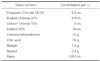
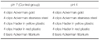
Before starting the mechanical fatigue testing, all materials under study were immersed in artificial saliva. The artificial saliva used in this study was produced in laboratory formula basis, per liter (Table 1).29
The 2 different pH values (pH 4 and pH 7) were obtained incorporating HCl in the base formula, in order to subject them to corrosion, for 30 days, according to the predefined distribution (Table 2).
It was done the placement of the attachments and bars inside glass vials closed containing artificial saliva pH 7 and pH 4, then heating the oven (Memmert®, Duesseldorf, Germany) at 37℃ to simulate the conditions of oral temperature and placement of all the vials containing the sample in the oven for 30 days. There was a control of the pH of the vials 3 times per week for 30 days, using a digital pH meter (ebro® Electronic PTH 810, Ingolstadt, Germany).
After removing the attachments of the oven, it was necessary to incorporate them into small acrylic cubes simulating its location at the base of the prosthesis. That was achieved by making a mixture of thermopolymerizable acrylic (megaCRYL® N, Megadental, Germany) with acrylizing within forms specifically designed to be incorporated in the test machine CS-Dental Testing Machine. Relief of the retention area of the attachments with pink wax (Anutex, Kemdent®, England) and Pan Placement for 10 to 15 minutes at 2 bar pressure.
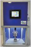
After this time, using the CS- Dental Testing Machine® (Idearum®, Igualada, Barcelona) (Fig. 1), 5400 insertionremoval cycles were simulated (5 years), and wear (removal/insertion values) was registered at 8 different times (initial, 1 month, 6 months, 1 year, 2 years, 3 years, 4 years and 5 years).
The attachment and bar were placed inside two brackets specifically designed for the testing machine. Then, it was run the entire protocol of driving and calibration of CSDental Testing Machine® and the test pieces were positioned inside the machine. Thereafter, it began the 5400 insertion and removal cycles of the attachments on the bar (5 years).
The testing machine has a USB port, allowing the collection of data by using a Pen Drive. Each file, during the execution of the 5400 insertion and removal cycles, registers about 250,000 read points transmitted by the load cell.
That sheet in Excel is treated by selecting the desired cycles for this study, ie, initial, 1 month, 6 months, 1 year, 2 years, 3 years, 4 years and 5 years.
The surface characteristics were also evaluated using an stereoscope (Olympus® SZ61, Tokyo, Japan) with a 90× magnifier.
Statistical analyses was performed to assess the influence of the factors (pH and attachment) on the average value of the insertion force at the eight evaluated times, it was performed ANOVA with repeated measures.
The assumption of normality was assessed using the Kolmogorov-Smirnov test, with a P>.05 for all evaluated time points, according to the pH and the type of attachment.
The assumption of sphericity was tested with the Mauchy test (P<.05), which rejected the sphericity of the data. Because the estimated value of epsilon was less than 0.75, it was used the Greenhouse-Geisser correction for the interpretation of the results for intra-subject effects. All the statistical analyses were performed using IBM® SPSS® Statistics version 22.0 (SPSS Inc., Chicago, IL, USA), and a P<.05 was considered significant.
RESULTS
The retention of the four types of attachments was evaluated by separately analyzing the values of the insertion and removal attachments.
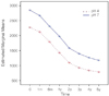
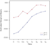
The analysis of the attachments insertion results revealed that there were significant differences in the average values of the insertion force at the different time points; that is, there was significant variation in the average value of the insertion force over time, with this value significantly decreasing over time (Fig. 2).
There were no significant differences in the average values of the insertion force due to the interaction of time and pH (F (1.503; 36.063) = 0.562, P>.05), indicating that the group averages (pH 4 and pH 7) were identical at the eight evaluated times, indicating that the average group values were identical at the eight evaluated times. These findings are reflected by the approximately parallel lines on the chart (Fig. 2).
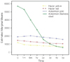
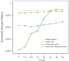
There were significant differences in the average values of the insertion force due to the interaction of time and attachment (F (4.508, 36.063) = 125.369, P<.05), indicating that the group averages (Hader Yellow, Hader Red, Ackerman Gold and Ackerman Stainless Steel) varied at the eight evaluated times. This finding is reflected by the nonparallel lines on the charts (Fig. 3).
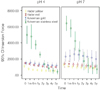
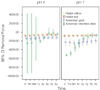
There were also no significant differences in the average values of the insertion force due to the interaction of time with pH and attachment (F (4.508, 36.063) = 2.064, P>.05), indicating that the average group values were identical at the eight evaluated times (Fig. 4).
An analysis of the attachments removal within-subjects effects revealed that there were no significant differences in the average values of the removal force due to the interaction of time and pH (F (1,065; 25.552) = 0.977, P>.05), indicating that the averages at pH 4 and pH 7 were identical at the eight evaluated times (Fig. 5).
There were significant differences in the average values of the insertion force due to the interaction of time and attachment (F (3.194, 25.552) = 0.977, P>.05), indicating that the group averages (Hader Yellow, Hader Red, Ackerman Gold and Ackerman Stainless Steel) varied at the eight evaluated times. The non-parallel lines on the charts (Fig. 6) reflect this finding.
Moreover, there were no significant differences in the average values of the removal force due to the interaction of time with pH and attachment (F (3.194, 25.552) = 1.018, P>.05), indicating that the group averages were identical at the eight evaluated times (Fig. 7).
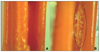
After being subjected to 5400 cycles of insertion and removal, all the attachments and titanium bars were observed with a stereoscope (Olympus SZ61) and a 90× magnifier to evaluate any changes in their surfaces.
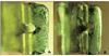
The Hader Yellow and Hader Red attachments, after the final cycle, showed visible wear in their retention loops, with the appearance of erosion zones on the Teflon (Fig. 8) but no apparent wear of the titanium CAD/CAM bar.
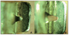
The Ackerman Gold attachments had polished surface zones at the end of testing, particularly at their side loops with the removal of small metal particles (Fig. 9), and the titanium bars had minor abrasions caused by the attachments.
DISCUSSION
In vitro fatigue tests are commonly used to determine the best attachment system for retaining overdentures over time. Pigozzo et al.15 evaluated the retentive strength and fatigue resistance of 4 overdentures bar/clip attachment systems. Forty bar/clip attachment system specimens were tested: Connection Bar Clip, Sterngold Hader Bar, 3i Gold Hader Clip and SIN Clipo. Specimens immersed in artificial saliva were tested for 5500 cycles using a servo hydraulic universal testing machine. Retention strength values were recorded initially and after 1100, 2200, 3300, 4400 and 5500 insertion and removal cycles during tensile testing. All attachments revealed a decrease in retention strength values during the fatigue testing after 5500 cycles of insertion and detachment. No relevant differences in retentive force were found in the groups using polymer clips and between the metal clip systems. The SIN Clipo system demonstrated the smallest retention strength values, which were significantly different from those of the other 2 attachment systems, the Sterngold Hader Bar and the Connection Bar Clip.15
Rutkunas et al.16,17 tested the fatigue of stud ERA Overdenture, Locator Root and OP anchor and magnetic attachments. Three samples of each attachment were used. Micro material testing machine (MMT-250NB-10, Shimadzu Co., Tokyo, Japan) was used to perform 2000 insertion detachment cycles. Retentive force was measured initially and after each 40 cycles.
The authors found relevant differences between the five types of attachments. Decrease of retention was characteristic for all attachments except OP. After fatigue LRP was most retentive. Magnetic attachments preserved maximum initial retention value at the baseline (98%). EO and EW attachments lost 75% and 63% of its initial retention.16,17
They concluded that, along the time, the attachments gradually lost their retention, and also, Stud attachments revealed to be more susceptible to fatigue than magnets.16,17
In our study, we introduced a new variable: the pH of the artificial saliva. Comparative tests were not found in the literature to compare our results with other in vitro studies previously carried out. However, some studies in the field of orthodontics served as a basis to highlight the influence of the pH of the saliva and other enhancers' factors of corrosion in metal ion release and degradation of the materials.
Gürsoy et al.28,30 compared new vs. recycled brackets and bands and found significantly higher release of Ni ions by recycled elements. Kuhta et al.31 investigated the effect of pH (3.5 and 6.8) on ions release in various types of wires produced by the same manufacturer: NiTi, NiTi Thermo, and SS. Dramatically it was found strong effect of pH on release of Ni. The concentrations of ions were 30-40 times higher at pH 3.5 when compared with 6.8. Gürsoy et al. found that recycled brackets released higher amounts of Cr ions. Kuhta et al.31 in the case of Cr confirmed that the mostly biocompatible material (in artificial saliva at pH 6.8 as well as in 3.5) was NiTi.28
All these facts included in these studies led us to deduce that the same corrosion phenomena also occur in attachments constituents of overdentures, causing changes in its durability and ability to retain, variable from patient to patient.
Our test results revealed that there were significant differences in the average values of the insertion force due to the pH, depending on the type of attachment, and in the average value of removal, due to pH and the type of attachment. The results revealed that, under acidic conditions, all the attachments revealed lower values of retention (insertion and removal). These findings should be seriously considered in the future when examining several attachments available on the market, especially in in vitro tests, because the durability of the attachment is influenced not only by mechanical factors of wear but also by chemical phenomena (e.g., corrosion) that vary in the oral cavity from person to person.
CONCLUSION
All the attachments, from both groups, lost retention (insertion/removal) over time.
The more acidic pH value (pH 4) caused a significant decrease in the average values of the insertion and removal forces of all the attachments.
The attachment with the most stable behavior was the Ackerman Gold.
The Ackerman Stainless Steel was the attachment with the highest values of insertion and removal initially.
The highest percentage of loss retention after 5400 cycles was observed in the Ackerman Stainless Steel attachment (pH 4), with 91.47%.
The clinical implications of this study are related to the necessity of shorten the overdenture attachments maintenance, when treating patients with poor hygiene habits, bad eating habits and diseases that decrease the oral pH.
 XML Download