

 PDF
PDF ePub
ePub Citation
 Print
Print


INTRODUCTION
As patient expectations about cosmetic dentistry have been increasing, dissatisfaction among patients with removable partial dentures is also rising because of the unpleasant appearance of metal frameworks, especially with regard to the exposure of a metal clasp. In response to this trend, the non-metal clasp denture was introduced as an alternative in which the clasp is fabricated with a thermoplastic polymer instead of the conventional heat-polymerized polymethylmethacrylate (PMMA) resin.
Non-metal clasp dentures are made of elastic denture base resins instead of conventional metals, and they have the following advantages: enhanced esthetic appeal, no metal-induced allergic reactions, no need for additional prosthodontic treatment or tooth reduction, and reduced chair time.1 Because of such esthetic and clinical benefits, they are preferred for the cases of anterior partial denture, growing patients, temporary dentures after implant surgery, and elderly or handicapped patients who are vulnerable to the complex procedure of conventional treatments.
Polyamide resins, polycarbonate resins, and polyethylene terephthalates have been introduced as representative thermoplastic denture base resin materials for the fabrication of non-metal clasp dentures.2 Of these, the polyamide resin, commonly referred to as nylon polymer, was introduced in the 1950s as a denture base material.345
Polyamide resin is produced from the polymerization of diamine and dibasic acid; while PMMA is amorphous, polyamides are crystalline polymers with properties suitable for high-quality elastic denture materials, such as low solubility, high thermal resistance and flexibility, high strength, and superior moldability.567 Despite these advantages, unlike common PMMA acrylic resins, polyamide resins have difficulties in reline and repair when relining becomes necessary because of resorption of the alveolar bone under the denture base.
To overcome this drawback, the thermoplastic PMMA resin, which is produced by using an injection molding method, has recently been introduced for the fabrication of non-metal clasp dentures. Acrytone, its key component, contains PMMA components that are found in auto- and heat-polymerized relining resins; this allows it to be applied for relining, thereby overcoming the drawback of existing elastic dentures made of polyamide resins.89
The required properties for good denture base materials include esthetic appeal, color stability, dimensional stability, low water sorption, ease of fabrication, easy to repair, and biocompatibility.8910 Of these, color stability has a long-term influence on the esthetics of denture base resins. Denture base resins prone to pigmentation and discoloration are often a cause of patient dissatisfactions and denture refabrication.11 Therefore, in order to ensure satisfactory long-term use of dentures, it is crucial to use denture base materials that can maintain color stability against external pigmentation factors in various oral environments.
Water sorption of a denture base resin can trigger discoloration and halitosis as well as dimensional instability, leading to internal stress and ultimately to cracks or failure of the denture.1213 In other words, a high water sorption rate tends to affect the material properties and consequently reduce the service life of a denture within the oral cavity; therefore, it is crucial to use materials with minimum possible water sorption rates.14
A denture base comes into direct contact with the oral mucosa, therefore, the use of biocompatible materials is essential for avoiding hypersensitivity reactions or the release of toxic substances. A serious problem associated with conventional acrylic resins was allergic reactions triggered by residual monomers after polymerization.15 To address this problem, materials and polymerization methods that reduce or eliminate monomers have been introduced, and the biocompatibilities of denture base materials is considered a very important quality.
These fundamental requirements for denture base materials also apply to the resins used for non-metal clasp denture fabrication to ensure the long-term success of the dental prosthesis and the oral health of the patient. This led to early research into the properties of the denture base materials for non-metal clasp dentures and their long-term stability in the oral cavity environment. However, the related studies still have very limited scope, and only a few studies have explored color stability, water sorption rate, and cytotoxicity in various oral cavity environments. Although the properties of these materials are currently unclarified, they are being used in clinical settings based merely on the preference or experience of the clinician.
Therefore, this study aimed to assess the feasibility of the clinical use of denture base resins for non-metal clasp dentures by comparing the color stability, water sorption rate, and cytotoxicity of the currently used thermoplastic acrylic resins with those of thermoplastic polyamide and conventional heat-polymerized acrylic resins.
MATERIALS AND METHODS
In this study, three types of denture base resins are used: 1) Paladent 20, a PMMA-based conventional heat-polymerized acrylic resin; 2) Bio Tone, a thermoplastic polyamide resin; and 3) Acrytone, a PMMA-based thermoplastic acrylic resin. The compositions of these materials are provided in Table 1.
Specimens fabrication: A total of one hundred five test specimens (35 specimens for each type of denture base resins) in a disc form with 20-mm diameter and 1-mm thickness were prepared according to the polymerization methods specified by manufacturers (Table 2). The fabricated specimens were polished according to the methods recommended for each denture base material type.
Color stability test: Twenty pairs of specimens for each type of denture base resins (total 60 specimens) were used for the color stability test. Specimens of each type were assigned serial numbers of 1 through 20, and the number was written on the side of the specimen that would not be used for the color stability test. These specimens were subdivided into 2 groups (n=10) and were immersed in two different beverages; coffee and green tea.
The coffee solution was prepared by filtering 250 mL of boiled distilled water mixed with 15 g of coffee powder (Maxim original, Dongsuh, Seoul, Korea) 10 minutes of stirring, and the green tea solution was prepared by steeping 5 green tea bags (Dongsuh, Korea) 10 minutes in 250 mL of boiled distilled water.
Specimens of each type of resin were immersed in 50 ml of each beverage in 200 mL glass beakers, while taking great care to avoid contact between the specimens within the beaker. The beakers with the immersed specimens were stored in a dark room at room temperature for 8 weeks.
The specimen colors were measured with a spectral colorimeter (SpectroShade™ Micro, MHT, Niederhasli, Switzerland). The color assessment steps were as follows: 1) baseline color measurement and recording prior to immersion; 2) interim color measurement and recording after 1 week of immersion, whereupon the specimens were removed, rinsed with distilled water, and dried with tissue paper, and 3) final color measurement and recording after 8 weeks, performed as described for 2). Each specimen was measured three times at randomly selected spots and the mean value was recorded to reduce potential error due to measurement at various areas. The differences between the baseline and post-immersion colors were measured according to the CIE Lab system. These differences are expressed as CIE color difference (ΔE) values. The method for calculating these values is described in the following section.16
The resulting color stability data were analyzed by using repeated measures ANOVA to validate the main effects and interaction among materials, solutions, and time dependence. The material-solution interaction effects on the results of 1-week and 8-week immersions were assessed by using two-way ANOVA. The material-dependent differences in color stability after 1-week and 8-week immersions were analyzed by using the Student's t-test. The solution-dependent differences in color stability after 1-week and 8-week immersions were analyzed by using one-way ANOVA, followed by the Newman-Keull's multiple comparison test.
Water sorption test: Ten specimens for each type of denture base resins (total 30 specimens) were used for the water sorption test. Prior to the test, the diameter and thickness of each specimen were re-measured by using an electronic digital caliper to calculate the precise volume (V). The weight of each specimen was measured with an electronic digital balance after storing the specimens in a drying chamber for 24 hours. The weight was recorded when the digital reading remained at a constant value (W0). After measuring the baseline volumes and weights, we stored the specimens in a constant-temperature water tank (37℃) filled with distilled water.
The weights were measured after 1 week and 8 weeks of immersion, whereupon 5 specimens for each type were removed from the tank, dried with tissue paper, and left to stand on the electronic digital balance for 30 seconds before reading the indicated value (W1). We performed a second measurement after the specimens had been stored in a drier for 24 hours after the first measurement was taken; the value was read when the balance remained at a constant value for 30 sec (W2). The water sorption rate was defined as the mass by which each specimen increased per unit volume after immersion,17 calculated as described in the following paragraph.18
The data on the time-dependent water sorption rates measured after 1 week and 8 weeks of immersion were analyzed by using repeated measures ANOVA to test the main effects and interaction of the material and time. The material-dependent differences in water sorption after the same immersion time (1 week and 8 weeks) were analyzed by using one-way ANOVA, followed by Tukey's multiple comparison test, and the time-dependent differences in the water sorption rate for each material were analyzed with the paired t-test.
Cytotoxicity test: Cell viability assay and cell attachment analysis were performed to verify the cytotoxicity of the denture base resins by using fifteen specimens (5 specimens for each). We pretreated the specimens by cleaning them an ultrasonic cleaner filled with pure acetone solution for 10 min, followed by washing with alcohol and rinsing with distilled water. Each sample was then separately wrapped and sterilized in a high-pressure high-temperature sterilizer in preparation for cell culture experiments.
Cell isolation and culture: Human gingival fibroblasts (hGF) were isolated from patient tissue that had been resected for a second-stage implant procedure, and cultured in Dulbecco's Modified Eagle's Medium (DMEM) containing 10% fetal bovine serum (FBS) and 1× antibiotic/antimycotic. The cells were cultured in a cell culture incubator (37℃, 95% humidity) containing 5% CO2. The media was renewed every 2 days. The 5th passage human gingival fibroblasts were used.
Preparations for cell viability test: The cells were seeded in a 12-well plate at a density of 1.8 × 10⁴ cells/well and were cultured for 24 hours in an incubator. The specimens were put into the wells (1 specimen/well) and were cultured for 1 day and 6 days. The cells were cultured in DMEM containing 10% FBS and 1× antibiotic/antimycotic. The EZ-Cytox Enhanced Cell Viability Assay Kit (Daeillab service co., Seoul, South Korea) was used to measure the effects of the test specimens on cell viability of the gingival fibroblasts. The test was performed in compliance with the guidelines provided by the manufacturer. After removing the specimens applied to the cells, 10 µL of EZ-Cytox reagent were added toeach well and cultured the cells for 4 h. Subsequently, the optical density (OD) at a wavelength of 450 nm was measured by using a microplate reader (Bio- Tek Instrument, Winooski, VT, USA).
Preparations for cell attachment analysis: In order to determine the time-dependence of the ability of the cells to attach to the surface of each specimen, the specimens were placed in a 12-well plate and the cultured cells were seeded in each well (6 × 10⁴ cells/well) that contained a specimen. The samples were incubated for 1, 6, and 10 days in DMEM containing 10% FBS and 1× antibiotic/antimycotic. At each measured point, the cells that had been incubated on each specimen were fixed with 2.5 % glutaraldehyde, and the extent of adhesion was observed with a FE-SEM (HITACHI S-4800, Tokyo, Japan) at ×1,000 magnification on 1, 6, and 10 days after incubation.
RESULTS
Results of color stability: The mean ΔE values, which indicate the color differences exhibited by the three types of denture base resin specimens when immersed in coffee and green tea after 1 week (T0T1) and 8 weeks (T0T2), are shown in Table 3.
As a results of the repeated ANOVA, there was no significant interaction effect among materials, solutions, and time dependence (P>.05). Under two-way ANOVA analysis, the material-solution interaction effect on the different times also showed no significant difference (P>.05). Therefore, material-dependent and solution-dependent differences after 1 and 8-week immersions were analyzed respectively by using Student T-test and one-way ANOVA.
Statistical analysis revealed no material-dependent color difference among the denture base materials after 1 week of immersion (ΔET0T1); however, a significantly greater color difference (ΔE) was observed for Paladent 20 and Acrytone in coffee to green tea (P<.001, P=.004 for each), whereas no beverage-dependent difference was exhibited by Bio Tone (P=.103).
In general, greater color differences were exhibited after 8 weeks of immersion. While no significant material-dependent color differences (ΔET0T2) were observed (P>.05), all three types of denture base resin showed greater color differences in coffee than in green tea (P=.003, P=.002, P=.022 for each).
Results of water sorption: The water sorption rates exhibited by the three types of denture base resin specimens after 1 week and 8 weeks of immersion in water are shown in Table 4.
As a results of the repeated ANOVA, there was no significant interaction effect between materials and time dependence (P>.05). Therefore, material-dependent differences after 1 and 8-week immersions were analyzed by using one-way ANOVA followed by Tukey's multiple comparison test,.
Acrytone showed a significantly lower water sorption rate than Bio Tone or Paladent 20 at both measured points (P<.001, P=.001 for each). Although Acrytone and Bio Tone showed a significant increase in the water sorption rate after 8 weeks of immersion compared to at 1 week (P=.045, P=.006 for each), no significant time-dependent difference was observed in Paladent 20 (P=.563).
Results of cell cytotoxicity
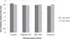
Cell viability: The cell viability test results for each of the specimens are shown in Fig. 1. The cell viability assay after 1 and 6 days of incubation were similar among the three types of resin and the control group, demonstrating unimpaired cell viability in the specimen groups and suggesting that all three types of denture base resin have a very low cytotoxicity. The comparison of the cell viabilities of each three types resin specimen after 1 and 6 days of incubation revealed increased viability in Bio Tone and Acrytone, demonstrating that their cell viabilities increased over time. In contrast, Paladent 20 showed a decreased absorbance on the 6th day, and its cell viability was lower than were those of Bio Tone and Acrytone on the 6th day.
Cell attachments: In SEM observation, Bio Tone showed the smoothest pre-test surface (Fig. 2) and the most efficient cell attachment on 1 day of incubation, while Acrytone and Paladent 20 showed moderate and poor cell attachment, respectively (Fig. 3), and the richest hGF cell attachment on 6 days of incubation (Fig. 4). On 10 days incubation, Bio Tone still showed the most abundant cell attachment, whereas Acrytone and Paladent 20 showed the similar degrees of cell attachment, with Acrytone showing a more stable pattern than Paladent 20 (Fig. 5).
DISCUSSION
Various methods are used for measuring color changes of denture base materials, such as visual assessment, digital image processing, slide projection, visualization of color spaces and assigning orders, and color comparison.1920 A spectral colorimeter was used to detect the color changes, which is a widely used digital image processing method for the denture base color change test because it can rule out errors due to observer bias.2122 Munsell and CIE Lab color systems are commonly used for color difference analysis. We opted for the CIE Lab color system because it is comprised of all colors, including light source colors; further, it is not mediated by human perception. This system is recommended for material color difference tests because it is the most scientific color system.1119
Denture base resins can undergo color changes over time due to intrinsic and extrinsic factors.232425 Intrinsic factors are related to material property alterations resulting from long-term exposure to the temperature and humidity conditions in the oral cavity environment.2425 Extrinsic factors for color change include absorption or adsorption of external materials.2426 Other causes of color change include stain accumulation, dehydration, water sorption, leakage of material components, roughness or abrasion of the material surface, chemical modification or degradation over time, and oxidation, among others.27 Several studies reported that extrinsic factors are more frequent causes of denture base color changes than intrinsic factors.272829 Therefore, we performed an extrinsic factor-related color stability test by selecting two beverage types as test solutions and observing the color changes resulting from the immersion of the denture base resin specimens in the solutions.
Some studies have investigated the pigmentation of resin materials induced by absorption or adsorption.2730 In particular, previous studies reported that acrylic resin or nylon denture base materials show color stability in water or air,1931 but lose color stability when immersed in beverages such as coffee, tea, or wine.9 The tannic acid (C14H10O9) contained in coffee and tea are water-soluble and are known to trigger brown pigmentation.32 The currently available studies on coffee- and tea-induced color changes have reported slightly varying results. A study noted that coffee causes less marked color change than green tea because its brown pigmentation has lower polarity than that of green tea,33 whereas another argued that coffee causes more marked color changes than green tea.29 Another study19 reported that the coffee-induced pigmentation of polymers is caused by the existence of brown pigmentation which has different polarity with that of green tea. Also, the study reported that the coffee-induced color change is more intensive because it involves complex interactions between adsorption and absorption facilitated by the affinity between colorants and materials, whilst the green tea-induced color change is primarily attributable to adsorption. Consistent with this finding, all specimens immersed in the coffee solution showed more intensive color changes than those immersed in the green tea solution at 8 weeks of immersion in our study. The different outcomes of the previous studies that investigated the beverage-related sensitivity to pigmentation presumably resulted from the different resin types and polymerization methods used in those studies, differences in specimen roughness or porosity, and different components or production methods of the beverages used their tests.
Other factors affecting the color stability of materials are hydrophilicity/hydrophobicity,3334 porosity, or surface roughness.135 Materials with high hydrophilicity are prone to pigmentation by the hydrophilic colorants in water-soluble solutions.33 Such denture base resins easily absorb coffee, tea, or wine along with water, resulting in color change. The porosity and roughness of a material surface are influenced by polymerization and polishing methods. In general, heat-polymerized resins can be more prone to pigmentation than thermoplastic resins because heat-polymerized resins have a higher porosity. According to previous studies on the surface roughness of resins, denture base resins of the PMMA acrylic group have a significantly smoother surface than resins of other groups35; further, if polished by using the same method, the polyamide resin shows a more intensive pigmentation than the PMMA acrylic resin because of the greater surface roughness of the polyamide resin. However, no such differences in surface roughness were observed in our study because each type of denture base resin specimens was polished with their own polishing burs according to manufacturer's recommendation, and some polyamide specimens even showed smoother surfaces. These complex issues presumably contributed to the observed lack of significant differences in color changes among the three types of denture base resin immersed in the same beverage solution. These results are consistent with those of previous studies that compared the color stability between heat-polymerized denture base resins and nylon denture base resins.19
The water sorption rate of materials affects their color stability as well as their physical properties. Acrylic resins absorb water for a prolonged length of time because of the polar property of the resin molecules, whereby the extent of water sorption is determined in proportion to the resin components with high polarity, which form hydrogen bonds with water molecules.36 The absorbed water infiltrates into the resin polymers and triggers reversible or irreversible bond breakages between weak molecular interchain bond, thereby causing deterioration of the mechanical properties of the materials, such as hardness, flexural strength, and fatigue limit, as well as dimensional stability.3437383940 Therefore, the International Organization for Standardization (ISO 1567:1999)41 specifies the maximum water sorption ability of heat-polymerized resin materials as 32 µg/mm3. Most of the previous studies on the water sorption abilities of acrylic resins have demonstrated a low water sorption rate of 10-25 µg/mm.316373842 All denture base resins used in our study also showed water sorption rates ranging between 17 and 25 µg/mm3, thereby meeting the pertinent ISO standards.
The earlier polyamide resins had limited properties regarding deformation, water sorption, surface roughness, and polishing.45 Polyamide-group denture base resins are subjected to water sorption between molecular chains due to the hydrophilicity of the many amide bonds that form the main chains of the resins, resulting in high water sorption rates. To overcome this drawback, recently available polyamide-type denture base materials were developed with the aim of reducing water absorption by controlling the amide-group concentration.143 As a result, recent studies using polyamide resins with lowered water sorption rates reported that no differences in mean water sorption rate were observed between heat-polymerized resins and nylon resins.44 Similarly, Bio Tone, of the polyamide group, was resistant to hydrogen bonding, thereby demonstrating water sorption rates similar to those of common heat-polymerized acrylic resins in our study. Further, the presence of residual monomers is reported to affect water sorption and expansion.45 In this regard, Paladent 20 is likely to have more residual monomers than Acrytone because the polymerization method for Paladent 20 employs compression whereas that for Acrytone employs pressure injection. This presumably explains why Acrytone showed a lower water sorption rate than Paladent 20 in our study.
Even polymerized denture base resins can release residual monomers or toxic materials such as formaldehyde, methacrylic acid, and benzoic acid, which can cause irritation, inflammation, or allergic reactions in the oral tissues.13 The amount of such residual monomers are known to be influenced by the type, curing method, and thickness of denture base resins; therefore, the tissue irritation induced by the denture base resin can be reduced by addressing these influential factors.464748 The majority of studies on the cytotoxicity of the resins currently used as denture base materials reported that these resins are not toxic49 and do not suppress cell growth.50 The three types of denture base resin materials tested in our study revealed that they have a negligible influence on cell viability and adhesion; hence, these materials are not believed to be cytotoxic.
This in-vitro study involved a limited analysis of color stability, water sorption and cytotoxic properties for the denture base materials used. For clinical application, further investigations regarding other mechanical and biological properties studies and warranting the long-term effect in vivo were still required.
CONCLUSION
The conclusion of this study indicates that thermoplastic acrylic resins used as materials for non-metal clasp dentures are applicable in the oral cavity environment because their color stability, water sorption rate, and cytotoxicity are similar to those of the thermoplastic polyamide and conventional heat-polymerized acrylic resins. However, to verify the long stability and elastic properties of thermoplastic acrylic resins in the mouth, additional in vitro studies are still needed.


 XML Download