

 PDF
PDF ePub
ePub Citation
 Print
Print


INTRODUCTION
Provisional restorations are an important part of prosthetic therapy procedures with fixed prostheses (i.e., crowns and bridges).1 Provisional restorations serve important roles during tooth preparation and until fitting, luting the final fixed restoration.1,2,3 These include pulpal tissue protection against physical, chemical, and thermal injuries; maintenance of positional stability and occlusal function; and provision of the prepared teeth with strength, retention, and aesthetics, which are essential to clinical success. Polymethyl methacrylate (PMMA) resins and composite-based resins (CBR) are the most common materials used to fabricate provisional fixed dental prosthesis (FDP).2,4,5 Their chemical natures differ; methacrylate resins use liquid/powder and are hand-mixed, and composite-based resins use paste/paste and are usually auto-mixed. The polymerization reaction of methacrylate resins initiates chemically (self-curing), while composite-based materials are available as both self-curing and dual-curing systems.
Fractures are a common cause of failure of provisional restorations. Although restorations should be designed to avoid failure, fractures can still occur. This may cause the patient discomfort and economic loss. Thus, the mechanical strength properties of provisional materials are important and should be considered to ensure the clinical success of provisional restorations.6 Incorrect occlusion, bruxism, undercontoured pontics, and trauma are potential reasons for restoration fractures during usage.3,7 Fractures can occur even during normal masticatory functions, especially when the patient has long-span bridges.8,9
Chemically polymerized materials available for provisional restorations using either PMMA or CBR have unique physical properties that are dependent upon the composition of the chemical monomer. Different monomers have different effects such as exothermic reactions, polymerization shrinkage, marginal fit, periodontal responses, color stability, and fracture strength.10,11 Fracture strength is related to the mechanical properties of the provisional restorative materials.9,11,12 Previous studies on the resins used for provisional restorations compared the mechanical properties of PMMA and CBR; however, the results were controversial.12,13,14
Chairside-fabricated temporary restorations are associated with some shortcomings regarding mechanical strength, surface texture, and fit15,16,17; certain mixing procedures and overfilling of the impression might lead to voids that compromise their mechanical strength.16 Furthermore, after producing these restorations, the flexural strength showed very low values.15
CAD/CAM technologies, which are used to fabricate temporary restorations, may solve some of these issues; resin-based blanks cured under optimum conditions exhibited increased mechanical strength and prevented porosities within the restorations.18 In addition, CAD/CAM-fabricated temporary restorations reportedly reduced the chairside time and produced superior results.19
This study investigated the influence of different fabrication methods and materials on the fracture strength of different provisional crowns.
MATERIALS AND METHODS
The fracture resistance of different materials and fabrication methods was evaluated in a laboratory assembly (25℃, 50% rel. humidity) on a Cr-Co alloy master model with a crown FDP. Table 1 shows the materials tested in this study. All specimens were prepared according to the manufacturers' recommendations.
A non-carious human upper left second premolar (no. 25) was used in this study. The tooth was embedded in a chemically cured acrylic resin block to 2 mm below the cementoenamel junction, and it was duplicated and cast using a Cr-Co alloy (Brealloy C+ B 270, Bredent GmbH, Senden, Germany). Shoulder preparation (convergence angle 6°) was used for the full crown preparation. The prepared tooth was also duplicated and cast using a Cr-Co alloy.
Six types of provisional restorative materials were directly fabricated using the over-impression technique. A vinyl polysiloxane impression material (Imprint™ 3 VPS Impression Material, 3M ESPE, MN, USA) was mixed, and it was placed in the casted master model. After setting, the impression was removed and controlled for any damage to the contour. Afterwards, the temporary crown was mixed according to the manufacturer's recommendations and placed into the over impression. After setting according to the manufacturer's recommendation, the over impression was cut into two pieces, and the temporary crown material was carefully removed. This procedure was used for all six groups (n=10).
The master model was scanned, and the data set was transferred to a CAD/CAM unit (Yenamak D50, Yenadent Ltd, Istanbul, Turkey) for the Cercon Base group. The FDPs were milled and carefully removed from the blocks.
The specimens were controlled for the existence of air bubbles. Inaccurate specimens were discarded and replaced. Ten crowns per experimental group were subjected to deionized distilled water at 37℃ for 24 hours, and then they were thermocycled for 1 week (TC, 5000×, 5-55℃; dwell time 30 seconds, transfer time 2 seconds).
The Cr-Co-alloy-prepared tooth model was placed in a universal testing machine (LS 500; Lloyd Instruments, West Sussex, UK) for fracture testing. The specimens were placed on a model. Specimens were subjected to a compressive load at a 90-degree angle to the center of the specimen until fracture at a crosshead speed of 1 mm/min Fmax was recorded. Fracture patterns were evaluated optically.
The influence of materials and fabrication methods was analyzed by the Kruskal-Wallis test (P<.05). Post hoc comparisons were calculated by Mann-Whitney tests (P<.05). All statistical analyses were performed using SPSS for Windows (12.0, SPSS Inc., Chicago, IL, USA).
RESULTS
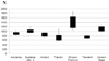
Both the materials and the fabrication techniques had a significant effect on the fracture strength (P<.05). Figure 1 shows the results of the descriptive analyses. As seen by the mean values of the CBR groups, Structur Premium showed the highest values (1392.1 ± 344.11 N), and Systemp c&b II showed the second highest (1009.0 ± 84.50 N). Acrytemp showed the lowest values of the CBR groups (910.05 ± 77.09). The PMMA groups Takilon (711.09 ± 179.18 N) and Temdent (745.23 ± 94.75 N) showed the lowest values. Imident showed the highest values of directly fabricated PMMA groups (843.71 ± 83.46). The only CAD-CAM PMMA group, Cercon Base, showed similar results (1106 ± 134.65 N) to the CBR Systemp c&b II group.
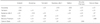
Table 2 shows the significance of the results of the Mann-Whitney U test. As shown, there is no significant difference between the CBR group Systemp c&b II and the CAD-CAM PMMA Cercon Base groups (P=.07), nor between the PMMA group Imident and the CBR group Acrytemp (P=.123). The other PMMA groups, Takilon and Temdent, also showed no significant difference (P=.290). The CBR group Structur Premium and the CAD-CAM PMMA group Cercon Base showed some significance (P=.049). The PMMA group Imident showed significant differences from the Temdent, Systemp c&b II, Takilon, Structur Premium, and Cercon Base groups (P<.05). The CBR group Acrytemp showed significant differences from the Temdent, Systemp c&b II, Takilon, Structur Premium, and Cercon Base groups (P<.05). The PMMA group Temdent showed significant differences from the Systemp c&b II, Structur Premium, and Cercon Base groups (P<.05). The CBR group Systemp c&b II showed significant differences from the Takilon and Structur Premium groups (P<.05). The PMMA group Takilon showed significant differences from the Structur Premium and Cercon base groups (P<.05). The CAD-CAM PMMA group Cercon Base showed highly significant differences from other PMMA groups (P<.05).
DISCUSSION
In the present study, the fracture resistance of seven temporary crown materials was evaluated. While this in vitro study may not reflect the oral conditions, strength values could be a useful predictor of clinical performance and helpful for comparing provisional materials tested in a controlled situation.
A temporary luting cement was intentionally excluded to omit an additional influencing variable. It was assumed that the luting cement would have increased the fracture strength; this subject should be investigated in further studies.
Restorations become weaker when they are exposed to compatible temperature changes (5-55℃) similar to those found in an intraoral environment. In this study, all specimens were placed in distilled water at 37℃ for 24 hours, and then were long-term thermocycled (5000×, 5-55℃, dwell time 30 seconds, transfer time 2 seconds).20,21 This TC is equal to 6 months of clinical use. Some studies have investigated the effect of different storage times on temporary materials.12,22,23 Balkenhol et al.15 found a positive correlation between storage time and mechanical properties. Koumjian and Nimmo9 found similar results and further discovered that dry storage showed higher transverse strength values than wet storage for all materials. Conversely, some previous investigations have shown relatively large increases in the mechanical properties of certain bis-acryl and PMMA interim resin materials between 1 hour and 24 hours storage times.15,24,25,26
Different chemical compositions can be responsible for differences in fracture strength. Conventional methyl methacrylate-type resins are monofunctional; they have a low molecular weight and are linear molecules that exhibit decreased strength and rigidity.27 Indeed, two of the materials that displayed the lowest fracture strengths were methacrylate resins (Temdent and Takilon).
Bis-acryl resin composite materials are difunctional, and thus they are capable of cross-linking with another monomer chain. This cross-linkage provides strength and durability to the material. Two of the materials with the highest fracture strengths were bis-acryl resins (Voco Structur and Systemp c&b II). The CAD-CAM group Cercon Base showed the highest fracture strength among PMMA groups and was higher than the Acrytemp and Systemp c&b II composite groups. The manufacturer of the Cercon Base PMMA stated that the material included highly cross-linked PMMA and was cured under idealized conditions. Thus, the CAD-CAM PMMA material is a more convenient temporary material than the other PMMA groups made by direct techniques.
The higher mechanical strength of acrylic-based temporary crowns compared to traditional monomethacrylates is in concurrence with the literature. Nejatidanesh et al.28 found that bis-acryl provisional materials showed higher flexural strength than methacrylate resins. Lang et al.29 compared two PMMA and four composite temporary materials in an artificial oral environment and found that the highest strength values were accompanied by low fracture rate in the composite-based group.
In this study, all the fractures were seen on the force applied surfaces and all the failures were recorded as catastrophic. Occlusal forces are usually measured in the incisors and molars in a stomatological system. The average values are 250 N in the incisor zone and 350 N in the molar zone, but the values are much higher in patients with bruxism.30 Some studies have shown that these values can rise to 720-815 N.30,31 Two of our PMMA groups (Temdent and Takilon) fractured within these values. For patients with bruxism, clinicians should choose the temporary crown and bridge material with care.
CONCLUSION
According to the findings of this study, composite-based materials offer more advantages than PMMA-based materials in regard to fracture strength. Thus, they should be preferred as a material for provisional restorations. PMMA-based CAD/CAM fabricated provisional crowns show higher fracture strength then directly fabricated crowns. Computer aided design and manufacturing might increase the strength of provisional restorations. Further studies will be supported by a greater variety of temporary CAD/CAM materials.

 XML Download