

 PDF
PDF ePub
ePub Citation
 Print
Print


INTRODUCTION
Bone dysplasias are characterized by the replacement of normal bone with fibrous tissue containing abnormal bone or cementum.1 Familial gigantiform cementoma, which is a subgroup of osseous dysplasias, is a rare condition of the jaw (Table 1).2,3,4 Its true incidence is unknown, as is the gender and ethnic predispositions. The etiology is also unclear, but it is believed to have a genetic component. The familial form is reported as an autosomal dominant trait with variable expression.5,6 The condition may be asymptomatic; in these cases, the lesions are detected radiographically as an incidental finding. The familial form presumably differs from the non-familial form clinically and pathologically.7
Ehlers-Danlos syndrome (EDS) is a group of disorders affecting connective tissues, causing primarily dermatological and joint disorders. The prevalence of the condition varies between 1:10,0008 and 1:150,000.9 EDS is an autosomal dominant inherited disorder, which can be primarily diagnosed on clinical findings and family history.10,11,12,13 The classical symptoms of EDS are joint hypermobility, skin hyperextensibility, fragile and soft skin, the presence of atrophic scars, and easy bruising.10 At least 15 subtypes of the syndrome have been described to date. EDS type VIII (periodontitis type) is characterized with severe periodontitis leading to precocious loss of permanent teeth and alveolar bone resorption.11 The periodontal problems begin with puberty and mostly lead to loss of teeth before the age of 30.10 The facies characteristics are hypertelorism, widening of nasal bridge, and narrow face.12
The present case report is the first known describing concurrent familial gigantiform cementoma and EDS in a single patient. The aim of this report is to discuss the oral management of patients diagnosed with both familial gigantiform cementoma and EDS.
CLINICAL REPORT
Case 1
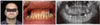
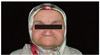
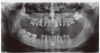
A 34-year-old man self-referred to the Istanbul University Faculty of Dentistry. His chief complaints were tooth loss, difficult mastication, poor esthetics, and periodontal disease. The extraoral clinical examination showed a difference between the proportions of upper and lower facial height and Class III malocclusion was also present (Fig. 1A, Fig. 1B). The examination also revealed temporomandibular joint hypermobility. Teeth 11, 15, 17, 25, 27 and 28 were missing. Tooth 12 was replaced with a premolar, and occlusal contact existed only between teeth 26 to 36. A reduced occlusal vertical dimension was observed by evaluating closest speaking space, proportional face measurement and interocclusal rest space. Panoramic radiographs showed multiple sclerotic masses with radiolucent borders in the mandible (Fig. 1C).
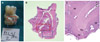
After performing dental prophylaxis, teeth 36 and 46 were extracted because of severe infection and were evaluated histologically. The mineralized material showed apposition and resorption lines, and spherical mineralized tissue (large pulp stones) were free-floating within the pulp chamber (Fig. 2). Based on the clinical and histological findings, the patient was diagnosed with familial gigantiform cementoma with Ehlers-Danlos syndrome type VIII.
The patient had a previous composite restoration on tooth 11 and mottled enamel on teeth 35 and 45. The old restorations were repaired, and the defects were restored with new composite restorations. After healing, irreversible hydrocolloid impressions were made; diagnostic casts and record bases with wax occlusion rims were also fabricated.
The maxillary record base covering all teeth was designed to assess lip support. The occlusal vertical dimension was established using visual observation of the space between the rims when the mandible is in its physiological rest position, judgment of the overall esthetic facial support and phonetic tests.13,14 The tests revealed that the occlusal vertical dimension needed to be increased approximately 9 mm to satisfy esthetic and functional requirements. The treatment options were discussed with the patient. Orthodontic treatment was not recommended because of possible root resorption, and orthognathic surgery was not recommended due to a surgical contraindication. In addition, it was impossible to restore the mouth with fixed prostheses.
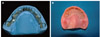
A tooth-supported maxillary complete denture15 to provide lip support and a mandibular overlay removable partial denture (ORPD) were selected as the treatment alternative. ORPDs, a subset of overdentures, are often referred to as a removable partial denture that has part of their components covering the occlusal surface of the abutment teeth to restore them into a functional occlusion.16 The mandibular framework was cast in a Cr-Co alloy with retention beads on the occlusal surfaces for the veneering material. (Fig. 3A).
After intraoral evaluation of the framework, veneering material was placed, and artificial teeth were arranged. Occlusal vertical dimension, esthetics, and maxillomandibular relationship were examined. A heat-processed silicone liner (Molloplast-B) served as the retainer for the tooth supported complete denture (Fig. 3B). It also compensated for the resilience difference between the teeth and soft tissues. The patient used the overdentures for one year with regular reevaluations (Fig. 4). No muscle tenderness, tooth sensitivity, or temporomandibular dysfunction was observed during this period.
Case 2
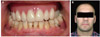
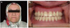
The 38-year-old sister of the first patient also selfreferred to the Istanbul University Faculty of Dentistry. Unlike her brother, she did not have skin hyperelasticity on clinical examination. Head and neck examination showed no abnormalities or facial asymmetry, but the temporomandiular joint did demonstrate hypermobility. A difference between the upper and lower facial height and Class III malocclusion were diagnosed (Fig. 5). Early-onset periodontitis was also noted. Teeth 14, 17, 18, 26, 38, 41 and 45 were missing, besides teeth 36 and 37 were impacted (Fig. 6). The incisors and canines had mottled enamel, and there was only occlusal contact between teeth 15 and 46. Panoramic radiography showed radio-opaque masses scattered throughout the mandible and maxilla. Like her brother, she was diagnosed with familial gigantiform cementoma with EDS type VIII.
All tooth defects were treated with composite restorations. The occlusal vertical dimension needed to be increased by approximately 12 mm. to satisfy esthetic and functional requirements. New tooth-supported complete dentures providing lip support and rehabilitating the decreased occlusal vertical dimension were fabricated (Fig. 7).
In this case, acceptable retention was achieved without requiring soft liner; the dentures were fabricated as described above. The patient used the dentures for one year with regular re-evaluations. No muscle tenderness, tooth sensitivity, or temporomandibular dysfunction was observed during this period.
DISCUSSION
This is the first known case report describing familial gigantiform cementoma associated with EDS.
Few cases have been reported because EDS type VIII is a rare disease17; among these reports, only diagnosis and routine dental treatment were discussed, thus, dental treatment options for patients with EDS remain unestablished. In EDS type VIII patients, loss of the occlusal vertical dimension (OVD) has not been reported, but this may reflect the sparse literature, particularly describing the oral manifestations. Notably, our patients' father also had a Class III malocclusion, and the findings may only be coincidental; more cases are needed to confirm our suspicions. Also notable, the clinical manifestations overlap between the different EDS types, and it is a highly variable clinical entity, presenting a broad clinical spectrum that may also include an increased risk for malocclusion.18
One of the most significant oral features of the syndrome is early-onset periodontitis1,9 which results in premature loss of primary and permanent teeth.8,10 Histopathology suggests that this may be caused by reactive or dysplastic changes in the periodontal ligament7, but radiographically, the lesions adjacent to the teeth appear to have little possibility of originating from the periodontal ligament.7 Although antibiotic prophylaxis was administered in present cases, post-surgical hyperpigmentation and fibrous nodules were detected after the extractions. In the absence of clinical symptoms, re-evaluation with panoramic radiographs every 2 or 3 years is adequate.5
In these cases, the reduced occlusal vertical dimension and negative horizontal overlap were restored with overdentures and overlay removable partial dentures. This is a simple, reversible, non-invasive, and cost-effective solution that resolves the esthetic and functional concerns. Overdentures can be fabricated without any tooth preparation. However, there are several disadvantages associated with them such as increased risk of framework and veneer material fracture.19
Overall, a non-invasive treatment approach was considered the best and most effective treatment option because it resolved the patients' esthetic concerns, improved mastication, and improved speech function.
In the two cases reported, the familial form of OD was symptomatic. This is a very rarely encountered condition in clinical practice; however, the diagnosis is simple, relying on adequate clinical, histological and radiographic examination. The dentist should be able to easily diagnose in order to manage treatment satisfactorily.
 XML Download