

 PDF
PDF ePub
ePub Citation
 Print
Print


INTRODUCTION
For prosthetic treatment, abutments are made of titanium, which is the same metal used in the fixture. However, the final restoration is made of another metal with better castability. Recently, there has been increasing interest in new base metal alloys because of high costs of noble metals such as gold and palladium.1 Base metal alloys have their advantages in terms of strength and costs, but they have a lower corrosion resistance than precious metal alloys. Accordingly, for selected alloys, it is important to evaluate their biocompatibility and corrosion resistance.2
Corrosion can occur in any dental prosthesis, and it may be accelerated by the use of high proportion of base metal, the formation of multiphase microstructures and segregations of elements.3,4 Because each metal has a different electrode potential, if dissimilar metals are placed in contact with an electrolyte, an environment accelerating galvanic corrosion may be created. Then, galvanic corrosion occurs more actively and many metal ions are released if metals have a larger potential difference or poorer corrosion resistance.5
The release of metal ions into the oral cavity can be harmful to the cells of the adjacent tissues, and they may cause side effects including cytotoxicity, allergies, and mutagenesis. Wataha.6 reported that these side effects are influenced by the exposure time and show dose-independent patterns. Schmalz and Garhammer.7 reported that local side effects, for example, gingivitis and periodontitis, occur more frequently when a large amount of metal ions is released. Particularly, elements of base metal alloys such as nickel, chromium, cobalt, and aluminum may cause an allergic reaction in sensitive patients.8 According to Moffa,9 women show higher sensitivity to nickel than men. In the study by Roach,10 allergic skin reactions to metal alloys were observed in 10 to 20% of the patients. Bumgardner and Lucas.11 reported that non-beryllium alloys had a higher corrosion resistance than beryllium alloys and the released metal ions reduced the proliferation of human gingival fibroblasts.
Therefore, the purpose of this study was to evaluate the metal ion release caused by electrochemical corrosion due to contact between metals and to assess the cell toxicity effect. For this purpose, a condition was assumed, in which a prosthesis was made of a base metal on the titanium abutment using three types of Ni-Cr alloys with different components and compositions.
MATERIALS AND METHODS
The three types of Ni-Cr alloys and titanium alloy used in this study are shown in Table 1. A total of 30 Ni-Cr alloy specimens were prepared from three base metals using the following method. The baseplate wax pattern (Daedong industry, Daegu, Korea) measuring 10 × 10 × 1.5 mm in size was prepared and invested in phosphate-bonded investment (CB-30, Ticonium, Albany, NY, USA) and then casted using the conventional lost-wax technique. Fifteen specimens of titanium were cut to the same size with that of base metal. All of the specimens were sandblasted with 250 µm sized aluminum oxide and the process was sequentially finished at 20,000 rpm using tungsten carbide bur (HP194GH50, Bredent, Senden, Germany), stone point (Dura-Green stones, Shofu, Kyoto, Japan), polishing wheel (Polisoft, Renfert, Hilzingen, Germany), and silicone point (Greenie HP PC025, Shofu, Kyoto, Japan). Afterwards, the specimens were ultrasonically cleaned for 15 minutes, sterilized, and stored at room temperature.
The samples were divided into 6 groups of five specimens according to the type of base metal and the contact with titanium (Table 2). In the experimental group (NB+Ti, NT+Ti, N+Ti) the alloys were in direct contact with titanium; but in the control group (NB, NT, N), the alloys were not in direct contact with titanium. The samples were immersed in 6 ml of culture medium - Dulbecco's modified Eagle's medium [DMEM] (Gibco, Co., Grand Island, NY, USA) and kept at 37℃ under an atmosphere of 5% CO2 in air for 48 hours. After 48 hours, the released metal ions were detected by using an inductively coupled plasma mass spectrometer (Agilent 7500A, Agilent Technologies, Santa Clara, CA, USA). The mean concentration of detected metal ions was recorded as parts per billion (ppb).
In this study, mouse fibroblast cells (L-929 mouse fibroblast CCL-1, American Type Culture Collection, Manassas, VA, USA) were used for the evaluation of cytotoxicity.12 The culture medium was made by adding 10% fetal bovine serum, 10 µg/mL gentamycin, 500 unit/mL penicillin and streptomycin into DMEM. Cells were cultured for 48 hours and each aliquot of 1.0 mL was seeded onto 24-well plates by counting 5.0×10⁴ cell/mL per well. After the cells were allowed to settle onto the bottom, the culture medium was removed and 1.0 mL of the solution containing the released metal ions was added to 10 wells for each group. Cells with 1.0 mL of DMEM were the negative control and DMEM containing 10% dimethyl sulfoxide (DMSO, Duksan Pure Chemical Co., Ltd. Ansan, Korea) was added as the positive control. The cells were incubated for 48 hours at 37℃ and 5% CO2.
After the incubation period, the cytotoxicity of the specimens was assessed using the 3-{4,5-dimethylthiazol-2yl}-2,5-diphenyltetrazolium bromide (MTT) assay.13,14 1.0 mL MTT diluent solution was added to each specimen and removed after 4 hours. The resulting formazan crystals were dissolved and the optical density (OD) was measured at a wavelength of 570 nm using a plate reader (Spectramax plus, Molecular Devices, Sunnyvale, CA, USA). The relative cell growth rate (RGR) was expressed as percentage using the following formula:
To compare the amount of metal ions released by galvanic corrosion in each metal, the nonparametric Kruskal-Wallis and Mann-Whitney test were performed after the normality was tested using SPSS statistical software (SPSS 20.0 for Windows, SPSS Inc., Chicago, IL, USA). For the evaluation of cytotoxicity caused by corrosion, the statistical significance between groups was analyzed by two-way ANOVA. Multiple comparison Tukey's test was used for the post-hoc analysis. The statistical significance was verified at a confidence level of 95%.
RESULTS
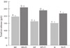
The types and amounts of metal ions released due to galvanic corrosion in each group are shown in Table 3. The ions detected were Ni, Cr, Mo, Be, and Ti; and a small amount of other ions was also detected. The amount of ions released in the experimental group was the largest in the NB+Ti group (265.7 ± 11.0 ppb), followed by the NT+Ti group (236.3 ± 7.4 ppb), and the N+Ti group (216.3 ± 7.9 ppb). Regardless of the type of alloys, a larger amount of metal ions was released in the experimental group than in the control group (P<.05) (Fig. 1). The amount of released Ni ions was increased after corrosion, and the largest amount of Ni ions was released in the NB+Ti group (242.5 ± 12.5 ppb), followed by the NT+Ti group (212.7 ± 7.3 ppb), and the N+Ti group (193.1 ± 4.9 ppb). Among the detected metal ions, a greater proportion of Ni and Be ions was released compared to their composition in the alloys; but the other ions were detected less than their composition.
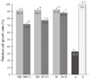
Table 4 summarizes the results of the two-way ANOVA, which showed that the effects of alloy type on MTT activity depend on the galvanic corrosion. On analyzing the difference between the groups by two-way ANOVA, the groups in which the alloy was in contact with titanium or without showed a statistically significantly lower RGR compared to that in the negative control group, but they showed a higher RGR than that in the positive control group (P<.05) (Fig 2). The RGR was significantly lower in the group that showed galvanic corrosion due to contact with titanium than in the other group in which the alloy was not in contact with titanium (P<.05). The lowest RGR was measured in the NB+Ti group (71.8 ± 1.7%), followed by the NT+Ti group (78.4 ± 3.4%) and the N+Ti group (87.9 ± 1.4%). The RGR was not significantly different in other groups in which the alloy was not in contact with titanium.
DISCUSSION
A metal is destroyed by an electrochemical reaction in certain environments, and this procedure is referred to as corrosion. Oxidation and reduction reactions take place at the anode and the cathode, respectively. Corrosion mostly occurs through very complex different processes and it is influenced by the components, microstructure, surface conditions of the metal, electrolyte, concentration of the solution, and temperature. In other words, if dissimilar metals are in contact with each other, there is a wide surface area for contact and there are multiple elements of electrolytes; hence, corrosion takes place more actively.15 Galvanic corrosion occurs more frequently when the potential difference is larger between metals. Moreover, it can be accelerated if the metal is covered with a mucous film or placed under relatively high temperatures and humidity.16 In the oral cavity, a temperature of 37℃ and high humidity are maintained and electrolytes are always present in the saliva. Also, the amount of salivary electrolytes or pH is likely to be changed according to the food intake. Therefore, it can be said that a dental prosthesis is always exposed to a corrosive environment.
Corrosion plays a key role in bone destruction around an implant, reduction of buffering capacity of the surrounding tissue, weakness of restoration, and physiological side effects caused by metal ion release.17,18 Venugopalan and Lucas19 demonstrated the side effects caused by galvanic corrosion in Ni-Cr alloys that were in contact with titanium, and Wylie et al.20 reported a decrease in fibroblast proliferation.
According to these results, a larger amount of metal ions was detected when the alloy was placed in contact with titanium and this may be due to galvanic corrosion. A higher proportion of Ni and Be ions was released in all of the groups than the weight ratio presented by the manufacturer. This finding tended to be similar to the study results for the corrosion character of the metal surfaces.21 In this study, alloys containing Be released a larger amount of ions than non-Be alloys. Beryllium was added to improve both the alloy castability and adherence of veneering porcelain.20 However, it significantly decreases the alloy corrosion resistance owing to the formation of a chromium-depleted Ni-Be eutectic phase.22,23 In addition, high Cr alloys tended to release small amounts of metal ions. This is considered to be because of the addition of Cr which improves the corrosion resistance by forming a protective oxide film on the surfaces.21,24
In all of the groups in which the alloys were in contact with titanium, the rate of cell proliferation was decreased than that in the groups in which the alloys were not in contact with titanium, and this occurrence was regarded to be due to release of ions caused by galvanic corrosion. On considering the corrosion resistance and cytotoxicity, a large amount of ions were released and high cytotoxicity was observed in the Ni-Be alloy with a relatively low corrosion resistance. Conversely, high Cr alloys with a high corrosion resistance showed lower cytotoxicity. The effect of Ni and Be ions on cytotoxicity has already been demonstrated in many studies.25,26 Since less amount of Cr ions was released than that of the other ions and Mo ions have lower cytotoxicity, the cytotoxicity would not be greatly affected.27 These facts support the results of this study that metal ions released by galvanic corrosion can affect the cytotoxicity.
This study had limitations in that the complete intraoral environment was not examined, including saliva, microorganism factors and the presence of adhesive material between the abutment and prosthesis. Also, long-term corrosion was not considered. Taken together, the release of metal ions was increased by galvanic corrosion between different metals and it affected the cytotoxicity, thereby causing side effects on tissues around the implant. In clinical situations, the amount of metal ions released will increase if corrosion progresses over a long period of time. Further research is needed with respect to this issue.
CONCLUSION
Within the limitations of this in vitro study, following conclusions were drawn: The amount of metal ions released was increased by galvanic corrosion in all of the groups in which Ni-Cr alloys were in contact with titanium. Cytotoxicity was significantly increased in all of the groups in which Ni-Cr alloys were in contact with titanium as compared to that in the group in which Ni-Cr alloys were not in contact with titanium. After galvanic corrosion, the amount of metal ions released and cytotoxicity of Ni-Cr alloy with beryllium were significantly larger than other Ni-Cr alloy which not contain the beryllium.


 XML Download