

 PDF
PDF ePub
ePub Citation
 Print
Print


INTRODUCTION
Composite resin restorative materials are widely used in dentistry. Their exposure to the oral conditions requires significant durability. One of the most important problems is degradation of composite restorations in oral conditions.1 The composite resins are degenerated by the effect of pH, saliva, temperature variance, and a wet environment over time.2,3,4 This degradation promotes superficial loss, cohesive fractures, color changes, loss of brightness, and restoration staining5,6 and causes long term clinical failure and esthetic dissatisfaction. Removing and replacing the restorations is the traditional treatment method for the failed (defective, discolored) composite restorations, but the disadvantages of this method are the removal and loss of healthy tooth structure, widening the cavity.7 Crown restorations can be an alternative treatment modality for the failed restorations and composites resins can be used as a core or restoration material for large, defected vital or devital teeth8,9 and teeth, with new or aged composite resin restorations, can be used as a base for crown or bridge restorations such as zirconia.
Zirconia (ZrO2) is used as an alternative to traditional dental porcelains and for the fabrication of posterior fixed partial dentures owing to its good mechanical and aesthetic properties.10,11 It has been widely used in dentistry for fixed partial denture and full crowns, orthodontic brackets, posts, and implant abutments as a core material.12,13 Zirconia has high flexural strength (1000 MPa) and also has optical advantage including color adjustment in which it requires a minimum layering porcelain thickness (compared to conventional ceramics) to obtain the required color.14,15
The bond strength between composite core-resin cement and resin cement-zirconia affects the long term success of the restoration.16 Although having superior mechanical properties (strength, toughness, and fatigue resistance), there are some basic problems associated with zirconium such as cementation.17,18,19 The cementation technique, cement types and surface characteristics of zirconia are important factors for the successful long-term results20 and high bond strength between the zirconia and resin cement is required for better marginal adaptation, retention and high fracture resistance.21,22 In previous studies12,20,23,24,25 it was stated that application different priming agents, cement type, cementation technique, surface treatment methods and surface characteristic of zirconia have effect on the bond strength between tooth and zirconia. However, there is no data about the effect of aged direct composite restorations and surface treatments, applied to composite restorations, on the bond strength between core material and resin cements. For these situations, the bond strength between aging composites and resin cement must be investigated.
The aim of the present study was to evaluate effect of the aged composite restorations and surface treatments, applied to composite restorations, on the bond strength of resin cements. The null hypothesis of the study was that aged composite-resin cores and surface treatment methods do not affect bond strength between the composite resin cores and luting agent.
MATERIALS AND METHODS
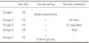
Seventy-five resin composites were prepared with a microhybrid resin composite (Clearfil APX, Kuraray, Kuraray Medical, Osaka, Japan) using a cylindrical mold (diameter: 10mm and thickness: 2 mm). For the 75 specimens, composite material was filled into the mold with one increment of Clearfil APX using a plastic device and composite discs were prepared according to manufacturer's recommendations. Fifteen resin blocks served as a control (intact specimens) (Group 1) and thermocycling (10000 cycles and 5 to 55℃) was applied to the other 60 composite resin blocks for simulating the thermal aging in the oral cavity. After aging, the 60 samples were divided into 4 groups (Table 1).
Group 1: Intact composite: specimens haven't been exposed to any surface treatment (replaced and never been treated composite).
Group 2: Air-flow powder (3M Espe AG/Germany) was applied for surface treatment in all specimens at 90° angle with an air-flow device (Air-Flow Master; EMS, Nyon, Switzerland).
Group 3: 35% phosphoric acid (ScotchBond, 3M ESPE, Seefeld, Germany) was applied for surface treatment according to manufacturer's recommendations, and specimens were dried.
Group 4: Er:YAG laser (Smart 2940D Plus, Deka Laser; Florence, Italy) (2.94 µm wavelength at 150 mJ, 10 Hz, 1.5 W, and 700 pulse duration) was applied for surface treatment and irradiated specimens. Water and air cooling were used during the laser irradiation of the samples.
Group 5: Aged composite: specimens haven't been exposed to surface treatment (Control Group).
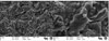
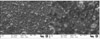
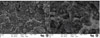
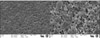
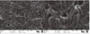
A flexi mold was used for embedding the discs into autopolymerizing acrylic resin (Meliodent, Bayer Dental Ltd; Newbury, UK). After surface treatment, one sample per group was randomly selected and analyzed with a scanning electron microscope (SEM, Noran Instruments JSM 6400; Middleton, USA) at 500× and 2000× magnification.
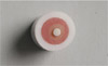
Seventy-five commercially available zirconium core materials (Cercon, DeguDent, Hanau, Germany) were selected for this study. Zirconium oxide specimens (diameter: 2.5 mm; thickness: 3 mm) were manufactured and sintered. Discs were kept in an enclosed condition. Zirconia discs were cemented to the composites discs (Fig. 1) with a phosphate monomer (MDP) based resin cement (Panavia F 2.0, Kuraray, Co. Ltd.; Osaka, Japan) using a cementation jig and 10 N load was applied for 0.5 mm/min.26,27 The curing period was finished according to manufacturer's recommendations.
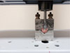
Prepared specimens were kept in distilled water (37 ± 2℃) for 24 h. Specimens were fixed to an universal testing machine (Shimadzu AG-X; Tokyo, Japan) and loaded to failure with a crosshead speed of 0.5 mm/min (Fig. 2). The fractured surface was classified according to one of the 3 types: (1) adhesive failure, (2) cohesive failure and (3) adhesive and cohesive failure.
The data was submitted to Levene Statistics (P<.05) and Shapiro-Wilk Statistics and these tests showed that there was no variance in homogeneity. Therefore, non-parametric tests Kruskal-Wallis and Bonferroni correction Mann-Whitney U test (P<.05) were used to comparison of data. The comparison of failure modes among groups was made with Chi-square test was used to analyzing data. SPSS 20 (IBM, Armonk, NY, USA) for Mac statistical program software was used for data analysis.
RESULTS
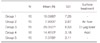
Statistical analysis revealed significant differences in the bond strength values of the groups (P<.05). The mean baseline bond strength values were between 7.07 ± 2.11 and 26.05 ± 6.53 N (mean ± SD). The mean SBS values of the groups and results of multiple comparisons are listed in Table 2. The highest bond strength of 26.05 ± 6.53 N was obtained with Group 3. Group 5 showed the lowest value of bond strength (7.078 ± 2.11 N).
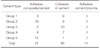
Specimens' failure modes were evaluated. Table 3 shows the distribution of failure mode for the different adhesive systems. Groups 1, 3, and 5 specimens showed cohesive failures. Groups 2 and 4 specimens showed adhesive failures. There were no significant different modes among the groups (P>.05).
SEM images (500 and 2000 magnification) of all composite samples are presented in Fig. 3, Fig. 4, Fig. 5, Fig. 6, Fig. 7. Composite surfaces treated by air polishing, acid etching, and Er-YAG laser are showed in Fig. 2, Fig. 3, Fig. 4. The surfaces treated with Er-YAG laser and acid etching showed irregularities which may provide mechanical retention. The control group (Group 5) has the same surface irregularities and air polishing surfaces the shallow pits remained.
DISCUSSION
The null hypothesis of this study, aged composite-resin cores and surface treatment methods have no effect on the bond strength between the composite resin cores and luting agent, was rejected. Statistically significant changes occurred in the bond strength of the resin-composite cores with aged composites. Different surface treatments of the composite restorations influenced adhesive bonding that occurs between the aged/new composite and luting agent.
In-vitro bonding testing, after long-term oral simulation, is necessary to provide clinical recommendations. In laboratory studies, different methods can be applied for the aging of composite resins. Thermal cycling is the commonly used method for artificial aging in-vitro studies.28 Therefore, in this study, the effects of thermal cycling on the bond strength between the composite-resin cores and luting agent were evaluated. Thermal aging period is controversial in the literature and number of thermal cycles which must be used is unclear.29 In this study, 10,000 cycles were applied for aging of the composite specimens.
The bond strengths were evaluated using a µSBS because it provides a common and simple measurement of the maximum possible stress at the bonding interface.30 The µSBS test performed without sectioning procedure, which may have induced early micro-cracking, so that this method have advantages over the microtensile bond strength.26
Various factors affect the bonding of the aged composite resin including surface roughness, cement type, repair material, and also time after repairing.31
Various surface treatment methods have been applied before cementation and repairing to obtain higher bond strength of the restoration. The more surface roughness results in the better mechanical interlocking. In addition, it is more probable to observe residual free carbon bonds throughout the surface area by increasing the surface roughness.32 In the current study, aged composite + laser surface treatment applied specimens exhibited the highest bond strength compared with the other groups. At the same time, the intact composite restoration exhibited higher bond strength than air polished or acid etched specimens. The specimens which were aged but had no surface treatment exhibited the lowest bond strength. Aged composite + laser surface treatment applied specimens and intact specimens showed more cohesive failure. These results may indicate that with surface treatment adhesive bonding (composite/cement) were improved.
Aged resin composites have a minimum number of free carbon bonds to adhere to a new layer of resin.30 This indicates that surface treatments should be applied on aged composites for optimum bond strength. The bond strength between aged and new composites reduced about 25-80%.33 In our study, the bond strength of the new composite (Group 1) and aged composite (group 5) showed a 60% reduction.
A smear layer has a negative effect on the bond strength of the restorations. Kimyai et al.34 reported that laser applications do not create smear layer and laser irradiation also provides a higher bonding strength after roughening of the substrate surface in which the surface energy and wettability of the adhesive increase. At the end of the laser irradiation, morphological alterations occurred on the surface of the material. These surface alterations can be varied by laser energy, structure, and chemical composition of the composite.35 Furthermore, Cho et al.30 reported that the Er,Cr: YSGG laser did not increase SBS. The differences in the results might be attributed to the differences in the type and mechanism of the lasers used in the two studies. In our study, the laser treatment significantly improved the bond strength of the specimens when compared to the control group. This may result from the increased surface roughness after laser irradiation.
Shimizu et al.36 reported that air polishing causes increasing the surface roughness of the composite. In a previous study, Rinaudo et al.37 concluded that air polishing cannot remove the smear layer; therefore, the bond strength of the restorations decreases. Structure of the powder particles also has an effect (positive or negative) on the adhesion surface. In the present study, the air polishing treatment has no significant effect on the SBS. Spraying time, distance, and type of abrasive powder can have an effect on the surface treatment and surface characteristics.38
Acid etching is a commonly used method for surface treatment of composite resins. However, its effectiveness is controversial. Swift et al.39 stated that acid etching treatment has no effect on the bond strength of composite. Acids with different concentrations and types have been used in studies and various result have been obtained.31,32,33,34,35,36,37,38,39,40,41 Surface treatment with acid alone did not produce remarkable changes in the superficial texture of the composite compared with that of an untreated sample; it seemed to only have a cleaning effect.29 In the present study, acid etching of the surface increased the bond strength of the aged composite. Application method and type of composite resin may affect this result. Burnett et al.40 investigated the effects of laser, air abrasion, acid etching, and silane application surface treatments on the bond strength of composite restorations and concluded that the laser was the most effective for the improving bond strength.
It was stated that the repair of aged and defective composites is a more conservative and economic treatment option.41,42 In addition, Gordan7 reported that replacing composite restorations caused a loss of the tooth structure and widened the cavity. In the present study, the bond strength value of the renewed composite is the highest after laser roughening. However, it is not desired to renew the composite because of the reasons described above. In the clinic, the worst bond strength results from including the composite to the restoration without any surface treatment.
The Er-YAG laser is a conservative treatments option in dentistry35,40,43,44 and laser treatment may be used as surface treatment methods for composite restoration.35 Lizarelli et al.43 reported in their study that composite resins treated with a laser had their polymeric matrix removed, leaving behind an area occupied by the reinforcement particles. Increasing the energy of the laser pulse does not promote general aspect changes, but a bigger alteration occurs in the polymer. In laser applications, the type of the composite influences the outcome of the surface treatment. Hybrid composites are reported to be more convenient for laser application and bond strength than others.44 In the current study, Er:YAG laser-treated specimens and renovated composite specimens mostly cohesive failure, in aged composite and air polishing treated specimens adhesive failures were occurred.
Few laboratory studies evaluating the bond strength between tooth-luting agent and zirconia-luting agent were performed under clinical conditions.18,20,21,22,23,24 Using in vitro tests to evaluate the bond strength of restorative materials is one way to assess their effectiveness. However, shear bond strength test has limitations with regard to obtaining information on the internal behavior of the tooth-restoration complex before failure. The test standards and conditions are not identical to the clinical situation; they allow for comparison of different materials within a given standard. The clinical significance of these findings remains to be determined. Additional in vitro and in vivo studies are required to demonstrate long-term results.
CONCLUSION
Within the limitations of the study, it can be concluded that aged composite restorations and different surface treatment methods have effect on the bond strength between composite-resin cores and luting agent. Surface characteristics of core material are important factors for the successful long-term results and high bond strength between the composite-resin cores and resin cement is required for better marginal adaptation, retention and high fracture resistance. Improved bond strength could be achieved by the different surface treatments and the highest bond strength was achieved with laser surface treatment which applied on aged composite surface. Appropriate surface treatment method should be applied to composite restorations or aged-composites restorations should be replaced for the optimal bond strength and the clinical success.
 XML Download