

 PDF
PDF ePub
ePub Citation
 Print
Print


INTRODUCTION
Resin cements are now widely used in clinical dentistry and improved or new versions are constantly being introduced which are claimed to offer advantages over their predecessors.1,2 The ability of luting multiple structures together, high resistance, less solubility in mouth liquids and colour options makes resin cements an alternative in cementation of esthetical restorastions.3 Adhesive cementation systems have been considered the best option for luting ceramic restorations and the application of resin cement on prepared teeth has demonstrated good biomechanical behavior, particularly with ceramic restorations.1
Due to their chemical structures, resin cements adhere to tooth structure. The bonding of resin to dentin is complex; it's achieved through penetration of hydrophilic monomers to partially demineralized apatite structure of etched dentin. Hence, adhesion is created via micromechanical interlocking of resin to hybrid layer or resin diffusion zone.3
The mechanism of modern adhesion is currently believed to be based on micromechanical interlocking rather than on primary chemical adhesion. Resin infiltration into demineralized dentin permits formation of hybrid layers and resin tags thus producing micromechanical retention.4 Resin tags are formed in the opened dentinal tubules which are intended to contribute to the final dentin bond strength.5 The contribution of resin tags to bond strength relative to the role of intertubular dentin, depends on the orientation of dentinal tubules and dentin depth of tested materials. While the penetration of resin tags into dentinal tubules is believed to contribute little to final bond strength, the adaptation to inner tubule walls probably contributes significantly much more to bonding efficiency. Hybridization by resin interdiffusion into the exposed dentin collagen layer, combined with attachment of resin tags into dentin tubules, appeared to be essential for a reliable dentin bonding.4,5 Current literature showed several contradictory interpretations regarding the formation of tags: some researchers found no correlation between bond strength and formation of resin tags,6,7 while others appreciated that the resin tags may contribute about 30% to the total strength of adhesive-dentin bonding8,9 or at least that the resin tags are a major factor influencing the bond strength.10 Also, it was reported that micromechanical retention of the dentin surface is not adequate without resin tags.11 From that point, it may be thought that resin tags are essential for improving microshear bond strength.
Residual cement and debris might impair etching quality of tooth surface, infiltration of adhesive system, or may even inhibit the polymerization of resinous monomers and thus the fit and final bonding of restoration.12 If proper cleaning of the dentin surface of abutment teeth is not carried out, the interaction between resin cement and dentin may be weakened or lost, resulting in bond failure.
Although several investigators have studied methods for the removal of remnants of provisional cement in vitro,12 there is no research related to the techniques in cleaning of permanent resin cement residuals on dentin in dental literature.
A great number of cleaning agents for dentinal surface, which are based either mechanical or chemical methods have been reported.13,14,15,16 The most common techniques for chemical cleansing include the use of clorhexidine digluconate, sodium hypochlorite, hydrogen peroxide and ethylene diamine tetra acetic acid (EDTA).13 In addition, Endosolv R has been improved for cleaning of resinous materials and resinous remnants from dentin canals by its softening/dissolving ability on resins.17 So, it may be effective on eliminating resin remnants from dentinal tubules in repetitive cementations.
Laser etching of dentin has been reported to yield an anfractuous surface and open dentin tubules, both apparently ideal for adhesion.18 The member of Erbium laser family, the Erbium, Chromium:Yttrium-Scandium-Gallium-Garnet (Er, Cr:YSGG) hydrokinetic laser system has been useful for preparing tooth surfaces for adhesion.11,19,20 Although its efficiency was reported, bond strength of composite resin cement to tooth substrate prepared by Erbium laser are often confusing and contradictory.18 Some researchers noted that lasers may be used to increase shear bond strength (SBS) by treating dentin surface in prosthodontic practice19,21,22 while others found similar bond strength values with or without the application of Er, Cr: YSGG laser.20,23
Considering the previous experiments with Erbium lasers in composite resin removal and roughening of the surface,24,25,26,27 the use of these systems may be applicable for removing adhesive cement remnants from dentinal tubules in repetitive cementation. The purpose of this study was to examine the effect of chemical agents and Er, Cr:YSGG laser operated at different outputs on microshear bond strength between dentin and resin cement in repetitive cementation via eliminating or decreasing the amount of adhesive cement residuals from dentin tubules.
MATERIALS AND METHODS
This study was conducted after approval of Research Ethical Committee of Near East University (Approval no: 79/2013).
A total of 90 non-carious, intact human molar teeth which were extracted within 3 month period, were selected and stored in distilled water at 4℃. Prior to use, teeth were washed under running water to eliminate storage solution residues.
The teeth were embedded in an acrylic resin (Heraeus Kulzer Ltd, Newbury, London) with the occlusal surface of the crown upwards and parallel to the base of resin block. Each sample was sectioned at a level below occlusal pit and fissure level perpendicular to the long axis of the tooth with a diamond blade saw (Precision Sectioning Saw, Isomet 1000, Buehler, IL, USA) on an automated sectioning device under water irrigation, to have superficial dentin cross sections in 1 mm. Then, exposed superficial dentin surfaces were polished with a 600 grit silicone carbide paper (ZiBo Sisho MT Coated Abrasive CO, Ltd, Shandong, China) to create a flat surface with standardized smear layer formation. Afterwards, all dentin cross sections with mesio-distal width facing upwards were fixed on a chemically cured acrylic resin blocks with 2 cm diameter and 1.5 cm height, with cyanoacrylate adhesive (Zapit Base, Dental Ventures of America, Corona, USA).
The dual cure resin luting cement (Variolink N Professional Set, Ivoclar Vivadent, Schaan, Liechtenstein) was applied to the surface of sliced teeth with the use of cylindrical tubes with dimension of 4 mm × 4 mm, according to the manufacturer's instructions. The cement was light cured for 40 seconds with halogen light source which has 450-500 nm wave length and 500 Mw/cm2 power (Hilux Dental Curing Light Unit 250, Benlioğlu Dental Inc., Ankara, Turkey). Two parallel resin cylinders were located on each sample at least 1 mm far from dento-enamel junction (Fig. 1A).
Bonding areas of resins were defined with a permanent marker to help performing cleaning techniques and placing repetitive resin cylinders in appropriate area. Initial debonding was performed, after 24 hours which is required duration for complete polymerization, by removing of resin cement mechanically with a carving instrument without harming any teeth tissue until dentin surface appeared macroscopically clean (Fig. 1B).
Samples were randomly assigned to 6 groups of 30 specimens each, according to cleansing procedures on removal of adhesive remnants which is listed in Table 1.
Any surface treatment was not applied to samples in Group 1 (control group). The samples in Group 2 and Group 3 were treated with organic solvents. 17% concentrated ethylene diamine tetra acetic acid-EDTA (Henry Schein Inc., NY, USA) or Endosolv R (Septodont, Cedex, France) which is consist of 66.5 g formamide and 33.5 g phenylethyl alcohol, was applied all over the marked areas of the samples. Following step for either Group 2 or Group 3 was rinsing of organic solvents and drying teeth with absorbent paper to avoid dentin dehydration.
An Er, Cr:YSGG laser (Waterlase MD, Biolase Technology Inc, CA, USA) with wide output power range between 0.1-8 W, operating at a wavelength of 2,780 nm was used for Group 4, 5 and 6. The laser energy was delivered through a fiber-optic system to sapphire tip terminal 600 µm in diameter (MMG6, Biolase MD Technology Inc., CA, USA). Samples were lased at 1.25 W (Group 4), 2 W (Group 5) and 3.5 W (Group 6) output power for 15 seconds in non-contact mode with the angle of 85°-95° to the flat specimen surface with a 1 mm fixed distance from the laser tip. A sweeping motion was used to achieve an even coverage of surface by overlapping the laser impact. To standardize the distance, 1 mm acrylic disc was used. Holes, 4 mm × 4 mm in size were constructed on the surface of 1 mm acrylic disc. The acrylic disc was placed on the teeth surfaces for all specimens which were irradiated, to avoid unnecessary laser irradiation. Also, all laser treatments were performed by same operator.
Investigation of treated dentin surfaces was performed by Scanning Electron Microscopy (SEM) (QUANTA™ 400F Field Emmision SEM, Tokyo, Japan). Two samples for each group were selected randomly.
The specimens had their surfaces desiccated and coated with Au-Pd alloy for SEM examination at × 1,500-2,000 magnification.
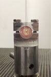
A tube with an inner diameter 1 mm and height of 1 mm was attached to each of the treated or untreated (control group) dentin surfaces. Resin cement (Variolink N, Professional Set, Ivoclar Vivadent, Schaan, Liechtenstein) was applied and polymerized for 40 seconds with halogen light source (Hilux Dental Curing Light Unit 250, Benlioğlu Dental Inc., Ankara, Turkey) (Fig. 1C, Fig. 2). Manufacturer's recommendations were strictly followed during bonding procedures. Repetitive cementation was performed in all samples same as the initial application. After the cementation was completed, the tubes were carefully removed.
Prior to Microshear bond strength (µSBS) test, the specimens were stored in distilled water at 37℃ in an incubator for 48 hours. µSBS tests were performed by using an universal testing machine (EZ-test 500 N Shimadzu, Kyoto, Japan) and a computer program (Nexygen Software, Lloyd Inc., Leicester, UK). Samples were loaded to failure under shear using a wire loop loading (Fig. 2) head at a cross head speed 1 mm/min until the composite resin cylinder was dislodged from tooth surface. Samples were oriented in a holding vise so that the direction of shear testing was parallel to the flat surface and the loading head is in contact to the bonded tooth surface. The maximum load to failure was recorded digitally with a computer for each sample. µSBS was calculated according to the following formula and expressed in MPa: Stress = Failure Load (N) / Surface Area (mm2).
After µSBS testing, failure modes were assessed for each specimen under a light microscope (Olympus SZ 40, SZ-PT, Tokyo, Japan) at × 100 magnification and classified as: adhesive, cohesive and mixed (both adhesive and cohesive) failure.
All statistical analysis were performed with Statistical Package for Special Science SPSS 18.0 software (SPSS Inc., Chicago, IL, USA). Descriptive statistics, including the mean, median, standard deviation, minimum, and maximum, were calculated for each of the six groups. After analysis of the normal distribution of µSBSs of samples with Shapiro-Wilk (sig) test and test of homogeneity of variances, comparisons of the medians of µSBS in six groups were carried out with Kruskal Wallis H Test with Bonferroni correction. Statistical significance was considered as P<.05.
RESULTS
The topography and morphology of treated dentin surfaces were investigated by SEM. The control group showed scratches, fundamental smear layer and plugs in the SEM observations of untreated dentin (Fig. 3A). SEM revealed, diffuse cement layer particularly removed in small insular areas in Group 2. Also, some smear was observed in these areas (Fig. 3B). In Group 3, there were sporadically closed dentin tubules (Fig. 3C). Cement was perforated by laser in few points in Group 4 (Fig. 3D). But there were areas which had not interacted with laser. Laser is seemed to be locally efficient on those areas. Fig. 3D shows particularly opened dentin tubules by laser. Fig. 3E presented sequent circular areas which were formed by laser for Group 5. When the circles were examined, some areas were found to be perforated up to enamel border and also dentin tubules were open. In addition, even dentin and opened dentinal tubules without the presence of cement remnants were appeared. Laser almost exposed even dentin and few open dentinal tubules in Group 6 (Fig. 3F).
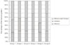
Mean microshear bond strength ± standard deviation (MPa) for each group was 34.9 ± 17.7, 32.1 ± 15.8, 37.8 ± 19.3, 31.3 ± 12.7, 44.4 ± 13.6, 40.2 ± 13.2 respectively.
When the two by two comparison had been done among 6 groups (P<.05), Group 5 revealed statistically significant difference from Group 1, Group 2 and Group 4. Also, comparison indicated that Group 6 has statistically different microshear bond values from Group 4.
No statistically significant difference was found between remaining groups (P>.05).
DISCUSSION
Bond failure during prosthetic treatment may be relatively frequent and undesirable. It is important to better understand what to expect when a tooth is debonded more than once. In a study, findings indicated that shear bond strength during second bonding/debonding sequence significantly decreased approximately 61% due to cement residues.27 In present study, the effectiveness of Er, Cr:YSGG laser in repetitive cementation for cleaning of adhesive cement remnants from dentin tubules was compared with some other methods. This comparison was held with µSBS test results.
Bond tests are used for examining resistant of material by imitating the possible tension forces which the restorations would be exposed in oral circumstances.28 Shear bond strength testing with bonded cross-sectional areas of 1 mm2 or less is also referred to as microshear bond strength. This relatively simple test permits efficient screening of adhesive systems, regional and depth profiling of a variety substrates and conservation of teeth. In addition, testing of multiple specimens from a single tooth conserves teeth and allows research design which is not possible using conventional macro methods29 that is why microshear bond test was preferred rather than shear bond test in this study.
Generally, there is variability among the dentin bond values reported by various researchers.19,20,22 This may be attributed to different testing methods and conditions, the varying nature of dentin substrate and the composite adhesive used. Morphological and structural variations in dentin may influence the bond strengths of the adhesive systems to dentin.30 Researchers noted that most of the adhesive systems gave higher microshear bond strength to superficial dentin and they mentioned progressive decrease in bond strengths to deeper dentin.31,32
In addition, it was shown that age factor may affect bond strength.33,34,35 For this reason, superficial dentin layers from the teeth which were collected from the patients with the age group between 20 and 30 were used in present study for both standardization and higher bond strength. Also, teeth were cut with micro saw under water cooling with diamond burs to avoid sudden increase in temperature rate and samples were kept in distilled water at 4℃ until usage.36
Various techniques involving organic solvent application were used for removing cement residues. In a study, EDTA was effective in removal of the remnants of provisional cement, so EDTA was considered to create appropriate physical and chemical interactions between the resin cement and dentin.15 With regard to this finding, EDTA was preferred as one of the dentin cleansing agents in this study.
According to some researchers, 17% EDTA usage did not significantly alter or decrease the µSBS values14,37 while others concluded that dentin treatment with EDTA is effective in improving bond strength.15,38
However, in the present study, results indicated that EDTA did not show significant difference in µSBS from control group so it was found ineffective in dentin cleansing. Moreover, concurring with previous study14 samples in EDTA group showed more adhesive fracture pattern compared to the untreated (control) group in failure of mode analysis.
On the other hand, Endosolv R group showed higher µSBS than control group and also comparable mean to 2 W and 3.5 W laser groups. Also in a study, it was conducted that Endosolv R prevented deterioration of resin-dentin bond strength17 while others concluded it had no positive effect on the bond strength.39 Therefore it may be conceivable to use this organic solvent rather than laser systems which have much incommensurable cost. In addition, usage of Endosolv R is easier than laser application. Although Endosolv R was found to have superior penetration40 and eradicate residual materials,41,42 manufacturer suggested two-visit application for better utilizition40 which is a time consuming. With respect to micromorphological changes in lased dentin surface, SEM analysis figured out better findings than Endosolv R group which 2 W and 3.5 W laser groups were completely lacking out of smear with opened dentinal tubule orifices without any widening. Moreover, it is prudent to point out that the formamide which is one of the co-solvent of Endosolv R, is a potential teratogen. Thus, female workers/patients of child-bearing age must be made aware that a known teratogen is being used and appropriate 'right-to-know' compliance policies be adhered to by providing them with data from appropriate animal studies.17,43 Besides, there are limited numbers of clinical reports which conducted on usage of Endosolv R, in the dental literature so further investigations are mandatory.
Currently, many studies have been focused on the evaluation of the effectiveness of Er:YAG and Er, Cr:YSGG lasers on bond strength between dentin and cement.11,19,21,22,23,24 Er:YAG and Er, Cr:YSGG lasers can selectively effect on the surface of dentin, resulting in an irregular surface pattern that may potentially improve the micromechanical retention of adhesive systems to dentin, when used at appropriate doses. The laser system used in this study was Er, Cr:YSGG which is a hydrokinetic one. The main advantage of lasers was avoiding immediate increase in temperature which may result in an inflammatory pulpal response. With this system not only could the temperature be suppressed, but also cutting efficiency could be increased. Laser energy is delivered through a fiber optic system to a sapphire tip terminal.19,22,44 The average output can be varied from 0.1 W to 8 W. For dentin irradiation, several investigations have suggested irradiation outputs varying from 1.25 W to 5 W can be used.19,21,22,38,44 In a study it has even used the maximum output power of 5 W,19 providing increased value of energy density and as a consequence, a higher efficiency of laser ablation. However, use of higher values of energy density has also been related to a more harmful thermal effect on dental hard tissues, particularly when laser irradiation is nor accompanied by a continuous air water spray. The presence of micro cracks under partially attached dentin particles may provide a weakened substrate, which is more prone to the occurrence of fractures during bond strength testing.44 To avoid these negative effects, we used continuous air-water spray continuously at 65% and 55% percentages, respectively.
In the study, in order to irradiate the dentin, 1.25 W, 2 W and 3.5 W outputs were used. The varying power outputs produced different patterns. Laser irradiation with high power outputs show higher µSBS means while lower laser outputs demonstrated significantly lower µSBS and closed dentinal tubules.
In laser groups, Kruskal Wallis H Test detected significant difference between the µSBS means of 1.25 W lased dentin surfaces and other 2 groups, while there was no any significant difference between 2 W and 3.5 W laser application.
1.25 W laser irradiation showed statistically no significant µSBS value than the control group. Commercial dental laser manufacturers claim that enamel and dentin can be successfully etched at lower power settings with the Erbium lasers. Apel et al.45 investigated the ablation threshold of Er:YAG and Er, Cr:YSGG lasers used when preparing tooth structures and noted that there is a possibility of micro cracks developing in dental structures below the ablation threshold. These cracks act as starting points for fracture and failure, which may reduce or eliminate the possible positive effect of Erbium laser irradiation. What is more, researchers considered power setting of 1.5 W is the lowest power setting that is able to thoroughly treat the dentin surface, as assessed by light microscopy.19 The µSBS values of 1.25 W laser applied group which significantly differ from the other 2 laser groups may be explained by this theory.
The literature had shown that surfaces irradiated by 2 W Er, Cr:YSGG laser displayed rough surface.46 In the present study, it may be thought that laser act as an etching agent to make rough dentin surface and therefore cause an increase in the µSBS values of Group 5. However, in previous studies, researchers claimed that laser irradiation with 2 W, 3.5 W and similar doses had no positive effect on bond strength between dentin and cement,11,22,47 in other words they pointed out that there was no increase in etching. Moreover, Moretto et al.48 concluded that Er, Cr:YSGG laser irradiation interacts with the dental hard tissue resulting in a specific morphological pattern of a dentin and collagen fibrils that negatively affected the bond strength to resin. At this point, it may be concluded that, the procedure done in this research primarily was not etching with laser, it was cleaning of resin cement remnants from dentin tubules that led new resin tag formation in repetitious cementation which increased µSBS.

 XML Download