

 PDF
PDF ePub
ePub Citation
 Print
Print


INTRODUCTION
Studies on the facial morphometry and mandibular movements are an essential element in a scientific approach for treating temporomandibular dysfunction and are useful in the practice of orthodontics, restorative esthetic dentistry, maxillofacial surgery, and prosthodontics. In prosthodontics, measurements of the face are often used as a guide for tooth size, and are helpful in achieving the desired amount of face and lip support, tooth exposure, and sometimes in determining the correct vertical dimension of occlusion.1,2,3,4
The size and the form of the face have been shown to have a positive relationship in many instances to the size and form of the teeth. On 555 dentulous subjects, House and Loop2 found that the greatest bizygomatic width of the skull divided by 16 gave an estimation of the width of the upper central incisor, and when divided by 3.3 gave an estimation of the width of the upper six anterior teeth when carded flat. The importance of seeing the face in its normal proportions has been emphasized.5 The neoclassical canons are known to most plastic surgeons. The neoclassical canons of the face are, the forehead height (tr-n), nose length (n-sn), and lower face height (sn-gn), are same length.6 Farkas et al.7 studied the lower face in young Caucasian adults and reported that the lower lip occupied one third of the lower face. Measurement of the face has been performed both direct and indirect anthropometry. Current technology includes digital image analysis systems, threedimensional space range-camera techniques, stereo-photogrametry, laser scanning, and optoelectronic systems supplying the digital coordinates and landmarks of interest, non-invasively.7,8,9,10,11
The occlusal plane represents the planar mean curvature of the opposing occlusal surfaces. Establishing the correct occlusal plane and proper vertical dimension of occlusion are very important procedures in the fabrication of complete dentures. Several different reference points have been suggested to help determine the location of the occlusal plane. Nagle and Sears12 suggested parallel to and midway between the residual ridges. Lejoyeux,13 advocated parallel with the resting upper lip and parallel with Camper's line. Ismail and Bowman14 suggested the upper third of the retromolar pad parallel to the lateral border of the tongue. Hickey et al.15 advocated parallel with the interpupillary and ala-tragus lines.
The range of mandibular movement is an important measurement in the examination of patients with suspected functional disorders of the masticatory apparatus. Also, in healthy subjects with normal function, the determination of mandibular mobility is considered to be an essential part of any physiologic study of the stomatognathic system.16,17 Posselt18 defined the border movements of the mandible. Functional mandibular movement differs from border movement. Determination of the maximum jaw openings and excursions is a valuable and simple method for assessing proper function of the masticatory system. Visser et al.19 described a relationship between the opening capacity of the mandible and the function of masticatory muscles. There are many reported studies regarding the maximum jaw opening and excursion capacities.20,21,22,23,24,25
This study measured several recognized facial landmarks and determined mandibular excursion capabilities on a large group of young dental students hoping to provide them with a meaningful clinical experience.
MATERIALS AND METHODS
Measurements were performed on each other by 316 firstyear dental students at The Ohio State University during 1969-70. Six percent were females. Their average age was 22.5 (20-33) years, average height was 180 cm (155-203 cm), and average weight was 76 kg (57-107 kg). Eighty (25.3%) had undergone orthodontics. Thirty-seven (11.7%) had minor bruxism, fifty-nine (18.7%) exhibited crepitus, and eighteen (5.7%) reported minor temporomandibular joint discomfort during maximum jaw openings. For most measurements, the subjects were seated in a chair with the examiner sitting in front of them. The head of the examiner was level with the head of the subject. All measurements were carefully verified by the authors.
Facial measurements
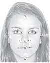
The dental students recorded 5 vertical and 6 horizontal facial measurements on each other's face (Fig. 1). The five vertical measurements were: ① trichion to gnathion (height of the face, tr-gn), ② endocanthion to subnasale (en-sn), ③ subnasale to stomion (upper lip height, sn-sto), ④ stomion to gnathion (height of the mandible, sto-gn), and ⑤ subnasale (sn) to central incisor edge (upper lip height plus amount of incisor exposed below the lip). A sixth vertical distance from subnasale to gnathion (height of the lower face, sn-gn) was calculated from the other measurements because it is often referred to in prosthodontics.
The six horizontal distances recorded were: ① widest skull base width (t-t), ② face width (zy-zy), ③ distance between the center point of the pupils (interpupillary distance, p-p), ④ nose width (al-al), ⑤ width of the mouth at rest (ch-ch), and ⑥ distance between the cusp tips of maxillary canines.
A vernier caliper was used to measure the nose width, the distance between the canine cusp tips and for verifying the interpupllary and some other distances. Face widths and lengths were recorded using a facebow.
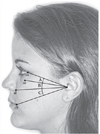
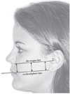
The angles between exocanthion (ex), orbitale (or), ala (al) and cheilion (ch) from the tragus were measured on the left side of the subject's face using a protractor (Fig. 2). Marks were placed on the tragus and lower border of the ala and a line connecting the dots was drawn on the face with a very soft lead or finely pointed indelible pencil. Then each subject placed a tongue depressor between the teeth on the left side so the wooden depressor extended about 25 mm forward between the lips. A second line was drawn on the side of the face continuous with and parallel to the protruding tongue depressor. The distance between two lines was measured three places; near the ala, at a midpoint, and just anterior to the tragus (Fig. 3).
Measurements of mandibular movements
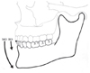
Measurements of mandibular movements involved recording the amount of horizontal and vertical overbites of both the central incisors and the canines on dental stone casts. A plastic millimeter ruler was used and the distances were recorded to the nearest half millimeter. The amount of maximum normal and hinge opening (Fig. 4) was recorded between the subject's upper and lower incisal edges with the jaw open and then adding this amount to the vertical overlap of the incisors. The maximum protrusive movement of the mandible was measured between the facial surfaces of the upper and lower central incisors in the mouth (Fig. 5) and then adding this amount to their horizontal overlap for the total. The maximum amount of lateral excursion on each side was measured between the facial surfaces of the upper and lower canines with the mandible on that side. Then the horizontal canine overlaps on each side, was added to the distance recorded in the mouth on that side, to determine the total lateral movement (Fig. 5).
All measurements from the 316 dental students were compiled for the average and highest and lowest recorded distances (range). Once the data had been studied, (Table 1, Table 2, Table 3, Table 4) the interesting and practical results were reported back to the students as part of their freshmen course in occlusion.
RESULTS
Facial measurements
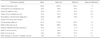
Six vertical and six horizontal dimensions of the face were summarized in Table 1. Trichion to subnasale (tr-sn) was 120.0 mm and endocanthion to stomion (en-sto) was 68.8 mm. Subnsale to gnathion (lower face, sn-gn) was 70.0 mm and endocanthion to gnathion (lower half of the craniofacial height, en-gn) was 114.2 mm. The minimum to maximum ratio of upper lip length (sn-sto) measurements was as much as 2.3 times the average whereas it only varied 1.3 times on the interpupillary distances measured (p-p).
The mean size of the angles (Fig. 2) between exocanthion, orbitale, ala and cheilion were given in Table 2. Angle A (ex to or) averaged 11.9, angle B (or to ala) 13.6, and angle C (ala to ch) 13.6 degrees. The average distances between the ala tragus to line and the occlusal plane line was 31.3 mm near the ear and 29.9 mm by the nose (Table 3). The distance by the ala was recorded as largest on 19.2% and the tragus distance was largest on 58.7% of the subjects. One unique finding on two students was that they found the ala distance to be 12.0 mm longer than near the tragus, meaning that their Camper's lines were less parallel to the occlusal plane than for the majority of their classmates.
Measurements of mandibular movements
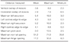
The average measurements of the vertical and horizontal overlaps of the incisors (3.6 mm and 2.9 mm respectively), and the maximal mandibular excursions were summarized in Table 4. Maximum normal jaw opening at the incisors averaged 51.2 mm or 3.0 times more than the maximum hinge opening (17.2 mm). The combined maximum left side (9.4 mm) and maximum right side (9.2 mm) lateral excursions gave a total side-to-side mandibular movement of 18.6 mm, which was 2.3 times as far as the average maximum forward protrusion (8.0 mm) of the mandible. Forward movement of the mandible so that the upper and lower central incisors were edge-to-edge was on the average 36.3% of the maximum protrusive movement. Movement of the mandible so the canines were edge-to-edge averaged 45.2% of the maximum lateral excursion on the left side, and 44.0% of the maximum right lateral excursion.
DISCUSSION
The face is the most variable part of the body. It permits distinction between races, ethnic groups, sexes and even members of the same family. Variability is manifested by the different sizes and shapes of individual features and even more by the relationships of features to each other.26
In dentistry, restoring normal facial esthetics is one of the main goals as well as fine-tuning occlusal function. There are many traces from the Greek era trying to establish esthetic criteria of the face.10 Da Vinci27 had divided the face vertically into two equal parts by the endocanthion (v-en = en-gn) and divided the face vertically into the three equal parts (tr-n = n-sn = sn-gn). In the author's study the average of two measurements (tr-en) and sn-gn) differed by only 1.2 mm (44.2 and 45.4), and (tr-sn and en-gn) differed by only 5.6 mm (119.8 and 114.2 mm). Francesca28 divided the lower face into the three equal parts: upper lip, lower lip, and chin (sn-sto = sto-sl = sl-gn). In the current study, the average length of the lower face (sn-gn) 70 mm, was 37% of the height of the face (tr-gn)189.8 mm, and was 2.85 times larger than the upper lip length (sn-sto) of 24.6 mm. These results are similar to the neoclassical canon and Francesca's opinion on the criteria of facial esthetics.28
Farkas et al.7 reported the craniofacial norms in nineteen to twenty-five year-old North American Caucasian men and women (109 male, 200 female). Among these norms: the width of the face (zy-zy) was 139.1 mm to 130.0 mm (male to female), the height of the face (tr-gn) was 187.2 mm to 173.3 mm, the height of the lower face (sn-gn) was 72.6 mm to 64.3 mm, the height of the mandible (sto-gn) was 50.7 mm to 43.4 mm, and the lower half of the craniofacial height (en-gn) was 117.7 mm to 102.7 mm. The width of the nose (al-al) was 34.9 mm to 31.4 mm, the width of the mouth (ch-ch) was 54.5 mm to 50.2 mm, and the length of the upper lip (sn-sto) averaged 22.3 mm to 20.1 mm. In comparing these measurements with our current study, most of Farkas et al.'s measurements were very similar. However, the height of the face (tr-gn), the height of the upper lip (sn-sto), and the width of the nose (al-al) were comparably larger than Farkas et al.'s. In the current study, the average length of the face 189.8 mm (tr-gn) was 1.44 times larger than the width of the face (zy-zy) 131.2 mm. They reported that the size and form of the artificial anterior teeth harmonized with the shape of the patient's face (ref 8-10). In the current study, the face width (zy-zy) 131.2 mm was 3.8 times larger than the distance between the maxillary canine cusp tips (34.8 mm).
An estimation of the position of the apex of the upper natural canine can be found by an extension of parallel lines from the lateral surfaces of the ala of the nose onto the labial surface of the upper occlusal rim.3 In this study, the width of the nose (al-al) 35.2 mm was as same as between the cusp tips of maxillary canines 34.8 mm. Although these two comparisons are frequently used to select the size of denture teeth, one dental student's nose was 29 mm wider, and another's was 11 mm narrower than the distance between the maxillary canine cusp tips. Consequently, even though the averages of these distances were almost equal (35.2 and 34.8 mm), such a comparison is relatively useless for selecting tooth size or for positioning upper canines on an edentulous patient. The average 24.6 mm upper lip height or length was the most variable dimension measured, whereas the 62.6 mm distance between the pupils was the most stable dimension among eleven facial measurements recorded.
In this study, the average length of the face 189.8 mm (tr-gn) was 1.44 times larger than its 131.2 mm width (zyzy). The average height or length of the face (tr-gn) 189.8 mm was 2.71 times larger than the 70 mm height or length of the lower face (sn-gn). The 70 mm average length of the lower face (sn-gn) was 2.85 times larger than the 24.6 mm upper lip length (sn-sto). The average length of endocanthion to stomion (en-sto) 68.8 mm was the same as the average length of the lower face (sn-gn) 70 mm. These two distances are often compared in selecting the proper vertical dimension of occlusion on edentulous patients. The position of the natural maxillary central incisors in this study averaged 1.2 mm longer than the upper lip. Many prosthodontists would agree that this amount of upper tooth exposure below the upper lip, is a useful guide in determining the upper tooth position and proper length on an edentulous patient.
In assessing the validity of Camper's plane (ala-tragus line) as a guide to determine the position of the occlusal plane line on edentulous subjects, the students drew two lines on the side of the face, and measured the distance between the lines three places: near the tragus, near the ala, and at the midpoint (Fig. 3). The mean of these three measurements was quite similar (Table 3). So, Camper's line or plane and the occlusal plan e are for all practical purposes parallel.
Three angles were established by drawing lines on the face connecting well known landmarks (Fig. 2), so they could be measured using a protractor. The average size and range in size of these angles are shown in Table 2. Angles A, B. and C, on the average were almost the same size (11.9, 13.6, and 13.6 degrees respectively). The smaller and larger angles seen under "minimum and maximum" in Table 2 merely represent normal variations of facial and structural features.
We measured both the maximum range and functional range of mandibular movement. A reduced range of movement of the mandible may be a sign of a disorder of muscular and/or temporomandibular joint function. In the current study, average vertical incisal overlap was 3.6 mm and average horizontal incisal overlap was 2.9 mm. These overlaps are similar to measurements reported by Woelfel at The Ohio State University (from dental casts of 796 dental hygiene students during 1974-1986.29 His measurements reported an average incisal vertical overlap of 3.3 mm and a horizontal overlap of 2.8 mm. On 318 male dental students, the vertical and horizontal overlaps were 3.6 and 2.9 mm respectively.26 Women's facial structures, dimensions, heights, and weights, averages about nine percent smaller than men's.8,9,10,26
Posselt20 reported a maximum opening capacity of the mandible between 50 and 60 mm. Ingervall21 found in a study on 60 women aged 20 years that the average maximum opening capacity was 51.3 mm. Sheppard and Sheppard22 studied a group of 61 enlisted military personnel between the ages of 21 to 30 and reported that the maximal opening average was 51.6 mm with a range of 36-68 mm. Theusner et al.23 reported a mean value of 50.2 mm with a range of 42-55 mm for an adult patient. Solberg et al.30 found a similar value of 50.9 mm. Reicheneder et al.'s study31 of 80 children ranging in age from 6 to 10 years has been compared with an adult group (average age of 32.5 years) with a mean opening capacity of 56.9 mm, concluded that maximum opening capacity increases with age.31 Our study found an average maximum jaw opening of 51.2 mm (35.6 mm-71.0 mm). These results are similar to Sheppard and Sheppard,22 Theusner et al.,23 Solberg et al.,30 but smaller than that Reicheneder.31 We report a mean maximum the hinge opening of 17.2 mm which is just 33.6% of the mean maximum opening 51.2 mm for 316 young adults. The protrusive path length reported by Posselt and Nevstedt24 was 10-12 mm from centric occlusion, which is four millimeters larger than in our investigation. Yatabe et al.25 found the mean total lateral condylar paths in adults were 19.5 mm which is almost the same as in this report (18.6 mm side to side, left 9.2 mm, and right 9.4 mm). Reicheneder31 found that the mean condylar path length was slightly larger on the left than right sides. This may be because the majority of people are right-handed and prefer to eat of their right sides thereby involving greater movements and functional activity in the left temporomandibular joints.
Limitations of the information and data from this 45 year old investigation would include the inability to do the statistical analysis, the mixture of 94% male and 6% female subjects, and also the fact that, eighty subjects (25.3%), had undergone orthodontics, thirty-seven (11.7%) mentioned that they had had some bruxism, fifty-nine (18.7%) noticed crepitus and eighteen (5.7%) said that they had minor temporomandibular joint discomfort during wide jaw openings. However, the collective comparisons of 316 young subject's facial dimensions and mandibular movements provides significant data on several commonly used procedures in the practice of clinical dentistry, and the results agree with findings in several previous publications.
Our dental faculty was favorably impressed with the very serious, exacting, and interested attitudes of the first year dental students as they followed directions closely, and in the majority of instances performed the measuring procedures quite accurately. The participating students seemed to enjoy participating in learning the exacting procedures and working closely with the dental faculty.
CONCLUSION
Three-hundred-sixteen first year dental students measured and recorded eleven facial dimensions and several specific mandibular movements on each other. The mean data from this large group compares favorably with studies conducted in several different countries. The upper lip length or height was the most variable distance measured with the interpupillary distance most stable among the eleven facial measurements. Three average facial distance comparisons were almost the same size: tr-sn = en-gn, en-sn = sto-gn, and ensto = sn-gn. An ala-tragus line drawn on the side of the face and the occlusal plane were, for all practical purposes confirmed to be parallel. The average size of three angles recorded on the side of the face below an exocanthion to tragus line (Fig. 2), were almost equal (11.9, 13.6, and 13.6 degrees, respectively).
The 51.2 mm average maximum opening of mandible was 3.0 times as large as the maximal hinge opening of 17.2 mm. The combined 9.4 mm maximum left and 9.2 mm maximum right lateral excursions constituted a mean total side-to-side mandibular jaw excursion of 18.6 mm or 2.3 times farther than the 8.0 mm maximum protrusion. This is in close agreement with Reicheneder.31 Movement of the mandible so that the upper and lower central incisors were edge-to-edge was on the average 36.3% of the total protrusive excursion. Movement of the mandible so the canines were edge-to-edge averaged 45.2% of the maximum lateral excursion on the left side, and 44.0% of the maximum right lateral excursion.


 XML Download