

 PDF
PDF ePub
ePub Citation
 Print
Print


INTRODUCTION
Color evaluation is a complex psycho-physiological procedure that depends on various parameters. Different thickness of translucent dentin tissue in changing proportions under the enamel is thought to be the main source of tooth color. The perceived tooth color is a result of returning light which are reflected from the enamel surface and transmitted inside the enamel and dentin.1
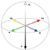
The Commission Internationale de I'Eclairage (CIE) color system is generally used to identify color changes. This system demonstrates the color parameters of L*, a*, b*, and ΔE color changes related with these parameters.2 The ΔE value reports whether or not there is a color variation that is noticeable to the human eye. If this value is more than 1, the color variation can be noticed visually by 50% of human beings. However, due to the uncontrolled factors around the mouth, values of 3.7 and lower are also clinically acceptable.2,3 All colors in the CIE L*a*b* system represent the relative mixture of primary colors of blue, green and red. The values of blue, red, and green are converted mathematically to the CIE L*a*b* scale and the color distance is calculated. The L* axis gives the coordinates of lightness and darkness and these coordinates change between 0 (extremely dark) and 100 (extremely light). The a* axis represents green and red coordinates chromatically; a decreased value of a* in the second measurement compared to the first measurement means a decrease in the red color. The b* axis represents yellow and blue coordinates chromatically; a decreased value of b* in the second measurement compared to the first measurement means a decrease in the yellow color (Fig. 1).4,5
The color of ceramic restorations varies according to many factors such as the thickness of porcelain,6 trademark,1,7 and condensation techniques,6 surface smoothness,8 degree of firings,7 dentin thickness,9,10 and number of firings.11,12 The external view of the layered ceramic may show a specific variability depending on the thickness of the core and veneer ceramic. It is difficult to find the ideal color in inadequate ceramic thickness.13 The translucency or the color shade of the ceramic depends on the type and thickness of the ceramic.1,14,15,16
The effect of the number of firings on color change was evaluated in some studies and no significant color difference was found after multiple firings.11,12,17,18 However, in other studies remarkable color changes after multiple firings were found.1,6,19 Varying degrees of color changes may be observed according to the type of the material used. Some specific metal ions such as palladium or nickel may affect the color of the porcelain.20,21,22 In a study using three different alloys Brewer et al.,8 reported a small degree of color change in the opaque porcelain stage; however, they noted a remarkable increase in color change according to the alloy type after dentin firing.
Pressable crystalline ceramics such as the IPS Empress 2 (Ivoclar Vivadent AG; Schaan, Liechtenstein) and the IPS Empress Esthetic (Ivoclar Vivadent AG, Schaan, Liechtenstein) are widely used in dentistry. IPS Empress 2 has lithium silicate containing high quantity crystalline glass matrix. Empress Esthetic is a leucyte-based material; however, its micro structure is more homogeneous compared to Empress 2 and includes small crystalline particles.23
All-ceramics have satisfying results as color and translucency due to having high permeability of the light; however, esthetically, it may not possible to obtain a perfect tooth-colored restoration.24 If the majority of the amount of light which passed through the ceramic could be transmitted and only a small amount of light is lost, the material might seem more semi-lucent.25 The amount of the light transmission and absorption depend on the amount of crystalline, chemical structure, and the size of particles in the core matrix compared to the wavelength of light.26,27 Heffernan et al.26 determined that the translucency of the core material is a primary affecting factor for the esthetics. Some ceramics have high in vitro resistance values because of having excessive crystalline structure.28,29 However, it causes not only ideal resistance but also high opacity.30,31
Surface roughness may also affect the color of ceramic restorations. Dental ceramics can have a fully smooth surface only by the help of the glaze or natural glaze method. The light reflects and scatters irregularly on rough and irregular surfaces, as a consequence the color of the ceramic restoration changes.
Color values of the surface of dental ceramics are affected by many factors. The aim of the study was to evaluate effect of type of the ceramic, method of glazing, multiple firings on the color change of dental ceramics. The null hypothesis of the study is that type of the ceramic, method of glazing, multiple firings have no effect on the color of dental ceramics.
MATERIALS AND METHODS
Three different types of dental ceramics were used in this study. These are IPS Classic metal-ceramic (MC), IPS Empress Esthetic (EE), and IPS Empress 2 all- ceramics (E2) (Ivoclar Vivadent AG, Schaan, Lichtenstein).
Twenty-eight (14 for each) disc-shaped MC and E2 specimens (2 mm thickness and 10 mm diameter) were prepared according to manufacturer instructions. Seven discshaped EE specimens (2 mm thickness and 10 mm diameter) were also prepared in the same way. Thus, a total of 35 specimens were prepared. In the metal-supported IPS Classic group, a traditional basic metal alloys (Colado NC, Ivoclar Vivadent, Shaan, Liechtenstein), composed of chrome-nickel (0.5 mm thickness), was used as the infrastructure. An IPS Empress Ivoclar EP 600 (Ivoclar Vivadent AG, Schaan, Lichtenstein) furnace was used for the preparation of the EE and E2 core specimens. Veneering porcelain with 1 mm thickness, suggested by manufacturer, was placed on the prepared core ceramics of E2 using the layered technique, then firing process was performed in the furnace of Ivoclar Program at P90 (Ivoclar Programmat P90, Ivoclar Vivadent, Shaan, Liechtenstein). The core ceramics of EE were prepared thicker than the E2 specimens according to manufacturer instructions, and the suggested specific finishing-staining technique were applied on their surface for smoothness.
The type of the material and glazing method were considered for creating the groups. According to this consideration, two different groups were prepared for MC and E2, including glaze and natural glaze methods. Only a glaze group was formed for EE, since the manufacturer does not suggest natural glaze method for EE due to its physical and microstructural form and it is not used routinely. Therefore, a total of 5 groups were formed, which were: Group 1: MC-Glaze; Group 2: MC-Natural glaze; Group 3: E2-Glaze; Group 4: E2-Natural Glaze; and Group 5: EE-Glaze (n=7).
Two different glazing methods as glaze method and natural glaze method were used in the study. For the glaze method, the glaze paste and liquid were mixed on a clean glass slab and applied to the specimen in a homogeneous texture. For the natural glaze method, the specimens were fired and polished with ceramic polishing kit (OptraFine Ceramic Polishing System, Ivoclar Vivadent AG, Schaan, Lichtenstein). These processes were conducted in accordance with the firing and polishing instructions provided by the manufacturer.
The first firings process, performed for preparing the specimens, was defined as control firings groups (C). Then, every specimen was subjected to repeated firings up to seven times. The glaze paste was smeared on the surfaces of the glazed specimens for each firings. Color measurements were performed and recorded for each specimen in the control (C), first (1), third (3), fifth (5), and seventh (7) firings.
A colorimeter (Minolta CR 321, Konica Minolta, Tokyo, Japan) was used for color analysis (Fig. 2). Color measurements were performed measuring from 3 different points of each specimen. The instrument calibration was evaluated after each measurement of each group and then the instrument was recalibrated. The CIE L*a*b* values of the measurements of each specimen were determined and recorded. Total color differences (ΔE) were calculated using the following equation32,33,34: ΔE* = [(ΔL*)2 + (Δa*)2 + (Δb*)2]1/2.
The color difference value represents the numerical distance between the L*a*b* coordinates of 2 colors. Under ideal observation conditions, a color match between 2 colors can not be judged when the ΔE value of 2 colors is less than 1 (ΔE<1). When the measured color differences are within 1 to 2 ΔE range, correct judgments are frequently done by observers. When ΔE values are greater than 2 ΔE, all observers can explicitly detect a color difference between 2 colors. The clinically acceptable limit of the color difference value is considered 3.7 ΔE units.
Statistical analysis of the results was performed using ANOVA test. The Tukey test was used to determine whether the differences originated by conjunction. ANOVA and Tukey tests were performed for each color parameter (L*, a*, b*) and color change value (ΔE).
RESULTS
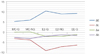
ANOVA test results for the changes occurring during multiple firings in the color parameters of ΔL, Δa, Δb, and the amount of color changes in ΔE were found to be statistically significant for the test conducted (P<.05). The confidence interval for the ANOVA test was identified as 95%. The number of sub-units in each groups was defined as n=7. Tukey tests performed to identify the origin of the difference between the groups were in concordance with ANOVA tests. The changes occurred in the ΔE, ΔL, Δa, and Δb values according to the material-method-firing interaction were evaluated by ANOVA and the P value was found to be less than .05, which demonstrated the statistical significance of the study. The Tukey test results supported the P value of <.05 and the results were parallel to the ANOVA test results. Color changes in all groups were above the critical acceptable level, MC-Glaze specimens demonstrated the relatively least change and the maximum change occurred in the E2-glaze specimens and the ΔL change in MC-Glaze and MC-Natural glaze specimens were lower when compared to the others (Fig. 3).
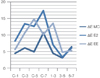
Color change was higher than 3.7 in all stages during the firing stages and generally ΔL, Δa, and Δb values were negative between firings according to type of the ceramics, the lightness (L), red (a), and yellow (b) components of the all materials increased (Table 1); only the color change between the third and fifth firing stages for MC and E2 was lower than 3.7 (3.06; 3.27) (Fig. 4).
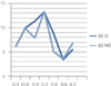
The ΔE values between the third and fifth firings were 3.44 in the glaze groups and 3.57 in the naturel glaze groups (Fig. 5); however, in the other firing steps, the ΔE value was higher than 3.7 which is the critical value for both groups. Generally ΔL, Δa, Δb values were negative for both groups, but for glaze group Δb values were positive and blue component of the specimens were increased (Table 2).
DISCUSSION
The null hypothesis of the study was rejected. Statistically significant changes occurred due to the type of the material, method of polishing, multiple firings (P<.05).
Rate of darkness-to-lightness in all ceramics tends to be in favor of lightness due to opacification, decrystallization, and devitrification when evaluated in terms of the number of firings and methods. The red component of the color increased due to negative Δa. The blue component increased in the glaze group, the b* value, and the yellow component increased in the rest of the groups.
All color changes except the color change between the third and fifth firings, were higher than 3.7. In the third and fifth firing, the color changes occurred according to the glaze method or type of ceramics smaller than 3.7 during the firings. Thus, the lightness, and yellow and green components of the color of the specimens increased.
Differences between control and firings groups, the ΔE value increased by almost two-fold in all ceramics groups. ΔL values tend to be negative, except the E2-Glaze group, which demonstrates an increased opacification in the porcelain surfaces. Though the Δa values during the seventh firings and control group did not show excessive numerical range, the Δb values shifted to negative for all ceramics. This shows that this color was deteriorating through the yellow component. In the MC specimens, surfaces tended to shift toward the red component of the color due to increased the a* value from the control to the seventh firing. The surface color shifted to the green component of the color due to decreased the a* value in the complete ceramics.
Additionally, the ΔL value in the EE and E2 specimens decreased considerably as the number of firings increased. This could be attributed to the damage that developed on the surface of the ceramic structure due to multiple exposure of the dense crystalline structure to high temperature firings.
In a study Yılmaz et al.34 reported that a significant color change for Vita In-Ceram specimens was not observed, the situation was the contrary to the Empress 2 specimens and firings significantly affected the color of opaque porcelain and in some stages the color variation could be easily seen. Mulla and Weiner,35 concluded that marked color changes occurs due to repeated firings compared to the initial firing. The results of this study, contrary to the previous reports,6,11,12,22 and in accordance with the studies of Yılmaz et al.34 and Mulla and Weiner.35; a remarkable color change was also noted in E2 and EE porcelains.
The light on a rough and irregular surface of a texture irregularly reflects and diffuses and color of the restoration changes.32 Kim et al.36 found that the texture of the surface changed the L* value. The L* value that measures the specular component excluded geometry (SCE) is lower on a glazed surface (seen as white) than on a polished surface. In present study, EE and E2 ceramics have a different micro-structure compared to the traditional dental porcelains; both ceramics are full of a high rate of crystalline particles. The quantity of transmitted and emerged light, in comparison with the wavelength of light, depends on the amount of crystalline particles in the matrix, their chemical structures, and the size of particles.26 So that the results obtained from colorimeter were different from those obtained from normal ceramics, and the color gradient increased noticeably.
As for the metal-ceramics, the ions in the metal-substructure may contribute to the color change in the porcelain after repeated firings. There are some studies which confirm these findings in the literature.2,20,37 The elements in alloys have different effects on the porcelain color.21 During the bulk transfer, elements migrate from the alloy to the porcelain throughout interface. The elements released from the metal structure spread to metal surface of the porcelain and reach the porcelain surface in the metal-porcelain joint. Lastly, vapor deposition is a mechanism in which the elements from the alloy vaporized and subsequently deposited onto the porcelain surface, resulting in discoloration.34 Nickel ions are colorants that produce a neutral gray color in sodium silicate glasses are likely associated with color changes in porcelain. In the present study, the substructure of the metal supported porcelain includes chrome-nickel basic metal alloy. One of the reasons of the changes in the porcelain color during multiple firings is thought to be detrimental effect of the elements of metal substructure to the porcelain surface.38
In the literature, there are various studies about the effect of firings on ceramic color. Several studies have suggested that certain metal oxides are not color stable after they subjected to firing temperatures, and color changes of surface colorants after firing have demonstrated pigment breakdown at firing temperatures.39 The results of Bachhav and Aras's study40 showed that the thickness of dentin ceramics and the number of firings definitely affect the color. Furthermore, the ΔE values of ceramics containing zirconium oxide increase when ceramic thickness increases; however, the value was not above 3.7.38,41 Barghi42 reported that the slight change in color after repeated firings may be attributed to the increase in density caused by the decrease of air bubbles trapped inside the porcelain. Porcelain has a high viscosity at its low liquid temperature, and diffusion of the ionic species in the molten glassy phase is consequently highly hindered. The results of this present study also demonstrated that multiple firings of complete ceramics, especially EE and E2, may increase the color changes of the structure significantly and thus more than one firing is not recommended for ceramics. In present study color changes may be occurred in metal supported ceramics, as was stated by Barghi,42 due to the ion diffusion to the softened glassy phase of the viscous porcelain.
The colorimeters are more economic and convenient devices compared to other devices such as the spectrophotometer.43 However, with the use of these small aperture devices, a considerable fraction of the light, entering the assessed material is lost because it emerges on the surface outside the aperture of measurement. This loss of light is termed as edge-loss effect, and may be a cause of color measurement errors.5,44 The edge-loss effect may occur particularly when translucent materials are used and the diameters of the evaluated materials are smaller than aperture of the colorimeter.34 The inadequacy of the colorimeter used in this present study is the result of light diffusion and use of translucent materials containing intense crystalline such as E2 and EE significantly affected the color changes. Variable results compared to clinical color measurements might have been obtained due to the in vitro nature of the study and the shape of used disc specimens. However, discshaped specimens are generally prepared in the measurements of surface smoothness and color changes. This type of geometry is sufficient and appropriate for surface smoothness and color change identifications tests. Celik et al.,38 also suggested that the disc-shaped specimens more accurate than crowns-shaped specimens. In conclusion, color change critics should be evaluated in other in vitro studies as well.
In the present study, the color change after the initial firing was remarkable, in accordance with the studies of Heffernan et al.,45 Mulla and Weiner,35 and Tylman.46 The increase in the L* value was likely due to opacification, and more yellow and red color characteristics appeared in the porcelain surfaces due to the decrease in the Δa and Δb values.
Natural glaze was not performed in EE specimens in this study due to both the composition and high temperatures of firing. However, it was seen that natural glaze did not cause less color change in E2 and MC specimens and it was observed that the natural glaze method, applied to MC and E2 specimens, did not cause less color variation. Regardless of the glaze or natural glaze method, a color variation was observed. Another reason for the color change can be considered as a detrimental effect of high firing temperatures of all ceramics containing leucite and lithium disilicate. It is likely that the continuous and/or high temperature firings of the porcelains caused pyroclastic stream with accumulation on the surface. Disc- shaped specimens lost their contours, recrystallization occurred, devitrification was observed, and as a result color changes exceeded the in acceptable limits.
The color of porcelain can also be affected by smoothness of the surface texture.8 On an irregular and rough surface, the light reflects and diffuses, altering the color of restoration.32 Kim et al.36 found that surface structure changed the L value. The L* value that measures specular component excluded geometry (SCE) is lower on a glazed surface (seen as white) than on a polished surface.
CONCLUSION
Within the limitation of the study, it is concluded that; ΔE values were above the 3.7 critical value. ΔL, Δa, and Δb values also tended to be negative. However, the metal-ceramic/glaze method was less affected during repeated firings. In E2 and EE specimens, surface color was significantly affected by repeated firings.
This study revealed that color changes occurred to some extent due to the insufficient smoothing of the natural glaze method of the all ceramics and metal ceramics because of the microstructural features.
Due to these factors, multiple firings should not be performed on dental ceramics; especially, all-ceramics of a high crystalline nature should be glazed at the ideal firing temperature and in compliance with the instructions of the manufacturer.


 XML Download