

 PDF
PDF ePub
ePub Citation
 Print
Print


INTRODUCTION
Mesenchymal stem cells (MSCs) mainly reside in bone marrow (BM). They can self-renew and differentiate into several cell lineages, including osteogenesis, adipogenesis and-chondrogenesis.1,2 As a result of these capabilities, MSCs play an important role in continuous preservation and repair most tissue types. Generally, it was accepted that the quantity and quality of MSCs decrease with aging, which is associated with the progressive failure of function of tissues and organs.3 Furthermore, MSCs become uncommonly found in bone marrow (- 0.001%). One of the main goals is still to optimize MSCs recovery and in vivo expansions so as to fulfill a great deal of functional cells in reasonable times.4 Apparently, the various methods for MSC isolation can lead to enrichment of different subsets of MSCs with different biological properties. Different methods can be customized to gain MSCs from the BM: plastic dish adhesion and negative (CD 45, Gly-A) or positive selection (CD133, Stro-1, and CD49-a).5,6,7,8,9 However, the adherence of MSCs to tissue culture plastic surfaces had remained the most traditional and customary of the various MSC isolation methods, and the non-adherent cell populations are generally discarded during the first media change. A little study have shown that non-adherent BM cells can lead to colony-forming unit-fibroblasts (CFU-f) in vitro10,11,12,13 and formed bone14 after in vivo transplantation. Various study have exhibited that fibroblast-like cells can be derived from peripheral blood and mobilized from BM by cytokines,15,16 hypoxia17 and acute skin damage.18 The observation imply the existence of non-adherent BM cell populations in BM and these cells maybe circulation when the useful time. Knowledge about the characteristics of non-adherent cell populations opens exciting perspectives in basic research on bone tissue formation and repair. Relatively few articles have been published on this issue, and the results obtained by several authors have suggested that, in non-adherent bone marrow stromal cell populations that may become adherent in vitro, begin to proliferate, and differentiate into several tissue lineages, as well as enhance bone formation in vivo.19 Recently, it was showed that the extracellular matrix (ECM) prepared from bone marrow progenitor cells advanced the proliferation of MSCs, facilitated master the portion of MSCs, and enhanced their capacity for stem cell-based therapy.20 This led us to investigate whether culture of MSCs on a quantification of ECM could improve their number and possession. The purpose of current study was to identify characteristic of low-adherent BMMSCs obtained by quantification of extracellular matrix and know the response of low-adherent MSCs on titanium (Ti) surfaces.
MATERIALS AND METHODS
C3H male mice aged 3 month, weighing 22 g were used in this study and fed a diet of standard commercial mouse chow. All of the animals were handled according to the "Recommendations for handling of Laboratory Animals for Biomedical Research" complied by the committee on the Safety and Ethical Handling regulations for Laboratory Animal Experiments in the college of Dentistry at Seoul National University (Approval number: SNU-120007-2).
BM isolation procedure based on the previous studies was utilized.21 Femur and tibia were carefully cleaned off soft tissue and BM were harvested by flushing with buffer consisting of phosphate buffered saline (PBS) containing 2% fetal bovine serum (FBS)(Equitech-Bio Inc, Kerrville, TX, USA), antibiotics. The BM cells were filtered through a 70 mm nylon mesh filter (Falcon, Franklin Lakes, NJ, USA). The collected BM cells were treated with Ammonium-Chloride-Potassium (ACK)(Lonza, Walkersville, MD, USA) lysing buffer at RT and centrifuged. The suspension was poured off, and the mononuclear cells were re-suspended with complete alpha-modified Eagle's medium (α-MEM) (Life Technologies, Grand Island, NY, USA) supplemented with 15% FBS (Life Technologies, Grand Island, NY, USA), glutamine (2 mM), 2-mercaptoethanol (55 µM), penicillin / streptomycin (Life Technologies, Grand Island, NY, USA) and L-ascorbic acid (100 µM)(Wako pure, Tokyo, Japan) and cultured for 14 days at 5% CO2 for 37℃ until the first medium exchange.
Constitution of ECM coated dishes was performed as previously described.20 BMMSCs were cultured on a dish and, after 1, 4, 7, 10 days of culturing, non-adherent cells were discarded, respectively. The once more adherent cells were then decellularized with 0.05% Triton X-100 at RT. The ECM coated dishes was washed with PBS and stored in a humidified atmosphere at 37℃ with 5% CO2.
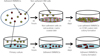
Non-adherent cells were reseeded on an ECM coated dish to acquire low-adherent BMMSCs according to the following steps. The first media change was made at 48hours after the initial seeding. For the first media change, the removed media containing cells was centrifuged and cells were replated in an 7 days culturing of adherent BMMSCs derived ECM coated dishes and then cultured in a humidified atmosphere at 37℃ with 5% CO2 for 14 days. After the 14 days in culture, the reattached cells were termed low-adherent BMMSCs and two additional passages were performed to obtain a sufficient quantity of cells, which were used to conduct the following experiments (Fig. 1).
The adherent BMMSCs and low-adherent BMMSCs proliferation was tested using the bromodeoxyuridine (BrdU) method following the manufacturer's instructions. Adherent BMMSCs and low-adherent BMMSCs were seeded on chamber slides (Nalge Nunc International, Rochester, NY, USA) and maintained in the medium. After one day, BrdU labeling reagent (Invitrogen, Frederick, MD, USA) were added in the cultures. After 24 hours, the cells were stained with a BrdU staining kit (Invitrogen, Frederick, MD, USA). Nine representative active images were used to calculate the number for BrdU-stained nuclei, using a microscope. The image was analyzed for determining the BrdU-positive staining percentage by manual counting.
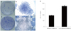
Freshly isolated BM cells were seeded on plastic dishes. After 48 hours of culturing, non-adherent BM cells were replated on ECM coated dishes. After 14 days, the cells were stained with 1% toluidine blue solution in 2% paraformaldehyde (PFA). A cell bunch over 50 cells was counted as a CFU-f.
Adherent BMMSCs and low-adherent BMMSCs were cultured under osteogenesis, and adipogenesis conditions in order to determine their capability to produce bone, adipose cell lineages. After induction, the cells were stained with alizarin red S staining and oil red O staining to detect bone and adipose lineages, respectively. Medium for osteogenic induction: α-MEM supplemented with 20% FBS (Becton Dickinson, Franklin Lakes, NJ, USA) L-glutamine (2 mM), 2-mercaptoethanol (55 µM)(Life Technologies, Grand Island, NY, USA), dexamethasone (10 nM)(Sigma, St. Louis, MO, USA), L-ascorbic acid (100 µM)(Wako Pure Chemical, Richmond, VA, USA), β-glycerophophate (2 mM) and penicillin & streptomycin (Life Technologies, Grand Island, NY, USA). Medium for adipogenic induction: α-MEM supplemented with 15% FBS (Becton Dickinson, Franklin Lakes, NJ, USA) L-glutamine (2 mM), 2-mercaptoethanol (55 µM), penicillin/streptomycin (Life Technologies, Grand Island, NY, USA), L-ascorbic acid (100 µM)(Wako pure, Tokyo, Japan), isobutyl-methylxanthine (0.5 mM) (Sigma, St. Louis, MO, USA), indomethacin (60 µM)(Sigma, St. Louis, MO, USA), hydrocortisone (0.5 µM)(Sigma, St. Louis, MO, USA), insulin (10 µM)(Sigma, St. Louis, MO, USA) was added to the confluent culture of cells.
Adherent BMMSCs and low-adherent BMMSCs were harvested after 3rd passage. 5 × 10⁵ cells were washed with fluorescence-activated cell sorting (FACS) buffer, incubated with 3% rat serum on ice for 30 min, and immunolabeled with 1 µg of R-Phycoerythrin (PE)-conjugated monoclonal antibody against mouse CD14, CD34, CD44, CD105, CD117, Sca-1, Oct-4, and CD29 on ice for 30 min. For immunolabeling with Oct-4, the cells were fixed and permeabilized with Fixation/Permeabilization working solution according to the manufacturer's instructions. The cells were then incubated with OCT-4 (Santa Cruz Biotechnology, Santa Cruz, CA, USA) on ice for 30 min and fluorescent secondary antibody on ice for 30 min. Analyses were executed by FACS Calibur (Becton-Dickinson, San Joes, CA, USA).
Total RNA was extracted with Trizol reagent (Life Technologies, Carlsbad, CA, USA) from untreated and differentiated adherent BMMSCs and low-adherent BMMSCs. For RT-PCR, cDNA was synthesized with SuperScript III First-Strand kit (Life Technologies, Carlsbad, CA, USA) using random hexamers, according to the instructions of the manufacturer. The reflection production were resolved by electrophoresis on a 1.5% agarose gel and viewed with SYBR Green I (Life Technologies, Carlsbad, CA, USA).
Both kinds of cultured cells were seeded onto 10 mg of hydroxyapatite/tricalcium phosphate (HA/TCP) powder (Zimmer, Freiburg, Germany) and incubated at 37℃ with 5% CO2 for 2 hours and subcutaneously transplanted into immunocompromised mice. After 8 weeks, the transplants were harvested, fixed, decalcified and paraffin embedded. The paraffin sections were stained with hematoxylin and eosin (H&E) staining.
In order to measure cell proliferation of adherent and low-adherent BMMSCs on machined and anodized Ti disc, an EZ-Cytox cell viability assay kit (Daeil Lab Service Co., Seoul, Korea) was used according to the manufacturer's instructions. Ti disc were made from pure titanium grade IV (Warentec Co., Seoul, Korea) with the dimensions of 25 mm diameter and 1 mm thickness. An anodic oxidation Ti disc was performed at 300 V in an aqueous electrolytic solution of 0.15 M calcium acetate monohydrate (Ca(CH3COO)2.H2O) and 0.02 M calcium glycerophosphate (CaC3H7O6P) and machined surface disc with no treatment.22,23 All samples were decontaminated with ethylene oxide gas. Both adherent and low-adherent BMMSCs were seeded on machined and anodized Ti discs. The optical densities (ODs) were assessed using a spectrophotometer at 405 nm, after 1, 4, and 7 days of culturing.
Test of normality and equality of variances were applied, and no violations of those basic assumptions were observed. Statistically significant effects were detected with t-test in SPSS 20.0. P-values less than .05 were considered significant.
RESULTS
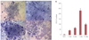
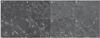
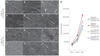
Our study investigated the capability of adherence cells for forming fibroblastic cells on a quantification of ECM coated dishes. Adherent BMMSCs were cultured on a dish and, after 1, 4, 7, 10 days, non-adherent cells were discarded by washing with PBS. In the present study, the isolated BM cells were adhered for 48 hours to plastic dishes, and then non-adherent BM cells were transferred and maintained on ECM coated dishes. After the 14 days, we obtained fibroblastic colonies with all of the adherence cells (Fig. 2A, Fig. 2B, Fig. 2C, Fig. 2D). However, the number of CFU-f cells was significantly higher when non-adherent cells were cultured on ECM coated dishes, which was made by 7 days of culturing adherent BMMSCs (P<.05)(Fig. 2E). SEM image of ECM coated dishes were showed in Fig. 3. The colony numbers were significantly increased in the 7 days culturing of adherent BMMSCs derived ECM coated dishes (Fig. 4). The ECM coated dishes which made by 7 days were used for further experiment.
To characterize the properties of low-adherent BMMSCs, we assessed the proliferation of low-adherent BMMSCs by BrdU incorporation. Our studies showed there was no different increased in BrdU uptake rate between adherent BMMSCs (Fig. 5A) and low-adherent. BMMSCs (P>.05) (Fig. 5B).
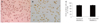
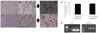
The properties of MSCs were their ability to differentiate into several tissues after induction. Our examination of differentiation potential, showed that low-adherent BMMSCs (Fig. 6C, Fig. 6D) have a similar potential to adherent BMMSCs (Fig. 6A, Fig. 6B) in terms of their formation of mineralized nodules under the osteogenesis inductive cultures (Fig. 6E)(P>.05), which was associated with expression of the runt-related transcription factor (RUNX2)(Fig. 6F). Furthermore, low-adherent BMMSCS (Fig. 7D, Fig. 7E, Fig. 7F) also resembled to adherent BMMSCs (Fig. 7A, Fig. 7B, Fig. 7C) in the formation of lipid droplets in adipogenesis inducing cultures (Fig. 7G) (P>.05), which was associated with the expression of lipoprotein lipase (LPL)(Fig. 7H).
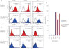
Expression of cell surface molecules on the isolated MSCs was evaluated by flow cytometry. Fig. 4 showed representative flow cytometry analysis for both MSC types. Both the adherent BMMSCs and low-adherent BMMSCs positively expressed CD29, CD44, and Oct-4 (Fig 8A). However, they were negative for CD34, CD117, and CD14 (Fig. 8B). We observed no differences in the percentage expression of any of the markers used for flow cytometry analysis (P>.05)(Fig. 8C).
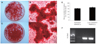
After 8 weeks of subcutaneously transplantation of BMMSCs into immunocompromised mice, newly formed bone tissues were seen in HA/TCP loaded with either adherent (Fig. 9A) and low-adherent BMMSCs (Fig. 9B). There was no significant difference bone formation ability between adherent and low-adherent BMMSCs in vivo (P>.05)(Fig. 9C).
The results of the (3-(4, 5-dimethylthiazol-2-yl)-5-(3-carboxymethoxyphenyl)-2-(4-sulfophenyl)-2H-tetrazolium) (MTS) assay are shown (Fig. 10). SEM image of adherent and low-adherent BMMSCs showed spindle-like morphology on Ti discs (Fig. 10 A-L). There was higher cell proliferation appearance in adherent BMMSCs and low-adherent BMMSCs seeded on anodized Ti discs than adherent and low-adherent BMMSCs seeded on machined Ti discs by time (P<.05)(Fig. 10M).
DISCUSSION
BMMSCs are usually obtained from BM stromal cells and appeared adherence and colony-forming unit fibroblastic on plastic culture dishes, followed by subsequent proliferation.24 The CFU-f assay has been thought to be an effective method to investigate the selection of BMMSCs. It was well known that MSCs lose their capability to proliferation as well as their differentiation potentiality upon long time culturing on plastic culture dish.25 In our study, we showed non-adherent cells cultured on ECM coated dishes made by adherent BMMSCs exhibited higher CFU-f number than freshly isolated BM cells cultured on plastic dishes. We confirmed in this study that a method of acquisition of low-adherent BMMSCs using ECM coated dishes were effective in increasing cell numbers and shortening the cell culture time and maintaining stemness of the stem cells. We also showed in this study that extracellular matrix coated dish made by 7 days culturing of adherent BMMSCs was appropriate for obtaining MSCs. A possible explanation for this mechanism was that 7 days culturing of adherent BMMSCs derived ECM may provide proper composition of the growth factors or cytokines to non-adherent cells. It has been reported that MSCs cultured on this ECM showed notable promotion of proliferation and retention of stem cell properties when comparing with cultured on uncoated plastic dish.26 From the point of clinical use, it is important step to obtain sufficient quantity of stem cells in primary cell culture stage. This study revealed a simple method to increase the number of acquired mesenchymal stem cells. We obtained abundant two types of BMMSCs which have similar characteristics for clinical application.
Previously study reported that the non-adherent cell suspensions of BM contained non-hematogenous cells which expressed osteoblastic markers.11,27,28 The non-adherent suspension cells contained more osteogenesis progenitors than adherent cells.29 the suspension cells from BM cultures were able to cause adherent stromal cells. It is possible that adherent potential cells existed in the non-adherent suspension cells and they could conduct osteogenic potential to clinical use. Up to date, there was no attempt to confirm the osteogenic ability of these non-adherent cells. In our study, we termed the non-adherent cells adhered to the ECM coated dish as low-adherent BMMSCs, because there was no consensus agreement of terming BMMSCs. More studies would be needed. This study was intended to report the characteristic of low-adherent MSCs acquired from non-adherent suspension cells which cultured on the ECM coated dishes. Our results showed that the non-adherent cells exhibited adherent morphology, multi-lineage potentiality, and also express MSCs surface molecular markers in vitro.
In addition, this study confirmed that low-adherent MSCs promoted bone formation in vivo. After 8 weeks of subcutaneous transplantation of MSCs in immunocompromised mice, many blood vessels were seen in MSCs loaded HA/TCP, indicating that adherent BMMSCs and low-adherent BMMSCs might enhance vascularization to induce new formation of bone tissue, which was found in both cell types, suggesting that low-adherent BMMSCs have osteogenic potential in vivo as do adherent BMMSCs. Our in vivo results may provide important progress in achieving critical clinical applications of using low-adherent BMMSCs. Thus, transplantation of autologous adherent BMMCSs or low-adherent BMMSCs obtained from adult BM may be able to reconstitute both the hematopoietic cells and their associate osteogenesis environment. Additionally, our results confirmed that low-adherent BMMSCs and adherent BMMSCs cultured on the anodized Ti discs showed higher cell proliferation rates than the machined Ti discs along with time. It seems that MSCs has affinity to the anodized Ti surface instead of the smooth surface as might have been expected.
CONCLUSION
In conclusion, 7 days culturing of adherent BMMSCs derived ECM was an appropriate microenvironment of obtaining non-adherent bone marrow cells. Low-adherent BMMSCs acquired by 7 days culturing of adherent BMMSCs derived ECM coated dishes can adhere, proliferate and differentiate into specialized cell lineages and have function in bone formation. It should be further proved that low-adherent BMMSCs may have important clinical therapeutic implication, therefore could be used effectively as adherent BMMSCs.

 XML Download