

 PDF
PDF ePub
ePub Citation
 Print
Print


INTRODUCTION
During the past few years, partially stabilized zirconia has been integrated into restorative dentistry.1 Yttrium-oxide partially stabilized zirconia (Y-TZP) has mechanical properties that are attractive for restorative dentistry; namely, its chemical and dimensional stability, high mechanical strength, and fracture-toughness.2,3
The Lava™ Zirconia System utilizes CAD/CAM technology to produce a densely sintered and high-strength zirconia framework with a 3% mol partially yttria-stabilized zirconia polycrystal content. The polycrystals have a tetragonal crystal structure and an average grain size of 0.5 µm or smaller. The equipments used for the Lava™ All-Ceramic System in a dental laboratory includes a special scanner (Lava Scan), a computerized milling machine (CAM)(Lava Form), and a sintering oven (Lava Therm) plus CAD/CAM software technology. Incorporating the positive material properties, the Lava™ Zirconia System (3M ESPE Dental Products, St. Paul, MN, USA) can be utilized to create all-ceramic crowns and FPDs for use in the anterior and posterior regions of the oral cavity.4
The marginal "fit" of any dental restoration is vital to its long term success. Lack of adequate fit is potentially detrimental to both the tooth and the supporting periodontal tissues.5 The internal accuracy and the marginal accuracy of a restoration are important for its longevity. The presence of marginal discrepancies in the restoration exposes the luting agent to the environment. The larger the marginal discrepancy, the more rapid the rate of cement dissolution.6,7 The resultant microleakage permits the percolation of the food, oral debris and other substances that are potential irritants to the vital pulp.6,8 The aim of this study was to measure the clinical changes on the marginal and internal adaptation of zirconia based anterior fixed partial dentures (Lava 3M ESPE, Germany) after the porcelain firing process.
MATERIALS AND METHODS
Twenty-eight patients (11 male, 17 female) with the indication for zirconia fixed partial dentures were selected for this study. The study was approved by the local ethics committee (MAR-YG-2009-0318). A total of 34 anterior FPDs (19 three units, 9 four units, 3 five units and 3 six units) were inserted (24 in the maxilla, 10 in the mandible). All abutment teeth were prepared in a standardized manner as follows: incisal/occlusal reduction of 1.5 to 2 mm; axial reduction of 1 to 1.5 mm with 6-degree taper; 1-1.5 mm wide chamfer finish line following the scalloping of the free gingival margins and located 0.5 mm subgingivally and rounded internal line angles. Impressions were made with additional silicone impression materials (Affinis, Coltene, Whaledent, Altstatten, Switzerland) using two stage impression technique. Working dies were fabricated with type 4 dental stone (Alpenrock, AmannGirrbach GmbH, Pforzheim, Germany).
Fabrication of frameworks with the Lava™ Zirconia System was performed with partially sintered zirconia blocks. Minimum connector surface area of 7 mm2 and a retainer thickness of 0.5 mm were provided. The thickness of the frameworks was measured and recorded with a custom-made digital caliper at the following points: (1) the retainers - midbuccal, midlingual, proximal (mesial or distal), and occlusally at the middle of the central fossa and (2) the connectors - occlusogingival height, and buccolingual width. The frameworks could have been colored in one of seven shades to correspond with the patients' natural tooth color before the sintering process (which lasted for approximately 7 hours) was started (Lava Therm; 3M ESPE). The frameworks were obtained.
Marginal and internal discrepancies were measured at two different times. The first measurement was done prior to veneering ceramic firing, while the second measurement was done after the ceramic firing to evaluate the influence of this process. As the veneering material (IPS e.max Ceram, Ivoclar Vivadent, Liechtenstein) with a coefficient of thermal expansion (9.5 × 10⁻⁶ K-1) adjusted for zirconia was used. No internal adjustments of the frameworks were done after veneering ceramic firing or at clinical try.
To evaluate and compare the gap dimension between the frameworks and abutment teeth the marginal and internal openings were measured by using a silicone replicas (Affinis, Coltene, Altstätten, Switzerland). The retainers of the FDPs were filled with light body silicone; then the frameworks were placed onto the abutment teeth and additional silicone impression material (Affinis, Coltene, Whaledent, Altstätten, Switzerland) were applied onto the frameworks with metal trays. After setting time, thin silicone film replicas, frame works and silicone impression were removed from the mouth. To stabilize the silicone films representing the space between abutment teeth and frameworks, a medium body silicone was injected on the light body silicone replicas and immediately before the setting time heavy body silicone (Affinis, Coltene, Altstätten, Switzerland) was used to cover the impression surface.
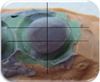
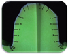
Silicone replicas were segmented two times in the mesio-distal and one times in the bucco-lingual directions with a razor blade (Fig. 1). Silicone replicas taken before porcelain firing process (initial) and after porcelain firing process (final) were examined under a binocular stereomicroscope (Leica Optic microscope, Leica Cambridge Ltd., Cambridge, England) at a magnification of ×31.13 to obtain marginal and internal discrepancy values and marginal adaptation types. From each segment, five different measurements for marginal discrepancy, marginal internal discrepancy, axial discrepancy and three different measurements for incisal discrepancy were made. A total of 78 measurements were obtained per abutment (Fig. 2). Marginal adaptation types were recorded (Fig. 3A and Fig. 3B).
Statistical analysis was performed using SPSS for Windows, version 15.0 (SPSS Inc., Chicago, IL, USA). Kruskal Wallis and Wilcoxon Signed Ranks tests were used for the statistical analysis of marginal and internal discrepancies and marginal adaptation types obtained before porcelain firing process (initial) and after porcelain firing process (final)(P<.05).
RESULTS
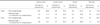
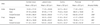
No statistically significant difference was found between initial and final marginal gap values (P>.05)(Table 1, Table 2, Table 3, and Table 4). At the internal gap measurements (Table 5, Table 6, Table 7, and Table 8), final marginal area values (59.54 µm) were significantly lower than the initial marginal area values (68.68 µm)(Fig. 4A and Fig. 4B) whereas no statistically significant difference was found between the axial and incisal area initial and final internal gap values (P>.05)(Table 7). The highest and the lowest internal gap values were observed at the incisal/occlusal area and at the marginal area, respectively (Table 5, Table 8). In addition, lower internal gap values were obtained for canines than for central incisors, lateral incisors and premolars at the incisal area (P<.05)(Table 6). However no statistically significant difference was found between marginal gap, marginal area internal gap and axial area internal gap values of different teeth groups (P>.05)(Table 6).
DISCUSSION
Despite careful preparation of a full coverage restoration and its precise cementation, a small gap will remain between the margin of the restoration and the finish line of the prepared tooth, predisposing the tooth to caries and periodontal disease.9,10 The closer the margin of restoration to the finish line of the preparation, the smaller the marginal gap and thickness of exposed cement layer at the margin.
Clinically acceptable marginal gap values had been reported in the range of 50-120 µm.11 Two in vivo and four in vitro study on the marginal deficiency at LAVA zirconia restorations had been published.12,13,14,15,16,17 Reich et al.12 compared the marginal and internal adaptation of four different systems (Lava, Cerec, InLab, Digident and conventional metalceramic) with the silicone replica method. Mean marginal gap values at Lava (n = 8), Cerec InLab (n = 8), Digident (n = 8) and metal-ceramic (n = 6) fixed partial dentures had been measured as respectively; 80, 77, 92, 67 µm. For posterior LAVA fixed partial dentures, Reich et al.13 reported mean 91 µm in vivo marginal gap values. In the in vitro fixed partial dentures study of Gonzalo et al.14, mean marginal gap values of Lava restorations before and after cementing were 67 µm and 71 µm, respectively; mean marginal gap values of Procera restorations were 26 µm and 12 µm. In the other in vitro study of Gonzalo et al.16, mean marginal gap values of Procera Zirconia, InCeram Zirconia, Lava and metal-ceramic were reported as respectively; 9 µm, 40 µm, 66 µm and 67 µm. However, Vigolo and Fonzi15 reported lower marginal gap values with Lava system (46.3 µm at core, 46.8 µm after veneering, 47.3 µm after glazing) than the Procera (61.1 µm at core, 62.5 µm after veneering, 63.5 µm after glazing) and Everest system (63.4 µm at core, 65.3 µm after veneering, 65.5 µm after glazing). In our study, 14.3 ± 18.8 µm mean marginal gap values before porcelain veneering and 1 ± 16.5 µm mean marginal gap values after porcelain veneering were detected. These are near the lower range of clinically accepted marginal gap values. Results of our study are parallel to the in vitro study of Beuer et al.17 Our marginal gap values are much lower than the values of previously published in vivo and in vitro studies.12,13,14,15,16,17 These lower marginal gap values can be explained with the update of Lava Scanner at 2008 (Lava Scan ST).
Reich et al.12 reported that the increase of internal gap would result in the retention loss and fracture of restoration indirectly. In the internal gap studies, number of replica sections and measurement points must be increased to take an exact data about the whole internal adaptation of restoration.12,13,17,18 Twenty-four different measurements per section and 78 different measurements per tooth were obtained in the presented study.
Mean axial and occlusal gap values observed in previous studies with Lava fixed partial dentures were 132 µm (axial), 215 µm (occlusal)12; 98 µm (occlusal), 102 µm (axial)13 and 71 µm (axial); 108 µm (occlusal).17 With recent literature, it had been detected that internal gap values were higher than the marginal gap values.10,11,12,13,17,19,20,21 In our study, parallel results were detected. Mean internal gap values before veneering at marginal, axial and incisal areas were measured as respectively; 68.68 ± 35.12, 85.38 ± 18.43, 134.83 ± 36.77 µm whereas after veneering measured as respectively 59.54 ± 54.11, 83.53 ± 18.84, 135.72 ± 38.43 µm.
Marginal and internal adaptation of CAD/CAM restorations are directly affected by the precise of scanning procedure. Recently recorded internal gap values were largest at incisal/occlusal surfaces whereas smallest at marginal areas.22 The reasons for this situation were the width of scanner tip leading insufficiency at some inclined surfaces and automatically adjusted cement gap. In our study, parallel results with Kokubo et al.22 were detected; internal gap values became higher from marginal area (initially 68 ± 35.12 µm, finally 59.54 ± 54.11 µm) to incisal surfaces (initially 134.83 ± 36.77, finally 135.72 ± 38.43 µm).
In the presented study, statistically lower internal gap values were obtained for canines (mean 111.77 µm) than for central incisors, lateral incisors and premolars at the incisal area. It is supposed to that very thin insical edges of central and lateral incisors affected the scanner tip adaptation and caused increase in internal gap measurements. On the other hand, morphological inclinations at the buccal surfaces of canines facilitated movements of scanner tip and led to lower internal gap measurements.
Changes at the marginal adaptation of restorations caused by firing process had been firstly reported by the studies of Castellani et al.23 and Balkaya et al.24 They were examined glass ceramics and copy-milling and In-Ceram Alumina respectively and observed vertical and horizontal distortion at the marginal area by the subsequent firing processes. This distortion would affect the fit and marginal adaptation of restorations. On the in vitro studies examined changes at the marginal and internal adaptation of zirconia based restorations during the porcelain firing process, different results had been observed.15,25,26,27 Vigolo and Fonzi15 reported no statistically significant difference after two veneering and one glazing cycles whereas Att et al.25 detected minimal increase after six firing cycles. In contrary, Dittmer et al.26 and Kohorst et al.27 after four firing cycles, observed a horizontal distortion by the veneer porcelain contraction towards the center of retainers that led marginal and internal gap decrease. Our marginal gap results were parallel to Vigolo and Fonzi,15 whereas internal gap values at the marginal area were parallel to Dittmer et al.26 In comparison with initial and final marginal gap values, no statistically significant decrease was detected after firing. However statistically significant decrease (68.68-59.54 µm) was observed between initial and final internal gap values at the marginal area. Cervical margin areas of the restorations were reported as more prone to horizontal distortion.26,27 The results of this study were able to compare with in vitro test results because no in vivo studies that measured the changes on the marginal and internal adaptation of restorations after the porcelain firing process were available.
Dittmer et al.26 reported that mismatch between thermal expansion coefficient of veneering porcelain and zirconia core caused detrimental stresses during firing process. Aboushelib et al.28 also detected that veneering ceramics should have slightly lower coefficient of thermal expansion than that of zirconia framework (- 0.6 × 10⁻⁶ K-1), resulting in a positive mismatch in thermal expansion coefficients. This positive mismatch is expected to induce beneficial compression stress on the veneering porcelain layer.27 Pospiech et al.,29 Raigroski et al.30 and Crisp et al.31 used Lava Ceram veneering ceramic which was compatible with thermal expansion coefficient of Lava zirconia framework (10 × 10-6 K-1). We preferred to use IPS e.max Ceram veneering ceramic (9.5 × 10⁻⁶ K-1) corresponding to the manufacturer's instruction, with 0.5 × 10⁻⁶ K-1 unit lower coefficient of thermal expansion than that of the Lava zirconia framework. In their in vitro study, Kohorst et al.27 used veneering ceramics that had lower coefficient of thermal expansion (8.8-9.2 × 10⁻⁶ K-1 and 9.4 × 10⁻⁶ K-1) than that of VITA In-Ceram YZ and Kavo Everest zirconia and observed statistically significant decrease between initial and final marginal/axial gap measurements. In our study, final marginal area values (59.54 µm) were significantly lower than the initial marginal area values (68.68 µm) whereas no statistically significant decrease was found between the axial and incisal area initial and final internal gap values, that was similar to Kohorst et al.27 It makes hard to compare these two studies that have differences with regard to conditions (in vitro and in vivo) and ceramic systems.
CONCLUSION
Marginal and internal gap values obtained after porcelain firing process with Lava zirconia fixed partial dentures were within the range of clinical acceptance. Minimal values of marginal and internal gap are important for the retention of the restoration. Related system could be used safely to prevent the abutment teeth from the caries and to support the health of periodontal tissues.


 XML Download