

 PDF
PDF ePub
ePub Citation
 Print
Print


INTRODUCTION
In oral rehabilitation procedures using prosthetic restorations, it is mandatory, yet difficult, to determine the prognostic factors for an individual tooth in order to predict its longevity and select the most clinically appropriate prosthetic abutments. These difficulties stem from multiple factors that are considered in the absence of definitive criteria, and the treatment plan is greatly influenced by patient preference and clinician experience.1
However, well-established prognostic variables include tooth mobility, alveolar bone support, root configuration, pulpal condition, opposing dentition, endodontic treatment completion and root/crown ratio (R/C ratio).2,3,4,5,6 The R/C ratio is a primary variable in the diagnosis of a tooth as an abutment for the various types of prosthetic restorations, such as fixed dental prostheses (FDPs) or removable partial dentures (RPDs) or overdentures.7,8,9,10
The R/C ratio is defined as the value between the root and the crown and may be classified as the anatomical R/C ratio or the clinical R/C ratio. While the anatomical R/C ratio uses the cemento-enamel junction as a reference, the clinical R/C value is defined by the alveolar bone level and provides information on the amount of alveolar support; therefore, unless otherwise specified, the term "R/C ratio" generally refers to the clinical R/C ratio that is determined radiographically.11 The exact measurement of the total periodontal-support area is more accurate than the linear estimate of the ratio; furthermore, the R/C ratio that is determined on radiographs has inherent limitations because it does not represent the actual condition of the total supported root surface of a tooth.12 However, the linear measurement is clinically useful for assessing the suitability of teeth as prosthetic abutments.1 Regarding the mechanical aspect, the R/C ratio represents the fulcrum of a Class 1 lever with its center of rotation in the supporting bone.13,14 The crown portion of the fulcrum acts as the effort arm, and the root portion of the fulcrum functions as the resistance arm. If the lengths of the crown and root change as a result of pathology or targeted therapy, then the center of rotation moves apically or incisally; therefore, the effects of lateral forces on the tooth might be harmful.3,9,13
The R/C ratios of normal dentition assessed from radiographs may act as reference values for various dental evaluations, including evaluations for prosthetic treatment, orthodontic treatment and surgical procedures.
Multiple studies have investigated the pathologic or clinical factors that influence the R/C ratio. However, many of the guidelines for the use of the R/C ratio for predictable prognoses tend to be less evidence-based and tend to use indefinite terms such as "favorable", "poor", or "appropriate". Few data exist regarding the R/C ratio for normal dentition on panoramic radiographs, and little information is available on the differences between ethnic groups; therefore, it may be difficult to apply the established data to other populations.
The purpose of this study was to elucidate the clinical R/C ratio as a useful guideline for the predictable diagnosis and prognosis of the status of the teeth by determining the absolute value of R/C from panoramic radiographs in a healthy Korean population.
MATERIALS AND METHODS
The present study was approved by the Institutional Review Board (IRB) of the Yonsei University Dental Hospital (IRB No. 2-2013-0019). Panoramic radiographs of 130 patients who visited from 2008 to 2010 and ranged in age from 16 years to 24 years were obtained. The young-adult patients included in the study had natural healthy dentition and a normal number of teeth. The medical and dental histories of the patients were assessed, and those who had several factors that might affect the growth of their oromaxillofacial structures were excluded. The exclusion criteria in this study were as follows: i) a history of orthodontic treatment (n = 23), ii) a history of trauma to the orofacial area (n = 1), iii) a radiograph presenting unclear reference points (for example, extensive caries or restorations, severe crowding, attrition, an underdeveloped tooth, or a low-quality radiograph)(n = 4), iv) a systemic disease (n = 3), and v) any type of periodontitis. All teeth were free of restorations and decay. In total, 99 patient radiographs (50 males and 49 females) were examined in this study, and 2,770 teeth were analyzed.
The teeth were measured by a method that modified Lind's measurements15 in the panoramic radiographs, using PACS tools.
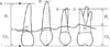
The lengths of the crown and root were calibrated in the following manner and were expressed in mm (Fig. 1).
The crown height (Crh) was defined as the perpendicular line from a point m to the incisal/occlusal reference line (i), 'm' was defined as the midpoint of the line connecting the mesial and distal proximal bone.
For teeth with one incisal tip or buccal cusp (i.e., the canines and most premolars), a line forming a tangent to the incisal tip or the buccal cusp tip was placed perpendicular to the long axis of the tooth.
For teeth with an incisal edge or several buccal cusps (i.e., most incisors and molars), a line was placed to follow the incisal edge or to connect the buccal cusps.
The root length (Rl) was measured from the point m perpendicular to the apical reference line (a) which was parallel to the incisal/occlusal reference line. For multi-rooted teeth, the length was measured to the apex of the longest buccal root. If only one root was visible, then that root was measured.16
To assess the intra-examiner reproducibility, 50 panoramic radiographs were re-measured after 2 months (measurement A2). To assess the inter-examiner reproducibility, another examiner measured the same teeth after being trained in the method (measurement B).
The statistical analyses were performed using the SPSS for Windows, version 9.0 (SPSS Inc., Chicago, IL, USA). To assess the intra-examiner reproducibility, the interclass correlation (ICC) between measurements A and A2 was calculated. To determine the inter-examiner reproducibility, the ICC between measurements A and B was calculated. If the ICC coefficient value was between 0.75 and 1.0, then the reproducibility was considered to be excellent; if it was between 0.6 and 0.74 the reproducibility was considered to be good, and if it was less than 0.6, it was considered to be unreliable.17 The R/C ratio was statistically analyzed according to gender, tooth and arch. The independent t-test was used to analyze differences between the male and female subjects, while the paired t-test was applied to compare the arches. The data were considered to be significant at P<.05.
RESULTS
The ICC coefficient between measurements A and A2, which indicates the intra-examiner reproducibility, was 0.732-0.931. The ICC coefficient between measurements A and B, which indicates the inter-examiner reproducibility, was 0.636-0.887. Therefore, the method employed in this study was determined to be both reliable and reproducible.
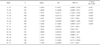
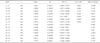
The mean R/C ratios varied from 1.29 to 1.89 (male: 1.28-1.84; females: 1.31-1.94). Detailed statistical data of the R/C ratios for the genders (males and females)(mean, SD, 95% confidence interval) are presented in Table 1, Table 2, and Table 3. In both arches of both genders, the highest R/C ratios were recorded for the mandibular canines (1.89), followed by the maxillary canines (1.79). The lowest R/C ratios were recorded for the maxillary second molars (1.31). In each gender, the highest R/C ratios were calibrated for the mandibular canines (1.84 in the males; 1.94 in the females), and the lowest R/C ratios were recorded for the maxillary second molars (1.28 in the males; 1.31 in the females). In comparison with the maxillary teeth (1.29-1.78), the mandibular teeth (1.47-1.89) yielded a higher R/C ratio, and this difference was significant in the females. The difference between the genders was not statistically significant, with the exception of the maxillary central incisors, mandibular canines and mandibular first premolars.
DISCUSSION
Bone support is one of the most widely used periodontal parameters for determining the prognosis of the teeth and is generally estimated based on radiographic analysis. In this study, the R/C ratios of all the teeth were determined from panoramic radiographs (PRG). PRGs are less accurate than periapical radiographs for measuring lengths and also have inherent problems, such as the overlapping of teeth and impaired visibility at the lateral regions of the maxilla.18 Some research has suggested that using radiographic means to detect periodontal osseous destruction is more accurate when periapical radiographs are used instead of PRGs.19 However, although other radiographic methods such as CT and periapical radiograph could provide more accurate assessment of R/C ratio, in the present study, we needed to measure mean values of R/C ratio of all teeth in a population by calculating the each ratio of the multiple teeth simultaneously; therefore, for this purpose, the assessment via PRGs might be a more useful and practical option. In terms of clinical application, the method based on PRGs is not only easy techniques without significant error under low radiation exposures but also more effective as these radiographs have been routinely taken in treatment planning procedure, evaluation and follow-up.
Assessing the R/C ratios via linear measurements is useful for radiographic examination. To a certain degree, changes in tooth angulation can affect the radiographic tooth length, but the resulting alterations in the R/C ratio are not significant.20 In the PRGs, the horizontal variables were unreliable, but the reproducibility of the vertical and angular variables was acceptable if the patient's head was placed in a holder.18 This phenomenon is a by-product of the horizontal scanning motion of the panoramic unit and is affected by factors such as the radius between the focal plane and the rotational center or the source-receptor and source-focal plane distances.21,22 Because the root and the crown usually lie in almost the same vertical plane, the magnification factor has no major effect on the R/C ratio.21 Therefore, we excluded the palatal roots from analysis in the present study due to their diverging inclination compared to the crown.16,21 Originally, Lind's method was used to study the short root anomaly by measuring the R/C ratio of the anterior teeth on periapical radiographs.15 Hölttä16 applied this method to assess the anatomic R/C ratio on the PRGs of Caucasian subjects with healthy dentition. In the present study, Lind's method was modified by moving the cervical reference line to the alveolar bone area and locating a new "m" point on the connecting line between the mesial and distal alveolar bone to calculate the bone-supported root length for quantitative assessment.
In the present study, the values of R/C ratios for the permanent teeth were acquired from healthy Koreans. There are many other previous assessments of R/C ratios but a variety of methods used make it impracticable to compare data between them and most of these studies were related to dental anomalies and site specific or gender specific values. Studies analyzing the R/C ratios of the full dentition in other races (except for the Caucasian population) have been very rare. The study of Caucasians has involved analysis of the anatomical R/C ratios.16 This provides no information on the level of alveolar support, such that these data cannot be directly applied to other populations.
In the Caucasian studies involving the anatomical R/C ratio, the general R/C ratio was remarkably higher than that of this study. Except central incisors of both arches and maxillary first molars, the mean R/C ratios were in the range of 1.9-2.46 while the mean R/C ratios in this study were less than 1.5 with the exception of canines, maxillary lateral incisors, and mandibular premolars. Although considering the difference between anatomical R/C ratio and clinical R/C ratio, the mean R/C ratios that varied from 1.86 to 2.44 (male) and from 1.78 to 2.46 (female) represented that Caucasian teeth had relatively longer roots than those of Korean. The highest mean anatomical R/C ratios were recorded for the maxillary or mandibular second premolars. The lowest anatomical R/C ratios were determined in the maxillary central incisors.16 Whereas the highest R/C ratios were calculated for the mandibular canines, followed by the maxillary canines and the lowest R/C ratios were determined for the maxillary second molars in this study. The R/C ratios of the maxillary central incisors, mandibular canines and mandibular first premolars revealed a significant difference between the males and females in the present study. According to Hölttä's study,16 substantial gender differences were observed in the maxillary central and lateral incisors, maxillary first molars, maxillary second molars and mandibular central incisors. As widely recognized, the mean lengths of both the crown and the root are longer in males than in females; however, gender differences in the R/C ratios exist for only a few types of teeth. These results suggest there are ethnic differences as well as a difference between the anatomical and clinical R/C ratio.
The results of both studies demonstrate that the mandibular premolars had comparatively higher R/C ratios and that maxillary molars had lower R/C ratios. Maxillary palatal roots were not considered in this measurement; therefore, for the more accurate R/C ratio of maxillary molars, an additional method should be revised. In comparison with the maxillary teeth, the mandibular teeth yielded a higher R/C ratio, as reported in previous studies.
The clinically ideal R/C ratio as an abutment for a FPD is considered to have a value of 2, but this ideal value was rarely observed in the present data. An R/C ratio of 1.5 is acceptable for abutment teeth, although teeth with a lower ratio may also be acceptable abutments when the periodontium is healthy and the occlusion is under control.7 Shillingburg et al.8 suggested that an R/C ratio of 1.5 is ideal for FPD abutments and a ratio of 1:1 is the minimum ratio for abutments under normal circumstances. The authors also proposed that if the antagonist consists of a tissue-supported prosthesis, then an R/C ratio of less than 1 might be accepted because of the reduced occlusal forces. Avila et al.23 recommended a novel decision-making process for tooth retention or extraction that a ratio of 1 is the minimum acceptable ratio when the periodontium is in healthy condition and the occlusion is controlled. Tylman24 recommended that teeth with normal alveolar bone support be used for abutments, but that teeth lacking one-third to one-half of their normal periodontal attachment might be successfully retained when carefully selected. When dentists determine the prognosis of the abutments, factors such as the tooth mobility, alveolar bone support, root configuration and angulation, opposing occlusion, pulpal state, history of endodontic treatment, and the remaining tooth structure should be comprehensively considered. Moreover, ethnic differences should also be addressed. Therefore, it is not practical to consistently apply a ratio of 2 or 1.5 as the standard ratio. In the present study, the mean R/C ratio for each tooth varied from 1.29 to 1.89, indicating that the R/C ratios rarely exceeded 2 and were mostly lower than 1.5, even in healthy conditions. Because of the various assessment methods, it is difficult to compare these data with the previous R/C ratio results. Therefore, to achieve exact comparisons, future studies should employ a standardized method for determining the R/C ratio; moreover, the radiographic analysis should include a consideration of ethnic differences. The assessment of the R/C ratio based on the PRGs in the Korean population is useful for evaluating developmental anomalies, determining the prognosis of teeth, comparing the pre- andor post-treatment conditions, and making comparisons with other populations, patient groups or individuals without referring to the values of other races.
CONCLUSION
Although the assessment of R/C ratios based on the PRGs has inherent limitation in accuracy, the mean values of R/C ratios of permanent teeth in a healthy Korean population were presented.
The mean R/C ratios varied from 1.29 to 1.89 (male: 1.28-1.84; females: 1.31-1.94).
The highest R/C ratios were recorded for the mandibular canines (1.89), followed by the maxillary canines (1.79).
The lowest R/C ratios were recorded for the maxillary second molars (1.31).
The mandibular teeth (1.47-1.89) yielded the higher R/C ratio than the maxillary teeth (1.29-1.78), and this difference was significant in the females.
The difference between the genders was not statistically significant, with the exception of the maxillary central incisors, mandibular canines and mandibular first premolars.

 XML Download