

 PDF
PDF ePub
ePub Citation
 Print
Print


INTRODUCTION
Dentin hypersensitivity (DH) is a painful and chronic dental condition, and it occurs when dentinal tubules on the dentin surface are open and patent to vital pulp, which is probably related to erosion.1 Although most erosive lesions are limited in enamel, deep lesions extending into dentin can occur if the erosive challenge persists.2 Such lesions require a restorative treatment and it is important to be aware of the bonding performance on this type of substrate.
Resin composite (RC) is a popular material for the restoration of abrasion, abfraction, or erosive (AAE) lesions and all other non-carious tooth defects.3 Although RC revealed good retention bonded to eroded dentin according to a 12-month-long clinical study,4 other investigations have shown that failure rates are high.5,6 Because of the poor outcomes of the restoration on AAE lesions, researchers have attempted to find a method to improve the bonding performance. Previous studies have evaluated the influence of different treatments involving roughing of dentin surface7 and the application of a fluoride mouth rinse8 on to bonding performance, however, it has not been determined whether desensitization procedures such as sealant application and laser irradiation affect micro-tensile bond strengths.
Therefore, the aim of this study was to investigate whether desensitization pretreatment influence the micro-tensile bond strengths of RC to eroded and normal dentin.
The null hypotheses were;
MATERIALS AND METHODS
This experiment was performed with an IRB approval of the Korea University Guro Hospital (NO. MD13024-001). Forty-two fresh human third molars without caries and restorations were extracted and restored in a 0.5% chloramine solution at 5℃ until use. The teeth were ground down to form a flat dentin surface from the occlusal side using 320-grit silicon carbide paper on a polishing machine (Kyung Do Precision Industrial, Korea), and the teeth were randomly subjected to an immersion in distilled water during the experimental period (sound dentin, n = 21) and an erosion pH-cycling challenge (eroded dentin, n = 21). Six cycles per day involving 5-minute demineralization and 60-minute remineralization per cycle were applied for pH cycling for 9 days. For erosive demineralization,8 a 0.05 M citric acid solution (pH 2.3, citric acid monohydrate) was used. After each erosion procedure, the teeth were thoroughly rinsed with tap water for at least 1 minute and kept in remineralization solution for 1 hour. The remineralization solution (pH 6.7)9 consisted of 4.08 mmol/l H3PO4, 20.10 mmol/l KCl, 11.90 mmol/l Na2CO3, and 1.98 mmol/l CaCl2. All chemicals were obtained from Sigma Aldrich (St. Louis, MO, USA). The pH values of solutions were checked with a pH meter (pH/ION Meter DP-880, Dong-Woo Medical System, Seoul, Korea) at the beginning and end of each experimental day.
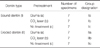
Afterward, teeth with sound dentin (I) or eroded dentin (II) were pretreated in three different ways (Table 1): a) application of Gluma® Desensitizer; b) irradiation with a CO2 laser; c) left untreated. Two dentin substrate types and 3 desensitizing pretreatments produced 6 groups with 7 teeth in each (Table 2).
After surface pretreatment, one molar from each of the 6 groups (Ia, Ib, Ic, IIa, IIb, IIc) was prepared for SEM to evaluate morphological surface changes.
Teeth from each group was treated with Scotchbond™ Etchant (LOT. 9AL, 3M™ ESPE™, St. Paul, MN, USA) for 15 seconds and rinsed. The total-etch adhesive Adper™ Single Bond 2 (LOT. N341785, 3M™ ESPE™, St. Paul, MN, USA) was applied according to manufacturer's instructions.
After bonding procedure, molars were built up in two layers of 2 mm each with a RC- Filtek Z250 (Lot. N453720, 3M™ ESPE™, St. Paul, MN, USA). Each layer of resin composite was light-cured for 20s, and all light-curing was performed with an LED light curing unit (LED curing light, Dong Yang Linuo Medical Apparatus Co., Ltd., China). And then, all specimens were stored in a water bath with distilled water (Water Bath, Chang Shin Scientific Co., Ltd., Korea) for 24 hours at 37℃.
Teeth were vertically serially sectioned (Minisaw, GLP Korea, Korea) perpendicularly to the bonded interface to produce several sections with 1 mm thickness. These sections were further sectioned perpendicularly to the adhesive interface to produce sticks with adhesive area approximately 1 mm2 according to the non-trimming version of micro-tensile technique.10 Six to seven sticks from each molar were obtained and four of them were randomly selected for the measurement of micro-tensile bond strengths (µTBS). In cases of pre-test failure, another stick from the same tooth was used as a replacement. The exact dimensions of each stick's bonding surface areas (BS (mm2)) were measured using a digital caliper (Mitutoyo, Kawasaki, Japan). The sticks were fixed by their ends to a test jaw mounted in micro-tensile tester (Microtensile Tester, Bisco Inc., Schaumburg, IL, USA) using cyanoacrylate glue (Zapit, Bisco Inc., Schaumburg, IL, USA). Then they were stressed in tension at a crosshead speed of 1.0 mm/min until fracture occurred, and the maximum force (Fmax (N)) was recorded. The µTBS values (MPa) were calculated using the formula, µTBS = Fmax/BS.
In addition, failure modes of debonded specimens were evaluated at 30× under a stereoscopic microscope (Model CX31, Olympus, Tokyo, Japan). Failure modes were classified as: (1) adhesive failures (failure at interface between adhesive resin and dentin), (2) cohesive failures (cohesive failure of dentin or resin), or (3) mixed failures (exhibits some cohesive failure and some adhesive failure).
After pretreatment, one molar per group was mounted on aluminium stubs, dehydrated in a desiccator (Freeze Dryer ES-2030, Hitachi Co., Tokyo, Japan) for 24 hours, and then sputter-coated with gold using a sputtering device (ION SPUTTER E-1045, Hitachi Co., Tokyo, Japan). Dentin surfaces were evaluated under an SEM (SEM S-4700, Hitachi Co., Tokyo, Japan), and photographs were taken at 1,000× and 5,000×.
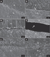
To analyze adhesive interfaces, one additional tooth from each group was prepared in a similar way as described for µTBS measurements. Samples were prepared for SEM and evaluated at 1,000× and 5,000×.
Micro-tensile bond strengths data were analyzed using two-way ANOVA, and Tukey's test was used as a post-hoc test to analyze difference between groups at a significance level of 0.05. The analysis was conducted using IBM SPSS Statistics (Version 19, IBM SPSS, Armonk, NY, USA).
RESULTS
Table 3 shows the µTBS means and standard deviations for all groups. Two-way ANOVA revealed that both the factors tested (dentin type and desensitizing pretreatment) and their interaction had significant influences on µTBS. Tukey's test further showed that for all three pretreatments, the mean µTBS to sound dentin were significantly higher than those to eroded dentin.
For sound dentin, the group of pretreatment with Gluma (Ia) exhibited the highest mean µTBS while the group of pretreatment with CO2 Laser (Ib) showed the lowest mean µTBS. There are significant differences (P<.05) between these two groups and the group without any pretreatment (Ic) in mean µTBS (Table 3).
For eroded dentin, the group of pretreatment with Gluma (IIa) presented significant higher (P<.05) µTBS than the other two groups (IIb and IIc), neither of which were different from each other (Table 3).
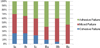
The failure mode percentages of the debonded specimens are shown in Fig. 1.
The overall failure types for specimens were "adhesive" and "mixed". However, more cohesive failures were observed in sound dentin groups (Group I) than in eroded dentin groups (Group II).
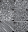
Fig. 2 shows the microstructure of the dentin (sound/eroded) surface after desensitizing pretreatment.
DISCUSSION
The prevalence of both DH and dental erosion has increased over the last few decades and their causalities appear to be linked.11,12,13,14 Increasing soft drink consumption contributes to the development of erosion.15,16 Initially, acid softens enamel and dissolves enamel crystals, however, the lesions formed can extend to dentin if etiological factors are not controlled, and then oral rehabilitation becomes necessary. The present experiment was designed to simulate the clinical symptoms of dental erosion and DH, and to investigate the effect of desensitizing pretreatments on micro-tensile bond strengths of RC to sound and eroded dentin.
The pH cycling performed in present study simulated erosion challenges, in terms of severity and time, that occur in patients at high risk of dental erosion, e.g., people who frequently consume acidic soft drinks.16,17 Such exposure can results in loss of minerals (around 90 µm),18 a broad zone of exposed collagen matrix, and increased tubule diameter.14 In the present study, SEM revealed that the smear layer was entirely removed and dentinal tubules were totally opened. After demineralization, peritubular dentin was apparent as some hydroxyapatite crystals only remain around dentinal tubules (Fig. 2D, Fig. 2E, and Fig. 2F). These surface changes induced by the artificial erosion process are in accordance with a previous report.8
The micro-tensile test was used to evaluate micro-tensile bond strengths, because it is believed to provide an accurate means of verifying bond performance. As compared with "macro" tests, micro-tensile bond strengths test share some advantage, that is, the conservation of teeth, fewer cohesive failures in substrate, and uniform distribution of stress to the material, all of which justify its choice. However, there are also limitations of µTBS testing, including the following: labor intensive; technically demanding; specimens easily dehydrate and easily damaged; post-fracture specimens adhere to the test jaw with glue can be lost or damaged during removal.3,19,20 To prepare the samples for micro-tensile test, we utilized the 'non-trimming technique' instead of the former 'trimming version' in which fabrication of dumbbell or hourglass specimens is needed. The resulting modified technique provides a uniform cross-sectional area and allows tensile stress to be applied to a beam uniformly.20
The adhesive system used in the present study (Adper™ Single Bond 2) is an etch-and rinse adhesive system and has been well investigated and well documented.7,21 Normally, this adhesive system is applied on sound dentin with presence of a smear layer. However, in the present study, we evaluated its bonding performance on demineralized dentin with an organic matrix.
It is well known that for etch-and rinse adhesive systems, bonding relies on the formation of a hybrid layer composed of residual hydroxyapatite, exposed collagen and infiltrated resin. After etching, resin monomers penetrate the spaces of the porous dentin collagen previously occupied by hydroxyapatite crystals. This etch-and rinse adhesive system has excellent bonding performance onto sound dentin surfaces, but its performance on erode dentin surfaces is much worse. This is in accordance with the findings of other studies regarding the lower micro-tensile bond strengths of etch-and rinse adhesives to demineralized dentin.7,8,22,23 Regarding the thick layer of exposed collagen on eroded dentin surfaces and its higher water content, it is conceivable that Single Bond 2 could neither infiltrate completely nor polymerize properly. Thus, the micro-tensile bond strengths values observed represent the strengths of an adhesive system that penetrated demineralized dentin incompletely. Accordingly, it is understandable that the micro-tensile bond strengths values were significantly lower than for non-erosively demineralized teeth, regardless of the pretreatment used.
The artificial erosion process did not only negatively influence micro-tensile bond strengths, but also changed the failure pattern. Higher percentages of cohesive failures (Fig. 1) were observed in the eroded dentin groups. This type of failure has often been interpreted as evidence that bond strength of adhesive to dentin is stronger than the cohesive strength of dentin/RC. However, the nature of fracture is far more complex than that. It has been suggested that failure mode is affected by the material properties of all components of bonded joints, that is, the RC, hybrid layer, and dentin.24 Thus, the presence of air bubbles can act as stress points in eroded dentin, which is softer and more porous than sound dentin, which would increase the likelihood of dentin cohesive fracture, especially in non-penetrate demineralized regions.
Moritz et al.25 reported the use of the CO2 laser for the treatment of DH for the first time, and demonstrated that its effects are related to its ability to occlude dentinal tubules. In the present study, micro-tensile bond strengths to sound dentin after irradiation were significantly lower (P<.05). The reasons for this are related to the existence of an acid resistant smear layer and the deficient diffusion of adhesive monomers within denatured fibrils.26 Laser irradiation can form different organic and inorganic compounds, such as, melted collagen fibrils, calcium pyrophosphate, calcium meta phosphate, and α- and β-tricalcium phosphates, which present different levels of acid solubility.27 Thus, it is likely that laser-modified dentin is not etched completely, and this could partially obstruct the micromechanical attachment of adhesives, and make it difficult for the bonding agent to penetrate dentin.28 The cohesive dentin failures, observed in the Ib and IIb groups, may suggest that the denatured surface of dentin is a fragile substrate that is not adequately infiltrated by resin monomer.
Gluma® Desensitizer contains 2-hydroxyl ethyl methyacrylate (HEMA) and glutaraldehyde and causes coagulation of dentin fluid protein in dentinal tubule, and thus, plugs tubules, which contributes to its desensitizing effect. In the present study, Gluma® Desensitizer had a positive effect on the micro-tensile bond strengths of etch-and rinse adhesive to sound dentin, which corresponds to the findings of other investigations.29,30,31 However, the new discovery was that bonding performance for eroded dentin was also developed after pretreatment with Gluma® Desensitizer. Since eroded dentin is characterized by exposed collagen fibrils, the etching process before bonding to eroded dentin can result in a wide, deep completely demineralized dentin zone.7 We supposed that the way of Gluma® Desensitizer to improve the bonding performance is related to the use of glutaraldehyde and HEMA. Application of glutaraldehyde can fix completely exposed collagen fibrils, binding and reinforcing it to the underlying structure.32 HEMA, on the other hand, can help resin monomer infiltrating into the collagen and blending hydrophobic and hydrophilic components.33 However, the durability of it is unreliable because of the hydrolytic instability of HEMA.34
Therefore, water ageing is needed to measure the long-term bonding performance to eroded dentin. It would add much more useful information regarding the clinical lifetimes of adhesive restorations.
CONCLUSION
Within the limitations of the study, the following conclusions were drawn;
Micro-tensile bond strength of resin adhesive to teeth was affected by dentin type and the surface treatments applied to dentin.
Erosion results in a significant decrease of bond strength compared to non-erosive demineralized teeth.
The application of Gluma® Desensitizer before bonding procedure is likely to promote the performance of the adhesive.
Irradiation with CO2 laser on sound dentin surface may reduce the bonding performance of adhesive.


 XML Download