

 PDF
PDF ePub
ePub Citation
 Print
Print


INTRODUCTION
There has been considerable effort to enhance osseointegration between bone and implant material. Especially, by modifying the surface properties of mechanical roughness, osteogenesis ability of the implant is enhanced. Not only topography but also chemistry of the TiO2 surfaces affect cellular attachment, proliferation, osteoblastic activity, differentiation, and formation of mineralized nodules on the surface. Therefore, a range of chemical modifications of titanium surfaces, such as fluoride treatment, anodic oxidation, NaOH treatment, heat treatment, and ion implantation have received attention.
Some studies1,2,3,4 demonstrated that divalent cations, such as magnesium (Mg) and calcium (Ca) ions play a momentous role in osteogenesis and bone remodeling. Mg ions in oxide layer migrate toward the amorphous immature bone layer, whereas P and Ca ions in the body fluid migrate toward the O2 layer of the implant.1,2 Magnesium ions play an essential role in the binding interaction between ligand proteins and cell surface receptors such as the integrin superfamily; vitronectin, fibronectin, fibrinogen, and some cell-cell adhesion receptors.2,3
It is necessary to understand the osteoblast and osteoclast responses on the titanium implant surface, which involves attachment, proliferation and differentiation, in order to understand the initial influence of the divalent cations. In this report, Magnesium ions were implanted into the titanium surface by micro arc oxidation (MAO) using a vacuum arc source filter. This study examined the initial cellular response of osteoblasts and osteoclasts to the titanium surfaces that had been implanted with or without Mg ions using MAO.
MATERIALS AND METHODS
Mechanical polishing was applied, and 40 specimens were grounded utilizing No. 800 silicon carbide sand paper to imitate machined surface on commercially made pure titanium plates 10 mm in diameter and 3 mms thick (Shinheung Co., Seoul, Korea). All the specimens were cleaned in detergent solution using ultrasonics and dried in an oven at 50℃ for 24 hours. Magnesium ions were implanted onto half of the specimens to induce bioactivity using the MAO method. Titanium disc and platinum were located on anode and cathode in magnesium electrolytic solution. The implantation field had an electric energy of 60-500 V. In this energy field, a micro arc was induced on the titanium disc and subsequently a magnesium titanate oxide was formed.
The Mg ions showed acceleration within the electric field located between the sheath and substrates. The electric energy in the implantation field was 15 keV and retention dose was 5.0 × 10¹⁵ ions/cm2.
All the specimens were enveloped and then sterilized with 30 kGy γ-radiation using a 60Co source at the UTR GAMA-Pi facility located at Greenpia in Yeoju, Korea (Fig. 1).
Using a scanning electron microscopy (SEM, JSM-5800; JEOL, Tokyo, Japan), the structure and surface morphology of the specimens were analyzed. And for the evaluation of hydrophilic properties of the titanium surface, surface contact angles were measured using the sessile drop method and a video contact angle instrument (Phoenix 150, SEO, Suwon, Korea)(Fig. 1).
MC3T3-E1 cells, osteoblast-like cells derived from mouse calvarias were used. The choice of culture medium was Dulbecco's Modified Eagle Medium (DMEM, Gibco, Rockville, MD, USA) containing 10% fetal bovine serum (FBS, Gibco, Rockville, MD, USA), 100 U/mL penicillin and 100 µg/mL streptomycin (Gibco, Rockville, MD, USA). Cell culturewas done in a humidified atmosphere containing 95% air and 5% CO2 at 37℃. Osteogenic medium, which contains 10% FBS, 10 µM dexamethasone (Sigma, St. Louis, MO, USA), 25 µg/mL L-ascorbic acid (Sigma, St. Louis, MO, USA), and 10 mM β-glycerophosphate, was added to the culture medium. This medium was changed every 2 days.
The experimental group was divided into the following 4 groups.
(1) NC group cultured on a machined titanium disc without the addition of cell differentiation media.
(2) PC group cultured on a plate with cell differentiation media.
(3) M group cultured on a machined surface titanium disc with cell differentiation media.
(4) MM group cultured on a Mg-incorporated surface titanium disc with cell differentiation media.
Osteogenic ability was evaluated by measuring the activity of ALP, which is a biochemical marker for osteoblasts. The MC3T3-E1 cells were cultured on discs within a 48-well culture plate. The initial seeding density was 1 × 10⁵ cells/mL/well in a humidified atmosphere containing 95% air and 5% CO2 at 37℃. By the 7th, 14th and 21st day, the medium was completely removed and washed 3 times with PBS and treated with 1 mL of 0.1% Triton X-100 (Sigma) in an ultrasonic processor for membrane dissolution. After centrifugation at 13,000 rpm for 10 minutes at 4℃, 200 µL of upper phase was added to a mixture of 50 µL diethanolamine buffer (pH 9.8, containing Magnesium Chloride, Sigma, MO, USA) and 50 µL p-nitrophenyl phosphate substrate, and then incubated for 1 hour. When enzyme corresponded with the substrate and appeared a yellowish color, 100 µL of 1 N sodium hydroxide was combined to stop enzyme reaction. Then absorbance measurements were made utilizing a microplate reader (Bio-Rad, Hercules, CA, USA).
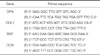
The cells were seeded on discs in 6-well culture plates with initial seeding densities of 1 × 10⁴ cells/well, and cultured for 7 and 14 days. From the cultured cells on 6 titanium discs, total RNA was extracted and quantified at each incubation time by utilizing RNeasy Mini kit (Qiagen, Valencia, CA, USA). First-strand cDNAs were produced by reverse transcription of 1 µg of extracted RNA using Superscript First-Strand Synthesis System (Invitrogen, Carlsbad, CA, USA). To determine various mRNA levels of osteopontin (OPN), collagen 1 (COL1), bone sialoprotein (BSP), and osteocalcin (OCN), real-time PCR reaction performance was obtained utilizing primers shown in Table 1. For this process, real-time PCR was perfomed by DyNAmo™ SYB® Green qPCR Kit (Finnzymes, Espoo, Finland) and ABI7300 Real-time Thermal cycler (Applied Biosystems, Forster, CA, USA).
Using alizarin red S staining, of the cell layer mineralization was observed. Triplicate plated osteoblasts at a density of 1 × 10⁵ cells/mL droplet were cultured on prefabricated titanium discs or tissue culture plastic. The 40 specimens were incubated at 37℃ in an incubator. After 21 days of culture, medium was then eliminated and cultured cells were gently rinsed 3 times with phosphate-buffered saline (PBS) and fixed in 70% ethanol for 1 hour at -20℃. After being fixed, the culture cells were rinsed with Nanopure water (no calcium ion) and stained with 2% alizarin red-S (AR-S, Kanto Chemical, Tokyo, Japan) for calcium at room temperature for 20 minutes. Cells were cleansed with Nanopure water. 10% cetylpyridinium chloride was added and absorbance was measured by spectrophotometer (Molecular Devices, Menlo Park, CA, USA) at 540 nm to evaluate the intensity of AR-S staining.
Mouse bone marrow-derived macrophages (BMMs) cells were cultured in a humidified atmosphere containing 95% air and 5% CO2 at 37℃. The culturing medium was Minimum Essntial Medium Alpha (alpha-MEM, GIBCO, NY, USA) containing 10% fetal bovine serum (FBS, GIBCO, NY, USA). This medium was renewed every 3rd day. 100 µg/mL RANKL and 40 µg/mL M-CSF were used for differentiation of the cells.
Staining actin filaments with rhodamine-conjugated phalloidin can detect actin rings of osteoclasts. The cells (bone marrow macrophages BMMs) were cultured for 6 days for the actin ring formation assay by being seeded in 48 well-plates with the presence of 25 ng/mL RANKL. At the end of incubation, cells were fixed in 3.7% formaldehyde for 10 minutes. Fixed cells were stained with rhodamin-conjugated phalloidin for 30 minutes. Osteoclasts staining with rhodamin-conjugated phalloidin for actin was initiated. Distribution of actin rings was visualized and detected using a fluorescence microscope. Cells were examined by a Zeiss Axiolab fluorescence microscope (FluoView FV300 Confocal Microscope, Olympus, Tokyo, Japan).
Total RNA was extracted from the cultured cells and isolated using RNA RNeasy kit (Qiagen, Valencia, CA, USA) according to the manufacturer's instructions, and cDNA was synthesized from 1 µg of extracted RNA using OligodT (Invitrogen, Carlsbad, CA, USA) by using bone marrow macrophages (BMMs) seeded in 6-well plate at densities of 1 × 10⁵ cells/mL/well. A specific primer was added to the synthesized cDNA to perform RT-PCR by PCR premix (Bioneer, Daejeon, Korea). The primer used in RT-PCR was synthesized from a part of tartrate resistant acid phosphatase (TRAP) gene, which was used as an indicator of osteoclast differentiation. The housekeeping gene, glyceraldehyde-3-phosphate dehydrogenase (GAPDH), was utilized as the standard control group (Table 2). After the initial denaturation step at 94℃ for 4 minutes, 32 PCR cycles were run at 94℃ for 30 seconds, 57-66℃ for 1 minute and 72℃ for 1 minute for gene amplification. DNA concentration was measured using ethidium bromide stained 2% agarose gel by a UV absorption spectrophotometer.
Statistical analysis was performed using the SPSS 14 statistical system (SPSS Inc., Chicago, IL, USA), which aided in calculating the standard deviation and mean of the data. One-way analysis of variance (ANOVA) with the Tukey's multiple comparisons test was executed to calculate differences among groups. Evaluation values of P<.05 were considered statistically significant.
RESULTS
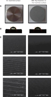
Fig. 1 shows titanium disc specimens with or without magnesium implantation. The size of each disc is 10 mm diameter × 3 mm thickness (Fig. 1A). Machined surface (M) is more hydrophobic than the Mg-implanted machined surface (MM) showing a bigger contact angle (Fig. 1B, Table 3). SEM images of the different titanium surfaces are shown (Fig. 1C). M shows the typical features of a titanium grinding surface containing parallel grooves. MM did not alter extremely, but changed slightly smoother after ion implantation in the micron scale. The reason for the change of surface morphology is due to ion bombardment during the Mg ion implantation process. This can be seen at a higher magnification. However, the surface roughness of the MM slightly decreased due to ion implantation.
After culturing, the morphology of the MC3T3-E1 of both groups was round and it spread partially over the experimental surfaces. The cells grew along the parallel grooves of the titanium surface and proliferated and covered the experimental surface. The cells were flattened and well spread, and there was little difference between M and MM.
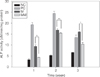
For investigating the cell differentiation, alkaline phosphatase activity was measured. An increasing ALP expression means a subsequent increasing of osteoblastic activity and bone formation. Fig. 2 shows the results of the ALP activity measurement. ALP activities showed an increase inincubation time in all the groups until the 14th day. After 21 days, ALP activities decreased or maintained stable. Only in the negative control group did the ALP activity always increase concurrently with incubation time. At the 7th and 14th day of culture, cells cultivated on culture plate with cell differentiation (positive control) showed the highest ALP activity and on culture plate without cell differentiation (negative control), the lowest ALP activity was seen. MM exhibited lower ALP activity than those of M (P<.01 at 7 and 21 days, P<.05 at 14 days). There was a significant difference between groups in ALP activity and differentiation. MM group showed relatively lower ALP activity at 7 days, but explosively increased at 14 days and relatively maintained stable at 21 days. Compared with M, ALP activities of the MM imply that magnesium incorporation did not promote differentiation of osteoblastic cells.
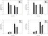
Quantitative real time PCR analysis of mRNA levels for osteopontin (OPN), collagen 1 (Col1), bone sialoprotein (BSP), and osteocalcin (OCN) in cells grown on different surfaces was performed on the 7th and 14th day of culture. Levels of osteoblastic phenotype gene expression are illustrated in Fig. 3. Enhanced alkaline phosphatase activity on M and MM on the 7th and 14th day resulted in simultaneous increases in the expression of marker genes (BSP and osteocalcin) for osteoblastic differentiation at 7th and 14th days. Enhanced expression of diverse integrins associated in cell grown on the MM at an early incubation time resulted in consecutive increase in BSP and OCN expressions on the 7th and 14th day. Expression levels of mRNA in BSP and OCN escalated in accordance with elapsed incubation time in all groups. OPN and Col1 mRNA expression were elevated in cells grown on both surfaces than negative control in every case. While the level of expression decreased as time passed, the patterns of OPN and Col1 mRNA expression were dissimilar to that of alkaline phosphatase activity on the 7th and 14th day of culture. Although MM surface did not exhibit a higher OPN, Col1, and osteocalcin mRNA expression levels on the 14th day, MM surface displayed relative stable mRNA expression on the 7th and 14th day. Results from replicated experiments showed similar gene expression patterns.
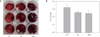
Alizarin Red-S stain for mineralization demonstrated that osteoblasts cultured on positive control, M and MM surfaces had higher levels of mineralization; more intense red staining which indicate calcium deposition was observed. When quantified by image analysis programs, significant (P<.05) enlargement in mineral amount was noticed in osteoblasts cultured for 21 days (Fig. 4A). But no significant difference among all the groups were observed (Fig. 4B). Therefore, we concluded that ostoeblasts increased mineralization on all surfaces without significant difference.
We examined the outcome of osteoclast maturation and differentiation ability to resorb bone during bone remodeling. Mouse bone marrow-derived macrophages (BMMs) cells were plated on each Ti disc. Cells were stimulated with 100 µg/mL RANKL and 40 µg/mL M-CSF. RANKL and M-CSF stimulated BMMs on M and MM surfaces and cells differentiated, suggesting that the bone resorption activity of cells made them into an active state resembling osteoclasts, which were produced by an actin ring structure. Immunofluorescence analysis was performed to find the effect on actin ring formation (Fig. 5). The majority of negative control (NC) group cells without addition of RANKL and M-CSF displayed no actin rings, while M and MM group cells revealed well-formed actin rings. As expected, M and MM group cells showed many full actin rings and disrupted actin rings with more than 50% intact compared to the NC group cells. No significant variation between M and MM group were observed, but the MM group showed relatively filled actin rings.
Phenotypes of differentiated mouse bone marrow-derived macrophages (BMMs) were characterized by investigating the gene expression by using RT-PCR (Fig. 6). Markers assigned to BMMs were TRAP to characterize osteoclasts. GAPDH expression wasdecided inorder to validate the usage of similar amounts of RNA for RT-PCR. Osteoclastic marker TRAP was detected. RT-PCR analysis showed the highest expression level for TRAP on MM surface.
DISCUSSION
Currently, biochemical bonding mechanisms have been observed utilizing a variety of chemical modifications of titanium surfaces; i.e. ion implantation.3,5 Plasma source ion implantation (PSII) was produced to implant ions into the Ti surface by changing the surface chemistry but not altering the surface topography. Changes in surface chemistry influence osteogenesis and bone remodeling. Previous studies have proven that Mg incorporated oxide surface manufactured by an anodic oxidation process makes stronger bone integration.6,7 Because a stronger bone response was investigated, Mg ion implantations have the possibility of higher osteoconductivity. Therefore it is necessary to understand the osteoblast and osteoclast responses on the Mg-incorporated titanium implant surface.
In this study, the surface topography which may be triggered by bombarding changed slightly after Mg ion implantation. Throughout this energetic bombardment of ions, the transfer of kinetic energy from incident particle to atoms on the surface of the specimen results in some of the atoms being removed from the surface. Recent studies suggest that these textures can also have an effect on the osteoblasts.8 This change can also provide a chance for stable cellular proliferation and differentiation because of good biocompatibility. Mg ion implantation has potential enhancement in the cell response at initial stage of osseointegration.9,10 In this study, the surface topography and surface chemistry of the substrates appeared to influence the initial cell response of osteoblast and osteoclast. But the SEM assay showed no significant difference between the surface morphology. Therefore, we can speculate that the initial cellular response was not affected by the surface roughness but that the surface chemistry influenced the cellular response through the initial and early stages, especially on osteoclasts.
By measuring the ALP activity of MC3T3-E1 cells cultured on M and MM, we found that Mg incorporation did not enhance the differentiation of MC3T3-E1 cells. But on both Mg surfaces that were incorporated and machined, MC3T3-E1 cells showed increased and maintained ALP activity and expression of various osteoblast phenotype gene which induce osteoblastic differentiation. Specific osteoblast phenotype genes (OPN, Col1, OCN, and BSP) appeared during active osteoblastic differentiation.11,12 Our findings conclude that the expression of osteoblastic phenotype genes, especially on BSP and OCN, parallels a similar induction to ALP activity which is in accord with previous literatures. BSP and OCN genes are responsive to osteoblastic differentiation. It is understood that magnesium ions aid the binding of the physiologic ligand to activated cation-binding A-domain.13 Therefore elevated levels of ligand-binding integrins activate integrin-mediated intracellular signaling pathways and can positively influence the consequent cell behavior on titanium substrates.14 Olivares-Navarrete et al.15 demonstrated that integrin a2b1 can mediate osteoblastic response to the microscopicsurface topography topography and chemistry of titanium substrate. Promoted differentiation and adhesion of osteoblastic cells induce the expression of transcription factor gene and osteoblast marker genes such as BSP and OCN.16 The improved osteoblast differentiation with incorporated magnesium ions in the titanium oxide layer may be accredited to increase magnesium ion-mediated binding to critical integrins activating consequent osteoblastic differentiation.9
Increased ALP activity and gene expression demonstrate the capability to differentiate into osteoblastic cells. Through the alizarin red S staining, mineralization was observed in every surface cell layer during osteoblast differentiation of the MC3T3-E1 cells. Therefore our data indicate that the MC3T3-E1 cells proliferated and differentiated in osteoblast cell on both M and MM without significant difference.
At the end of incubation, osteoclasts were stained with rhodamin-conjugated phalloidin for actin and DAPI for nucleus. A fluorescence microscope was utilized to detect and visualize the distribution of actin rings. Osteoclasts displayed full actin rings or disrupted actin rings with more than 50% intact.17,18 M and MM group cells revealed well-formed actin rings compared to the NC group cells which showed no actin rings. The expression levels of tartrate-resistant acid phosphatase (TRAP) mRNA were evaluated by semi-quantitive RT-PCR duringculture. As shown in Fig. 6, TRAP was also confirmed by quantitative RT-PCR. This data shows that MM has more affect in differentiation of the BMMs than M. In the BMMs, various intracellular signaling pathways are activated in osteoclast precursor cells and differentiating osteoclasts on MM. Previous studies showed that various intracellular signaling pathways involving ERK, JNK, p38, and Akt/protein kinase B (PKB) are involved in the differentiation of osteoclasts.19,20 In particular, osteoclast activity was higher in the MM group.
Such outcomes suggest that both machined and magnesium incorporated oxide surfaces enhance osteoblastic and osteoclastic cell differentiation, but Mg-incorporated oxide surfaces are able to mature and differentiate osteoclasts better. Although further detailed studies are in demand to find if this surface could also show in vivo bone healing-promoting effects, the limited outcomes from this in vitro study imply that the magnesium incorporated titanium oxide layer fabricated may have a synergistic effect by promoting osteoclastic differentiation.
The results obtained in this paper propose that the use of Mg-incorporated Ti oxide implant body can lead to improvements in bone remodeling by enhancing osteoclastic differentiation. From analysis of the response on the magnesium surface, there is anticipationof the magnesium ion implantation for application as a potential biochemical bond. Further studies are needed to examine the precise mechanism of magnesium ion implantation and the long term effects of Mg ion implants in humans.
CONCLUSION
Both machined titanium surface (M) and Mg-incorporated Ti oxide discs (MM) have a good effect on osteoblastic and osteoclastic cell differentiation, but MM may speed the bone remodeling process by substantially activating on osteoclast differentiation. Mg ion coating may be an effective approach in improving the osteoconductivity and bone remodeling of commercial titanium implants.


 XML Download