

 PDF
PDF ePub
ePub Citation
 Print
Print


INTRODUCTION
The introduction of osseointegration and the use of endosseous implants provide alternative treatment options to clinicians for all indications of edentulism.1 Implant-supported, fixed restorations are usually classified as screw- or cement-retained.2,3 The advantage of screw-retained restorations is the combination of a rigid connection between the restoration-abutment complex and its retrievability. However, these restorations are usually more expensive than cement-retained restorations because of the use of extra components and laboratory costs.4 Cement-retained restorations were introduced to compensate for problems of screw loosening and the lack of esthetics of screw-retained restorations.4 The lack of fastening screws in cement-retained restorations reduces the possibility of preload stress and screw loosening.5 The major advantages of cement-retained restorations are the passive fit of frameworks, enhanced esthetics resulting from lack of screw access holes, and reduced complexity of laboratory procedures and chair-side time.6,7,8 The disadvantages of cement-retained restorations include the requirement for extra time for cementation, removal of residual excess cement, limited design possibilities for superstructure, and the reduced possibility for modifying treatment in case of periimplantitis.9
The existence of residual excess cement in peri-implant sulcus is a common complication of cement-retained implant prostheses.10,11 If there is excess cement located in the soft tissue deeper than 3 mm, it might be difficult to observe and remove. Insufficient removal of excess cement may result in swelling, soreness, exudation or bleeding on probing, and can initiate a local inflammatory process, which is evidence of peri-implant disease and can ultimately lead to implant failure.12,13,14 Moreover, removal of excess cement may cause scratching and gouging on the implant surfaces when plastic and metal scalers are used.15 Several authors have reported on techniques regarding procedures used to assist in minimizing residual excess cement extrusion.16,17,18
This article describes a method of controlling cement flow, using stock or custom implant abutments, when cement-retained implant-supported restorations are utilized. The method can be used easily and quickly at chair-side by the use of daily restorative and laboratory materials. The use of die spacers results in a uniform space between the crown restoration and the implant abutment.
TECHNIQUE
Check the marginal fit of the crown restoration to the implant analog on the dental model.
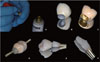
Apply die spacer into the intaglio surface of the crown restoration (Fig. 1A) according to the manufacturer's recommendations (Siladent Die Spacer 12 µm Gold, Dr. Böhme & Schöps GmbH, Germany).
Repeat applying the die spacer until the desired cement film thickness is achieved (application of the die spacer 3-4 times forms a film thickness of approximately 45-50 µm).
Completely fill the crown restoration with a bis-acrylic temporary restorative material (Luxatemp Plus, DMG Chemisch-Pharmazeutische Fabrik GmbH, Germany) and put a retention pin (Bredent GmbH & Co. KG, Germany) with a smaller diameter tip into the uncured material to form a handle (Fig. 1D) and secure the retention pin until the bis-acrylic material is cured.
Remove the crown restoration and check any discrepancies between the implant abutment and the bis-acrylic abutment. Check that there are no voids on the duplicate abutment, and that the finish line has been duplicated accurately (Fig. 1E, Fig. 1F).
Clean the intaglio surface of the crown restoration with air and check any residual die spacer.
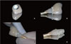
Mix a desired luting agent (Temp Bond NE, KerrHawe S.A., Switzerland) and apply to the intaglio surface of the crown restoration (Fig. 2A, Fig. 2B)
Place the crown gently onto the bis-acrylic abutment and wipe off the excess cement with a cotton swab (Fig. 2C).
Remove the crown restoration from the bis-acrylic abutment. Note that there is a residual cement layer on the bisacrylic abutment (Fig. 2D).
If there is a lack of cement layer, line the intaglio surface of the crown restoration with a thin layer of extra luting agent.
Place the implant restoration onto the implant abutment intraorally. There should be little or no excess cement.
SUMMARY
This article presents a method of minimizing the excess cement around implant-retained restorations. The advantage of the technique is allowing the control of cement flow by using a custom-made duplicate abutment that can be fabricated quickly, easily, and economically at the time of implant abutment/crown insertion.16 The major benefit of extraoral cementation is to allow the indirect removal of excess cement around the margins.
This clinical procedure is extremely important for avoiding the potential of peri-implant disease caused by residual cement left in the gingival sulcus. It is important not to form an oversized cement space when duplicating the implant abutment. The use of a die spacer provides a space of approximately 50 µm, which represents the ideal cement space, and may be used for both custom and prefabricated abutments.
The disadvantage of the technique is that it is a time-consuming procedure for routine clinical processes. Dumbrigue et al.16 stated that when the extraoral cementation technique is preferred, the luting agent must have a longer working time. When a custom abutment is to be used, the dental laboratory may be instructed to make an abutment analog using an acrylic resin, but this is time consuming for the technician and involves additional laboratory costs.18
 XML Download