Journal List > J Adv Prosthodont > v.5(1) > 1054164

Kumar, Garg, and Gupta: A determination of occlusal plane comparing different levels of the tragus to form ala-tragal line or Camper's line: A photographic study

Abstract

PURPOSE

The purpose of this study was to determine accurately the part of the tragus to be used to form the Ala-Tragal line or Camper's line in orthognathic profile patients.

MATERIALS AND METHODS

150 dentate subjects with age of 18-40 years with orthognathic profile were sampled. Life-size lateral digital photographs of the face with fox plane were taken in natural head position. Different angles between Eye-Ear plane and occlusal plane (OT1-OP), Eye-Ear plane and ala-superior border of tragus (OT1-AT1), Eye-Ear plane and ala-middle border of tragus (OT1-AT2) and Eye-Ear plane and ala-inferior border of tragus (OT1-AT3) were calculated using computer software package, AutoCAD 2004. From the three angles formed by the Eye-ear plane (OT1 or FH plane) and the ala-tragal lines, the one closest to the angle formed between Eye-Ear plane (OT1) and occlusal plane (OP) was used to determine the occlusal plane of orientation. The obtained results were subjected to ANOVA F test, Tukey's Honestly significant difference test, followed by Karl Pearson coefficient of correlation test. P values of less than 0.05 were taken as statistically significant.

RESULTS

The mean of base line angle i.e. OT1-OP angle (11.96 ± 4.36) was found to be close to OT1-AT2 angle (13.67 ± 1.93) and OT1-AT3 angle (10.31 ± 2.03), but OT1-OP angle was found to be more closer to OT1-AT3 angle. Comparison of mean angles showed that OT1-OP angle in both males (11.68) and females (12.51) is close to OT1-AT3 angle (males- 11.01, females- 11.95).

CONCLUSION

The line joining from ala to the lower border of the tragus was parallel to the occlusal plane in 53.3% of the subjects. There was no influence of the sex on the level of occlusal plane.

INTRODUCTION

In complete denture prosthetics, there are four fundamental requirements that every satisfactory denture should fulfill. They are esthetics, phonetics, mastication and comfort.1 These requirements are achieved by applying proper techniques in complete denture construction. Thus, the correct determinations of the artificial occlusal plane in the upper occlusal rim during jaw registration procedures play a vital role in achieving this objective. The orientation of the occlusal plane influences physiologic functions within the oral cavity. The proper height and width of the occlusal plane is essential for the adequate buccolingual exchange and control of food, speech articulation contacts, tongue space, esthetics and buccal soft tissue support. The occlusal table is a milling surface and strategically placed so that the tongue on the lingual side and the buccinator muscle on the buccal side are able to position the food bolus on to it and hold it in place while mastication takes place. Faulty orientation of the occlusal plane will jeopardize this interaction between tongue and buccinator muscle. If the occlusal table is too high, it will cause food collection in the sulci. If it is too low it will result in biting of the cheek or tongue.2 Occlusal plane should be oriented in such a way as to leave enough space for the tongue, as it plays a major role in speech. The posterior level of the occlusal plane is important for mandibular function and maintenance of the temporomandibular joint. There is a strong clinical indication that TMJ problems occur when the posterior position of the occlusal plane is farthest from center of the ramus.3 Boucher4 states that the teeth must be placed in exactly the same position as the natural teeth, which they are to replace. It is generally agreed that in the anterior region, the vertical height of the occlusal plane is governed by esthetics and less frequently by functional requirement. On the other hand, there are contrasting views in regard to the orientation of the occlusal plane in posterior region.5 Many investigations have been carried out to study its orientation and various conflicting reports published. A common concept is that the occlusal plane should be parallel to a line drawn from the lowest point of ala of the nose to the external auditory meatus or tragus. This part of the tragus is usually not mentioned. This line is known as the Camper's line after Petrus Camper, a Dutch anatomist,6 who in 1786 located on skulls and living heads a line passing from the "Ala of nose to the center of the external auditory meatus". Reliance upon this line is based upon a considerable number of years of clinical observation. Some dentists position the occlusal plane parallel to and mid way between the residual ridges.4,7,8 Still other dentists recommend placing the occlusal plane so that it terminates posteriorly at the medial 2/3rd of the retromolar pad.4,7,9 Thus, the differences of opinion exist today regarding the most appropriate location of the occlusal plane and its relation to the Camper's plane. Thus the aim of this study is to,
  • To determine accurately the part of the tragus to be used to form the Ala-Tragal line or Camper's line in Orthog-nathic profile patients.

  • To establish the relation between the Ala-Tragal line and the occlusal plane in Orthognathic profile patients.

MATERIALS AND METHODS

Different workers have used different methods to determine the occlusal plane and the Ala-Tragal line. In this study photography was used to determine the occlusal plane in dentulous patients with orthognathic profile. To fulfill the objective of this study and simplify the procedures, well-established landmarks, terminology and equipment have been used.
Landmarks used for this study (Fig. 1) are described briefly below:
  • Orbitale (O): the lowest point on the inferior border of bony orbit.

  • Tragion (T1): The rounded eminence anterior to external auditory meatus, the superior border of which is approximately on the level with the superior margin of tragus.

  • T2: The middle of tragus.

  • T3: The inferior margin of tragus.

  • Gnathion (Gn): The lowest and most anterior point of the body of the mandible.

  • Nasion (N): The deepest point of the bridge of the nose.

  • Subnasion (Sn): Point of junction between the nasal septum and the upper lip.

  • Ala: The lowest point of the ala of the nose.

  • Eye-ear plane (EEP): The plane passing through the orbital points and the tragion; comparable to the Frankfort plane.

  • Orbital plane (O-Plane): A plane passing through the orbital point (O) at right angles with eye- ear plane.

  • Nasion line (NL): A line dropped from the nasion (N) that runs parallel to the orbital plane (O-Plane).

  • Occlusal plane (OP): A plane through the incisal edges of the upper first incisors and the cusps of the upper first molars.

Simon's classification10 of maxillomandibular relations based on anthropometric landmarks was used to analyze facial profile from a structural standpoint. This classification is of value to prosthodontist because the landmarks are situated outside the area of changes, which occur following the extraction of natural teeth.
The Simon's classification in sagittal view is measured from the orbital plane and is divided into the protraction type - where the part of the jaw is too far forward and retraction type - where the part of the jaw is two far posterior. To determine the deviation of the maxillae, the nasion line (NL) is of great importance. The normal type, as proposed by Simon is a straight or flat type in which the orbital plane passes through the cheilion (corner of the mouth) and gnathion (Fig. 1). The subnasion lies between the orbital plane and nasion line, or coincides with the nasion line. In this study only normal types were considered.
This study was conducted on subjects who were out patients and students of Manipal College of Dental Sciences, Manipal. A total of 150 Indian subjects within the age group of 18-40 years were selected for the study. Selection Criterias were as follows:
  1. Straight or orthognathic profile.

  2. No previous history of orthodontic treatment.

  3. No congenitally missing or extracted teeth.

  4. Subjects had complete dentition without crowns, fixed or removable partial dentures or supernumerary teeth or retained teeth.

  5. No deciduous teeth.

  6. Subjects had regular alignment of teeth without any supra-eruption or drifting i.e. well formed occlusion.

  7. No congenital or acquired defects in the head region.

  8. Absence of advanced periodontal diseases and associated tooth mobility.

  9. Exclusion of TMJ disorder if any.

  10. A minimum of conservative treatment and that too not in incisors and molars.

The objectives and method of obtaining the photographs were explained to each subject and an informed consent was obtained from them.
In this study, a Sony digital camera model No. P200 with 3× optical zoom was used which stores the photographs digitally that can be later transferred to the computer. The camera has resolution of 7.2 Mega pixels, which is more than adequate for computer analysis. The in-built zoom lens with an auto focus range to infinity ensured that the image were of high quality. The camera was placed on a standard adjustable tripod stand. The arms and adjustable plates of the tripod stand were set so that camera was parallel to the horizontal. An adhesive strip, 1 mm wide and 12 mm long was placed horizontally on the face, with its mesial end coinciding with the right orbital point. A modified Trubyte occlusal plane plate (Fox Bite plane) was placed in the mouth in such a position that it touched the incisal edges of the first upper incisors, and the cusps of the left and right upper first molar. The plane was thus located in a position that is the equivalent of the occlusal plane of orientation used in the construction of complete dentures. The plane was held in position by pressure from the opposing teeth. The outer wings of the plate indicate the position of the occlusal plane and these are readily seen in the photograph. The dots on superior, middle and inferior margins of the right tragus and lower point of ala of nose were directly placed on the photo in computer (Fig. 2).
Each subject was asked to stand one meter away from the mirror. Photographs were taken with the subject standing and in their natural head position.11,12 Walking slightly on the spot and tilting the head backward and forward with decreasing amplitude, before standing still, helped the subject assume the natural head position. To prevent the subjects from swaying, it was found necessary to also define the feet position as "a comfortable distance apart and slightly diverging". The subject was then requested to look into the reflection of their eyes in the mirror (Fig. 3).
A plumb line was suspended in front of the subject, which was used to "bisect" the facial reflection and to minimize lateral head rotations (Fig. 3). The perpendicular distance between the subject's sagittal plane and photographic film was 1.5 meters. A life-size lateral digital photograph of the face with fox bite plane in mouth and adhesive strip with mesial end on right orbital point and patient holding the fox plane in position by pressure from the opposing teeth was taken.
The following points were then digitized on all the photographs on the computer (Fig. 2):
  • The superior margin of the Tragus (T1)

  • The middle margin of the Tragus (T2)

  • The lower margin of the Tragus (T3)

  • Lowest part of the ala (A) of nose.

The following lines were then digitized on all photographs (Fig. 4):
  • The Eye-Ear plane (EEP) was marked as a line connecting the orbital point (O) (the strip) to Tragion, rounded eminence anterior to external auditory meatus, superior border of which is approximately on the level with superior margin of the tragus (T1) i.e. OT1. It is comparable to the Frankfort plane.

  • Camper's plane or the ala-tragus line is a line drawn from the lowest part of the ala (A) to the tragus. Three points on the tragus were marked and three lines were drawn accordingly i.e.

    • - From Ala (A) to upper margin of tragus (T1) i.e. AT1

    • - From Ala (A) to middle margin of tragus (T2) i.e. AT2

    • - From Ala (A) to lower margin of tragus (T3) i.e. AT3

  • A line is drawn extending from the outer wing of fox plane, which is comparable to occlusal plane i.e. OP. Angles Measured were (Fig. 4).

  • Between eye-ear plane (OT1) and occlusal plane (OP) i.e. OT1-OP

  • Between eye-ear plane (OT1) and ala-upper border of tragus (AT1) i.e. OT1-AT1

  • Between eye-ear plane (OT1) and ala - middle border of tragus (AT2) i.e. OT1-AT2

  • Between eye-ear plane (OT1) and ala - lower border of tragus (AT3) i.e. OT1-AT3

Of the three angles formed by the eye-ear plane (OT1) and the ala-tragal lines, the one closest to the angle formed between Eye-Ear plane (OT1) and occlusal plane (OP) was used to determine the occlusal plane of orientation. The computer software, AutoCAD 2004, was used to calculate the angles. The above points were digitized two times and the averages of the two readings were calculated.
Comparison among the groups was done by ANOVA (analysis of variance) Fisher 'F' test. Inter comparison between the groups was done by Tukey's Honestly significant difference test. The correlation between the groups was found out by using Karal Pearson coefficient of correlation test. P value was used to find out level of statistically significance where P<.05-significant, P<.01-highly significant and P<.001-very highly significant. These were done using SPSS statistical package version 11.5.

RESULTS

Table 1 shows that mean of base line angle i.e. OT1-OP was found to be close to OT1-AT2 angle and OT1-AT3 angle but the OT1-OP angle was found to be more closer to OT1-AT3 angle.
Table 2 shows that there was very highly significant (vhs) difference (P<.001) between the values but it is found that mean difference between OT1-OP and OT1-AT3 was least among the others, which is 1.6484 and the correlation value ('r') was more between OT1-OP and OT1-AT3.
Table 3 shows the frequency and percentage of three angles coming closer to OT1-OP angle and it was found that OT1-AT3 had highest percentage.
Table 4 shows comparison of mean angles between male and female. From this it was found that angle OT1-OP in both males and females is close to angle OT1-AT3.

DISCUSSION

This study is a photographic evaluation of the border of the tragus to be used to form Ala-tragal line in orthognathic dentate subjects. The photographic technique used in this study is non-invasive and simple and the entire technique was standardized. The subject-to-camera distance (1.5 m) was set at approximately 10 times the maximum breadth of the subject (approximately 15 cm from ear to nose) reduces photographic distortion to less than 1%.13 With respect to the angular measurements from 2-dimensional image of a 3-dimensional object, 3 types of errors may arise.,14 namely; errors of projection, mechanical errors in drawing lines between points and errors of land mark location. Projection error is reduced by the use of angular measurements because the values of angular measurement remain constant regardless of the enlargement factor. Errors introduced in drawing and measuring lengths and angles by hand can easily be eliminated by machine computation, as done in this study, provided that the reproducibility of digitization of individual points is high. Precise positioning of the subject, especially with no external device is very difficult, resulting in a situation in which the true anatomic mid-sagittal plane coincides with the nominal mid sagittal plane at the focusing plane only rarely and by chance. To minimize this error, in the study, a plumb line was suspended in front of the mirror that was used to bisect the facial refection and to minimize lateral head rotations. This also served to standardize the mid-sagittal plane to film distance. The points that have to be digitized on the photographs are clearly defined to minimize the intraexaminer location error. Photographs were taken with the subjects positioned in the natural head posture (NHP). NHP is a logical orientation adopted for assessing facial profile as it relates to the patient's head posture in daily life. Subjects' natural posture was used as against a cephalostat to position the patients, as insertion of ear posts could itself be argued as positioning the subject unnaturally. Sutcher & Eliasson had concluded that insertion of ear posts altered the position of the condyle within the fossa and perhaps this could result in proprioceptive feedback altering the action of the muscles maintaining head posture.
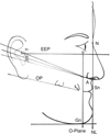
15 The Eye-Ear plane (EEP) i.e. OT1 which is comparable to Frankfort horizontal plane (FH plane) is taken as a standard reference plane as it is (FH plane) stable and is not affected by tooth loss. Also it is universally accepted as a fixed cranial plane.16 The subjects were selected between the age group of 18-40 years. By 18 years growth of the face ceases and there is no change in the relationship of camper's plane to the occlusal plane. The upper age limit was restricted to 40 years as at this age a dentition can be expected to remain normal without tooth loss and excessive attrition. For the ease of the comparison of the angulations, only orthognathic subjects were selected in this study.10 The angulation of the occlusal plane to the FH plane (OT1) is known as "cant of the occlusal plane" and was first enunciated by Downs.17 The mean cant of the occlusal plane (OT1-OP) as determined in this study is 11.96, which is comparable to the study done by Hartono18 who in case of normal type found to be 12°. Table-I shows that mean angle of OT1-AT3 (10.3167) is close to mean angle of OT1-OP (11.9651) i.e. the angle formed between FH plane and occlusal plane is closer to the angle between FH plane and ala-lower border of the tragus. This means that the occlusal plane is more parallel to the line drawn from the ala to the lower border of the tragus. This is in accordance with the studies done by Clapp6 (1910), Dalby6 (1912), Wilson6 (1917), Hartono18 (1967), van Niekerk19 (1985) and Karkazis and Polyzois5,20 (1986). They found out that the occlusal plane is parallel to the line drawn from the lowest point of the ala of the nose to the lower border of the tragus. The occlusal plane, as established by tooth arrangement should be located according to mechanical requirement for stability of denture and preservation of the supporting structures. It has been found (Nagle and Sears8-1962 & Swenson21-1947) in case of excessive resorption, the plane should be placed closer to the resorbed ridge to reduce the leverage. It should be perpendicular to the forces of mastication and should be developed parallel to the lower ridge. As the occlusal plane is parallel to the lower border of the tragus than the middle and superior borders the forces of mastication will be perpendicular to the occlusal plane and there will be less leverage on the lower residual ridge which is most commonly involved in resorption process (Jacobson and Karol6-1983) and results in denture stability. The occlusal plane placed high in relation to lower ridge result in additional leverage and denture instability (McGee22-1960). In this study comparison between angles OT1-OP (FH plane and occlusal plane) and OT1-AT3 (FH plane and ala to lower border of tragus) which is 1.6484, found to be close to the comparison between angles OT1-OP and OT1-AT2 (i.e. between FH-plane and occlusal plane angle and FH-Plane and ala-to middle border of tragus angle) 1.7093 (Table 1). This was due to the length of the Tragus. In the subjects with short tragus there was not much distance between lower and middle border of the tragus thus making both the lower and middle border almost parallel to occlusal plane. Length of the tragus was measured in these subjects. It was found that out of the 26% subjects showing occlusal plane parallel to line connecting ala to the middle of the tragus, in comparison 16% of the subjects had small tragus with very small distance between middle and lower border of tragus. Thus, these 16% subjects can also be included in subjects showing occlusal plane parallel to the line joining the ala to the lower border of tragus. It is found that there is no influence of the sex on the level of occlusal plane i.e. both males and females showed the occlusal plane parallel to the line joining the ala to the lower border of tragus. In this study it was found that in 20.7% of the subjects' upper border of the tragus is parallel to the occlusal plane, in 26% the middle border of the tragus and in 53.3% the lower border of the Tragus (Table 3). This might be due to the anteroposterior dimensions of the maxillary base, which governs angulation of the occlusal plane. The greater the distance between ANS (anterior nasal spine) and the hamular notch i.e. wider anterior cranial base, the more acute the angulation of the occlusal plane and conversely, the smaller the distance the more obtuse the angle will be. This is given by Sloane and Cook.23 This tendency has been confirmed by the cephalometric studies of L' Estrange and Vig24 and represents a phenomenon that may be explained by the "denture glass effect". It might also be due to the fact the steepness of the curvature of occlusal plane varies in different individuals and frequently a noticeable difference may be observed in the plane on the two sides of the arch in the same individual.25
As the prosthodontic intervention imposes most of its influence of the denture stability, due importance must be given in analyzing the occlusal plane level, which is the main contributing factor in denture stability. The position of the occlusal plane of orientation also forms the basis for ideal tooth arrangement and fulfills the necessary mechanical, esthetic and phonetic requirements and aid respiration and deglutition. Standard facial measurements are essential for establishing the level of occlusal plane. However, occlusal plane level analysis data apply to the ethnic group from which they are obtained. Data from one ethnic group may be misleading when applied to other ethnic group as different racial groups have different facial characteristics. Therefore, care must be taken when the norms found in standard textbooks are applied to non-Caucasian racial groups.

CONCLUSION

Following conclusions were drawn from this study,
  • The line joining from ala to the lower border of the tragus was parallel to the occlusal plane in 53.3% of the subjects. In 26% occlusal plane was parallel to the ala to middle border of tragus and in 20.7% occlusal plane was parallel to the ala to upper border of tragus.

  • There was no influence of the sex on the level of occlusal plane i.e. both males and females showed the occlusal plane parallel to the line joining the ala to the lower border of tragus.

  • In subjects with small tragus either middle or lower border of the tragus may be used to determine the level of occlusal plane.

Figures and Tables

Fig. 1
Landmarks used in study.
jap-5-9-g001
Fig. 2
References points digitized on the photograph.
jap-5-9-g002
Fig. 3
Setup for the photographic technique.
jap-5-9-g003
Fig. 4
lines digitized on the photograph and angles measured using AutoCAD computer software.
jap-5-9-g004
Table 1
Comparison of mean angle (in degrees) among four groups
jap-5-9-i001

F = 128.109, P<.001.

Table 2
Inter - Comparisons between the groups
jap-5-9-i002
Table 3
Frequency & Percentage of other three angles coming closer to OT1-OP
jap-5-9-i003
Table 4
Comparison of mean angles between males and females
jap-5-9-i004

References

1. Terrell WH. Fundamentals important to good complete denture construction. J Prosthet Dent. 1958. 8:740–752.
2. Monteith BD. A cephalometric method to determine the angulation of the occlusal plane in edentulous patients. J Prosthet Dent. 1985. 54:81–87.
3. Chaconas SJ, Gonidis D. A cephalometric technique for prosthodontic diagnosis and treatment planning. J Prosthet Dent. 1986. 56:567–574.
4. Boucher CO. Discussion of laws of articulation. J Prosthet Dent. 1963. 13:45–48.
5. Karkazis HC, Polyzois GL. Cephalometrically predicted occlusal plane: implications in removable prosthodontics. J Prosthet Dent. 1991. 65:258–264.
6. Augsburger RH. Occlusal plane relation to facial type. J Prosthet Dent. 1953. 3:755–770.
7. Hall WA Jr. Important factors in adequate denture occlusion. J Prosthet Dent. 1958. 8:764–775.
8. Nagle R, Sears VH. Denture Prosthetics. 1962. 2nd ed. St. Louis: The CV Mosby;134–136.
9. Yasaki M. The height of occlusal rim and the interocclusal distance. J Prosthet Dent. 1961. 11:26–31.
10. Simon PW. Fundamental principles of a systemic diagnosis of dental anomalies. 1926. Boston: Stratford Company;242.
11. Lundström F. Registration of natural head posture in children. Swed Dent J Suppl. 1982. 15:147–152.
12. Solow B, Tallgren A. Natural head position in standing subjects. Acta Odontol Scand. 1971. 29:591–607.
13. Gavan JA, Washburn SL, Lewis PH. Photography: an anthropometric tool. Am J Phys Anthropol. 1952. 10:331–353.
14. Baumrind S, Frantz RC. The reliability of head film measurements. 1. Landmark identification. Am J Orthod. 1971. 60:111–127.
15. Cooke MS, Wei SH. The reproducibility of natural head posture: a methodological study. Am J Orthod Dentofacial Orthop. 1988. 93:280–288.
16. DiPietro GJ, Moergeli JR. Significance of the Frankfortmandibular plane angle to prosthodontics. J Prosthet Dent. 1976. 36:624–635.
17. Downs WB. Analysis of dentofacial profile. Angle Orthod. 1956. 26:191–212.
18. Hartono R. The occlusal plane in relation to facial types. J Prosthet Dent. 1967. 17:549–558.
19. van Niekerk FW, Miller VJ, Bibby RE. The ala-tragus line in complete denture prosthodontics. J Prosthet Dent. 1985. 53:67–69.
20. Karkazis HC, Polyzois GL, Zissis AJ. Relationship between Ala-tragus line and natural occlusal plane. Implications in denture prosthodontics. Quintessence Int. 1986. 17:253–255.
21. Swenson MG. Complete Dentures. 1947. 2nd ed. St. Louis: The CV Mosby Company;177–180.
22. McGee GF. Tooth placement and base contour in denture construction. J Prosthet Dent. 1960. 10:651–657.
23. Sloane RB, Cook J. A guide to the orientation of the plane of occlusion. J Prosthet Dent. 1953. 3:53–65.
24. L'Estrange PR, Vig PS. A comparative study of the occlusal plane in dentulous and edentulous subjects. J Prosthet Dent. 1975. 33:495–503.
25. Landa LS. Practical guidelines for complete denture esthetics. Dent Clin North Am. 1977. 21:285–298.
TOOLS
Similar articles