

 PDF
PDF ePub
ePub Citation
 Print
Print


Introduction
According to the original protocol proposed by Brånemark, dental implant can be installed in a complete healing state after tooth extraction, and this procedure takes about 6 to 12 months.1,2 However, it was reported that the alveolar bone loss occurred in 23% during the initial 6 months after extraction and additional bone resorption during 5 years in 11%.3 To compensate the resorption of the bone, complicated soft and hard tissue regenerative procedure may be required. Immediate implantation was suggested as a complementary procedure against this sequela. This procedure reduces the number of surgical interventions4,5 and preserves the alveolar ridge.6-8 In addition, it is easier to determine the location of implant without surgical guide. However, immediate implantation is limited to the cases of sufficient bone quantity and good soft tissue condition. Indications of immediate placement areas follows; it must not have acute infection and any bone resorption around a fresh extraction socket. In addition, it should not have endodontic failure, root fracture and resorption.9
In immediate implantation, if an implant with large diameter is used to reduce the coronal gap, there might not be sufficient bone from the adjacent implant or the adjacent root. On the other hand, if an implant with a small diameter is used, successful osseointegration may not be achieved.10
It was stated that adequate apicocoronal placement influences the emergence profile of the superstructure and determines the establishment of the biologic width of the gingiva.11 Accordingly, for the determination of the natural contour, the adequate depth and location of the fixture installation is the aesthetically important factor during the immediate implantation. The aim of the appropriately positioned fixture installation is to get the easy fabrication of the prosthesis, aesthetically ideal results and stable occlusal dispersion.12 The best position for implant installation is determined by the amount of existing bone and the relative position of the alveolar bone to the tooth.13
In the process of the fixture installation, it is also important to get enough primary stability.14 Because implant success rate increases as the primary stability increases, thereby showing better prognosis.15 It was also suggested that the stability of implant consists of primary and secondary stability16 and primary stability is determined by the density and quantity of bone, the surgical technique, and implant design.
The purposes of this study were to elucidate the relationship between the dental roots and surrounding structures to verify the topography of the alveolar bones in maxilla for immediate implantation in the maxilla and to provide anatomical information of implant surgical site for selecting the immediate implant fixture which has proper diameter, shape, and location to get the ideal primary stability and marginal gap.
Materials and Methods
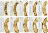
Jaw cross sections were analyzed in 20 maxillae (14 males, 6 females; mean age 66.1 years, age range 45-80 years). All specimens had normal occlusion and normal teeth alignment. Resin blocks were produced by dehydrating the specimens using a conventional method for 3 days before infiltrating them with a mixture of Technovit 7200 (No. 51000, EXAKT Co., Norderstedt, Germany) and 100% alcohol. The infiltrated samples were placed in an embedding mold and then polymerized using a light with 450 nm wave length in a light-curing unit (520 light polymerization unit, EXAKT Co., Norderstedt, Germany) for 1 day. The resin blocks were cut serially at 1 mm intervals from the cervical line to the root apex using Macro Cutting & Band System (300CP, EXAKT Co., Norderstedt, Germany). Images of each section were then obtained at a resolution of 600 DPI using a computer scanner (Perfection 3490 Photo, EPSON Co., Nagano, Japan) and stored in TIF format with high-quality compression (Fig. 1).
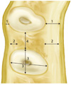
In the 200 sections of each maxilla, the following items were measured using an image analysis system (Image-Pro®Plus, ver. 4.0, Media Cybernetics Co., Bethesda, MD, USA) after performing a standard calibration (Fig. 2).
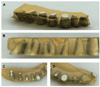
Each three maxillae nearest to the average measurements were chosen for the three-dimensional reconstruction. For the detailed 3-dimensional object, the image arrangement was performed using Photoshop program (Adobe Co., San Jose, CA, USA) and ten to thirteen images of the section were opened and superimposed on Photoshop program. To standardize the arrangement, the reference was determined by the dental root. In the cases of the multi-rooted regions, the reference was determined at the center of the trifurcation. After the arranged images were exported as a separate image, all the images were again imported to Mimics (Materialise Co., Leuven, Belgium) for the 3-dimensional object making. To distinguish the tooth and bone in the sections, those were selected independently by select tool. On the 3d studio max (Autodesk Co., San Rafael, CA, USA) program, 3-dimensional object was imported, and the final rendering was performed after establishment of material quantity and light setting (Fig. 3).
Results
In the maxilla, the facial and palatal plates began to exist 4 mm above the cervical line of the tooth. From the maxillary central incisor to the second premolar, the thickness of the facial and palatal plate increased and the palatal plates were thicker than that of the facial aspect. In every section of the maxilla, the thickness of the palatal plate increased from the cervical line to the apex, whereas facial plate tended to become thicker from the cervical line until 9 mm above the cervical line, where the thickness began to decrease, at the maxillary central incisor, lateral incisor, canine and 1st premolar. At the maxillary incisors, the labial and palatal plates of the central incisor were thicker than those of the lateral incisor. Especially, the labial plate of the canine was remarkably thinner about 0.1-0.6 mm than those of the central and lateral incisor (Table 1).
In the maxilla, the facial cortical bone showed a tendency to be thicker at the posterior interdental regions than at the anterior interdental regions, but the discrepancies in measurement were small (2 mm). The palatal cortical thickness of the maxillary interdental region was thicker in most regions (Table 2).
The root of the maxillary central incisor became narrower toward to the apex. The sections showing the root diameter less than 4 mm located from the level 10 mm and 8 mm above the cervical line in the labiopalatal and mesiodistal aspects, respectively. On the other hand, the sections showing the root diameter less than 4 mm located 10 mm and 5 mm above the cervical line in the labiopalatal and mesiodistal aspects, respectively in the lateral incisor. At the maxillary canine sections 12 mm above the cervical line, the root diameter became narrow less than 4 mm. At the first and second premolar sections, the diameter of the root became narrow less than 4 mm from the level 11 mm and 3 mm above the cervical line in the buccopalatal and mesiodistal aspects, respectively. In cases of bifurcated premolars, both the buccal and palatal roots became narrow less than 4 mm from the level 5 mm above the cervical line in the buccopalatal and mesiodistal aspects, respectively. At the maxillary first and second molar, the diameter of the mesiobuccal and distobuccal roots became narrow less than 4 mm from the level 10 mm and 8mm above the cervical line, and all the mesiodistal diameters were measured below 4 mm. On the other hand, the palatal roots became narrow less than 4 mm from the level 5 mm the above the cervical line in the buccopalatal aspect and from the level 10mm and 8mm, respectively above the cervical line in the mesiodistal aspects (Table 3 and Table 4).
The interroot distance on each sectioned specimen increased from anterior to posterior teeth and from the cervical line to the root apex in the maxilla (Tables 5). In the maxilla, the interroot distance was greatest between the second premolar and the first molar. The interroot distance at the maxillary anterior teeth exceeded 3 mm from 7 and 9 mm above the cervical line on the buccal and palatal sides, respectively. In maxillary posterior teeth, the interroot distance exceeded 3 mm from 3 and 2 mm above the cervical line on the buccal and palatal sides, respectively.
Discussion
In the present study, the horizontal cross sections of the maxilla were analyzed and three-dimensional reconstructions were obtained from the average values of the buccopalatal thickness of the alveolar bone of each root, the thickness of the interradicular alveolar bone, width of the root, and the interradicular distance measured. Based on the results, the selection of the shape, diameter, and position of implants to be placed in different regions of the maxilla can be recommended.
Since the average labiopalatal width of root of the maxillary central incisor 4 mm below the cervical line was 6.5 mm, a space of 2.5 mm in width is created when placing a 4 mm implant as far palatal as possible in this region. The maximum amount of space between implant fixtures and bone in which spontaneous healing can occur has been reported to be from 1.4 mm17 to 2.5 mm.18 Also, it has been reported that adding bone graft material to areas with thin facial bone such as the anterior region aids in maintaining the contours of the alveolar bone.11 Therefore, considering the fact that a 2.5 mm space would be gained according the results of this study, bone grafting may be considered as an option for preserving facial bone.
The amount of labial alveolar bone of the maxillary lateral incisors was similar to that of the maxillary central incisors, and while the labiopalatal width of the root at the cervical line was narrower, it was comparable to that of the central incisors 4 mm below the cervical line. The maxillary canine had less labial alveolar bone than the central incisors, and had a significantly lower amount 8 mm below the cervical line. Since the average mesiodistal width of the root of the maxillary central incisor was 5.4 mm at 4 mm below the cervical line, placing a 4 mm implant can create space mesiodistally as well as labiopalatally. Therefore, a 5 mm implant may be considered in order to achieve adequate initial stability. While in the past fixtures with large diameters were recommended for immediate implantation, such approach may cause thinning of the labial bone, leading to labial marginal bone loss. Therefore, it is necessary to consider a selective approach considering long-term esthetics. Also, placing an implant fixture that is substantially larger in diameter than that of the root in the maxillary incisor region may leave an inadequate amount of bone between the fixture and the root of the adjacent tooth, as the mesiodistal width of the alveolar bone of the maxillary incisors 4 mm below the cervical line is 1.8 to 2.3 mm.
It was suggested that an implant to be placed 3 to 4 mm below the facial cementoenamel junction of the adjacent teeth to restore the emergence profile, prevent soft tissue recession, and preserve the supporting tissue the adjacent teeth.19 The apicocoronal depth at which the mesiodistal width of the root becomes less than 4 mm was 8 mm below the cervical line for the maxillary central incisors, 5 mm below the cervical line for the maxillary lateral incisors. Therefore, placing a straight form implant with a diameter of 4 mm at the level of 4 mm below the cervical line would begin to gain the anchorage at 4 mm and 1 mm below the top of the implant fixture, respectively. On the other hand, placing a straight form implant with a diameter of 5 mm from the level of 4 mm below the cervical line for the maxillary central incisors and canines can get the anchorage at 2 mm below the top of the implant fixture in both regions.
Implants can be classified into straight or tapered by shape. While straight implants are placed so that initial stability is attained at 3 to 5 mm past the level of the apex,20 tapered implants conform to the contours of the extraction socket and thus achieve good osseointegration, obviating the need for placement 3-5 mm past the level of the apex. Also, because labial bone is thin in the maxillary incisor region and further decreases apically, incorporating tapered implants and placing them at a labial inclination may be a suitable approach.
The facial alveolar bone of the maxillary first premolars showed a decreasing tendency similar to the canines at 10 mm below the cervical line. The maxillary premolars had two roots buccopalatally with similar widths. Septal bone was detected 5 mm below the cervical line, and had a buccopalatal width of 1.1 mm coronally, increasing to 2.6 mm 10 mm apical to the cervical line. Because the maxillary first premolar has thinner buccal and mesial bone than the maxillary second premolar, a 4.5 mm implant would be more suitable than a 5 mm implant. Compared to the maxillary incisors, the maxillary premolars have sufficient bone around their roots on every side, possibly making them suitable sites for straight implants. Placing implants in the septal bone of the maxillary premolar region has been recommended, but the clinician must be cautious as the septal bone may be lost during the drilling procedure, thus increasing the dimension of the peri-implant defect and serving as a factor that interrupts healing.21
To avoid cantilever effects and produce esthetic results, implants must be placed in the location of the central fossa in the case of maxillary molars. We believe that to attain solid initial stability in the maxillary molar region, an implant of about 6.5 mm in diameter in the cervical region that tapers down to the furcation area after which it runs parallel like a straight implant would be the ideal form.
Ideally, immediate implantation in the molar region should be performed in the septal bone area to prevent the cantilever phenomenon and achieve good esthetics. However, when there is inadequate septal bone due to minimal root divergence, delayed implantation may be preferable to immediate implantation.
It had been reported that bundle bone was resorbed quickly whereas cortical bone resisted resorption in the resorption and remodeling phase of the extraction socket.22 Taking this into consideration, we attempted to measure the dimensions of both types of bone on the buccopalatal sides of the root, but failed to do so as it was not possible with the specimens used in this particular study. However, we believe that making a general prediction on the degree and pattern of resorption would be possible with either histologic specimens or methods which can selectively measure the two kinds of bone.
Because the present study measured dentulous regions from the normal cadavers, it is not applicable to teeth extracted due to chronic periodontitis. However, the measurements of the buccopalatal thickness of the alveolar bone of teeth, width of interradicular bone, and thickness of the cortical bone obtained in the present study may be employed as a clinical guideline for immediate implantation.


 XML Download