

 PDF
PDF ePub
ePub Citation
 Print
Print


INTRODUCTION
Patients with Class III malocclusion may have complex dentoalveolar problems, including anterior edge-to-edge relation or anterior and/or posterior crossbite.1 Class III malocclusion characterized by an anterior crossbite2,3 exhibits potential problems associated with esthetics, absence of centric contact on anterior teeth, lack of anterior guidance and in those patients the function is limited to vertical movements and they have a vertical function pattern.3 In addition clinically, these patients exhibit concave facial profile, retrusive nasomaxilla, remarkable lower third of the face and protruded lower lip respect to the upper lip.4
It has been stated that the subjects with malocclusion especially Class III malocclusions, have poorer masticatory performance.5 Maximum bite force is considered as the important indicator of the masticatory performance and malocclusions are often associated with reduced maximum bite force. Although decreased bite force in subjects with Class III malocclusion has been reported, it is not clear whether patients with other forms of malocclusion also have generally lower bite force.6 Therefore, the treatment of Class III malocclusion aimed to increase masticatory function related to bite force. Many of the patients with class III malocclusion need surgical and/or orthodontic treatment. However, due to the extent of elapsed time to complete orthodontic treatment, or avoiding surgical procedures with extended recuperation, restorative treatments might be an alternative treatment option to provide acceptable functional occlusion as an esthetic dentofacial appearance in patients with Class III malocclusion.3
Important consideration that should be made regards to restorative treatment of Class III malocclusion is the possibility of increasing the vertical dimension.2,3 Increasing of occlusal vertical dimension (OVD) should be within the range of neuromuscular adaptation.7 Adaptation of patients to changes in occlusal vertical dimension is usually confirmed by the use of diagnostic splint or provisional prosthesis.1,3 The present case describes the increase of OVD using an occlusal splint and then full mouth rehabilitation of a patient with Angle Class III malocclusion. Before and after the occlusal rehabilitation of patient, bite force was evaluated.
CASE REPORT
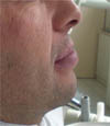
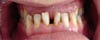
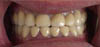
A male patient aged 33 years old was self-referred to the Faculty of Dentistry at University of Gazi. The main complaints of patient were difficulty in masticatory function and poor esthetic. The patient's medical history was reviewed and it was decided to be non-contributory. An extensive clinical examination was also made. On extraoral clinical examination, retruded upper lip with prominent lower lip was noted as usually seen in Class III malocclusion (Fig. 1). Cephalometric analysis of patient revealed that patient had Angle Class III malocclusion (ANB = -5). In dental examination, it was detected that Class III cuspidfirst molar relationship and anterior crossbite with the negative horizontal overjet in centric relation, diastemas in anterior teeth, and missed three molar teeth number of 17, 26 and 47 (Fig. 2). There was no caries, wear or restoration on teeth. The periodontal examination showed the presence of plaque and generalized staining. In the evaluation of temporomandibular joint, no history of dysfunction was reported and, no joint sound and pathology was revealed. Masticatory muscles, head and neck muscles were normal to palpation. The mandibular range of the motion and jaw opening were within the normal limits.
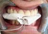
Before the treatment, maximum bite forces were measured from each side of the dental arch using two miniature strain-gauge transducers with stainless-steel cases (Model VLPB, Load Cell Central, Monroeton, PA, USA). Each transducer had a height of 4 mm and a diameter of 12 mm. The bite force was detected as a two-channel signal from each side with a biosignal acquisition device designed by Kardiosis (Tepa, Kardiosis, Ankara, Turkey). The force signals were monitored online and then measured on a PC screen, using a specific software program developed by the same company. Two transducers were placed bilaterally in contact with maxillary first molar teeth over metal plate. The transducers fixed with plaster (Betasan, Kocaeli, Turkey) to the metal plate. Then, metal plate and transducers were covered with a disposable latex finger coating to avoid contamination. During measurement, the subject was asked to clench teeth as forcefully as possible three times. The highest value of each clenching was recorded as kilogram (kg), and the mean value of the three highest clenching was considered as the subject's maximum bite force. The sum of the right and left bite force values was considered to be the maximum bite force of the subject. Maximum bite force of patient was measured as 42 kg before treatment.
Two following treatment options were discussed with the patient; (1) Orthodontic treatment and following surgical treatment, (2) Restorative treatment with increasing of OVD. The patient was informed about all the advantages, disadvantages and possible risks of both treatment options. In that patient, even if the orthodontic treatment is accomplished, the shape, size and color of present teeth would remain the same. Also orthodontic and surgical treatment would also require extended treatment time. On the other hand, as another option, restorative treatment would involve the preparations of all maxillary and mandibular teeth for improving occlusal relationships and also increasing of OVD. The patient's expectations from the treatment were a good esthetic and masticatory function with a less invasive way in a relatively short time. Therefore, increasing of OVD and then restoring the maxillary and mandibular teeth was decided to be chosen as the treatment option of patient. Full mouth rehabilitation with metal ceramic restorations was suggested to patient.
At first, periondontal treatment was performed and oral hygiene instructions were given. Then, irreversible hydrocolloid impressions (Hydrocolor 5, Zhermack, Zhermack SpA., Badia Polesine, Italy) of maxillary and mandibular teeth were made to obtain diagnostic casts. A face bow record (UTS Face-bow, Ivoclar Vivadent, Austria) and an interocclusal record in centric relation were completed and diagnostic casts were mounted in a semi-adjustable articulator (Stratos 300, Ivoclar-Vivadent, Austria). The new OVD was established using Niswonger method and facial measurements. Patient had increased interocclusal rest space (7 mm in the anterior teeth), therefore 3 mm rest space was pretended and actual increase were determined as 4 mm in the anterior teeth and 2 mm in the posterior teeth and the incisal guidance pin of the articulator was set.
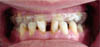
Diagnostic casts were evaluated and wax up of all anterior teeth with final contour and position was accomplished at the new OVD. Then an auto-polymerized acrylic resin (Panacryl, Arma Dental, İstanbul, Turkey) occlusal splint was provided to evaluate the adaptation of the patient to altered OVD. The splint was adjusted intraorally to ensure bilateral anterior and posterior simultaneous contacts in centric relation. Anterior region of splint prevented occlusion of splint, therefore excess of acrylic resin was removed from anterior region of splint (Fig. 3). Patient was instructed to wear splint during 24 hours, for 4 weeks. In clinical examination after the first week, no muscle tenderness and temporomandibular discomfort was found.
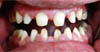
After the verification of functional adaptation to new OVD, anterior teeth were prepared for definitive treatment. In this session occlusal splint was used to check the preparation amount of anterior teeth and to take bite registration by placing the splint into mouth. Posterior teeth were occluded and anterior part of splint was supported with wax. Then posterior teeth were prepared. At this time anterior part of occlusal splint was occluded and posterior part was supported with wax. After preparation of all anterior and posterior teeth (Fig. 4), final impressions were made with polyvinylsiloxane impression material (Optosil Comfort/Xantopren VL Plus, Heraeus Kulzer, Hanau, Germany) and provisional restorations were fabricated using an auto-polymerized acrylic resin. The provisional restorations were used for one month, as a guide for the definitive restoration. During this period, the patient's condition and functions, such as muscle tenderness, temporomandibular joint discomfort, range of the mandibular movements, masticatory function, swallowing and speech were evaluated. Improvement in masticatory function, speech, and dentofacial esthetics affirmed the patient's tolerance to the new OVD. In the following session, metal substructures (Kera N, Germany) of restoration were adjusted. After the adaptation of metal substructure, the trial of ceramic restoration (Ceramco 3, Dentsply, Dreieich, Germany) were accomplished with minor occlusal adjustments. The occlusal sheme of definitive restoration was designed as mutually protected occlusion. During excursive mandibular movements the anterior teeth protected the posterior teeth from excursive force and posterior teeth supported the bite force in the maximum intercuspal position. Then definitive restorations were glazed and cemented with zinc polycarboxylate cement (Adhesor-Carbofine, Spofa-Dental, Germany) (Fig. 5). Oral hygiene instruction and regular check-up were administered.
One week later cementation of definitive restoration, maximum bite force was remeasured and recorded as 106 kg (Fig. 6). Patient reported increased masticatory efficiency with treatment.
DISCUSSION
Class III malocclusions that remained undiagnosed or untreated may be a result of genetic predisposition or occlusal premature and lead to abnormal closure patterns that contribute to functional and esthetic problems.8
In the present case, patient had retrognatic mandible, severe reduced overjet and intolerable discomfort. In such cases, alteration of OVD may improve dentofacial esthetics and facial proportion of patient, providing an important treatment modality for force management of the masticatory system. However, the alteration of OVD will conflict with the physiology of the masticatory system and the patient's ability to adapt.9 The patient's tolerance to the changes at the new occlusal vertical dimension is usually confirmed with the clinical evaluation of the patient using an acrylic resin occlusal splint, provisional restorations (i.e. direct bonded composite resins, or provisional fixed restorations) or base metal onlays (removable partial denture).10-12 Among these, the occlusal splints had easy laboratory procedures, low cost and allow for occlusal adjustment when needed. On the contrary, base metal onlays may cause speech interference, modest increases in laboratory procedures and high cost, typically more difficult adjustment due to the hardness of base metals, and difficult removal of onlays. Therefore, in the present case, occlusa11l splint was preferred for the evaluation of the increasing OVD.
There have been relatively few studies evaluating the relationship between malocclusion and maximum bite force.5,13,14 Although several studies indicate that adults with vertical deformities have lower bite force than normal15, it is not clear whether patients with other forms of malocclusion also have generally lower bite forces. It has been stated that the reduced maximum bite force in subjects with malocclusion is probably related more to the effect of occlusal contact and the biomechanics of the jaws and masticatory muscles.13
In our case, the patient had 42 kg maximum bite force initially, however it increased to 106 kg after full mouth rehabilitation of patient. This increased bite force might be due to increase in number and extent of tooth contact and increase in vertical dimension of jaw elevator muscles during clenching.16 Many studies reported that increasing of number and size of occlusal contacts provided the bite force increase.16,17 Based on this result, increased occlusal contacts with the restoration of maxillary and mandibular teeth might lead to increased bite force. On the other hand, the changes in OVD may alter the length of main jaw elevator muscles and the position of mandibular head in the fossa temporalis.18 The change in the length of the main elevator muscle fibers will in turn affect the bite force applied. It has been reported that when a muscle fiber is stretched beyond its resting length, more force is generated up to a point and further stretching results in reduced force generation.19 It was also stated that the maximum bite force increases as the jaw is opened which reaches a maximum level at 14 - 20 mm of interincisal distance, and then decreases as the jaw is further opened.20 Thus, this could be another reason for increase in bite force after rehabilitation of patient.
The present clinical report described the use of occlusal splint to restore occlusal vertical dimension and prosthetic rehabilitation of a patient with Class III malocclusion. Full mouth restorative treatment of a patient with Class III malocclusion could be an effective treatment approach to resolve esthetic concern and to improve masticatory function related to increased bite force.
 XML Download