

 PDF
PDF ePub
ePub Citation
 Print
Print


INTRODUCTION
The white spot lesion is the first visible evidence of a caries in the enamel, characterized by demineralized lesion underneath an intact surface.1 The lesion is caused by the accumulation of plaque and bacteria, developing among young adolescents with insufficient oral hygiene.2 The increased pore volume inside the demineralized lesion body leads to a different refraction index from the sound enamel.3 An inactive white spot lesion might act as arrested dental caries and impair only the esthetic appearance by displaying a milky white color from its interior opacity.4,5 Attempts have been made to conserve tooth structures with highly concentrated fluoride solutions to hyper-mineralize the surface of the lesion.6 Since these solutions do not thoroughly penetrate into the lesion, the opaque whitish aspect remains, compromising the appearance.6,7
For esthetic improvement of non-cavitated white spot lesions with remineralized surface, treatment may consist of tooth bleaching, , macroabrasion, composite resin bonding, prosthetic restoration or some combination depending on the severity of the lesion and its etiology.8 It remains unclear which treatment is most effective for white spot lesions. Tooth whitening is the most conservative approach. As a part of minimally invasive treatment, tooth whitening to mask the white spot lesion or achieve more uniform appearance has been reported.9-11 However, tooth whitening treatment alone to camouflage the white spot lesion seemed to be relatively unpredictable.12 Moreover, if tooth whitening is used in decalcified areas, the microhardness of the enamel surface may be reduced.13
The conventional treatment approach is based on restoration, which, in most instances, is quite invasive.14,15 Efforts should be made to achieve an ideal function and esthetics along with preserving the sound tooth structures as much as possible.16,17 Since most patients requiring treatment for white spot lesions are adolescents or young adults, minimally invasive treatments are needed to prevent excessive sacrifice of tooth material at an early age.
Recently, the resin infiltration technique was introduced as a type of minimally invasive treatment. The resin infiltration technique prevents further progression of the lesion using a low-viscosity resin with a high penetration coefficient, filling the enamel intercrystalline spaces.18-20 This technique has been reported to remove the whitish opaque color thereby changing the color and translucency of the white lesion.1,7 The purpose of this clinical report was to describe and illustrate a minimally invasive technique that improves the esthetic aspect of the white spot lesion adjacent to the restoring implant-supported prostheses via the multidisciplinary approach.
CASE REPORT
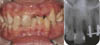
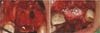
A 22-year-old woman who lost her maxillary right central and lateral incisor prosthesis presented to the Gangneung-Wonju National University Dental Hospital for treatment (Fig. 1A). The prosthesis was lost with severe secondary caries around the metal post. Each root canal was incompletely filled with a radiopaque resinous hard material, and the right lateral incisor root had apical rarefying osteitis (Fig. 1B). Extraction was necessary due to the large extent of secondary caries and the nonremovable canal filling material. A white spot was found in the maxillary left central incisor with poor oral hygienic control.
Because the patient refused tooth preparation, restoring the teeth with an implant prosthesis was planned. A diagnostic waxing of the teeth determined the installing site of implants to meet the esthetic needs and to fabricate a radiographic stent and a surgical stent. Six weeks after extraction, severe bone loss of the buccal plate was confirmed (Fig. 2A). In view of the insufficient implant primary stability, guided bone regeneration was performed as defects were filled with a xenograft material (Bioss; Geistlich Pharma AG, Wolhusen, Switzerland) and covered with a resorbable bilayer collagen membrane (Bio-gide; Geistlich Pharma AG) (Fig. 2B). Four months after guided bone regeneration, sufficient hard and soft tissue volume was acquired. A tapered internal hex titanium implants (Implantium; Dentium, Seoul, Korea) were installed, oriented by a surgical guide. An acrylic resin RPD was fabricated to replace the missing tooth during the healing period.
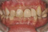
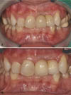
Six month after implant placement, a tissue-punch technique was used to place a screw-retained provisional restoration with a provisional plastic abutment (Plastic temporary abutment, Dentium). The treatment for the white spot of the left maxillary central incisor was also needed. Diagnosis for the white spot was performed with visual-tactile inspection evaluating change of the surface gloss and color after dessication. The lesion surface was hard and shiny, it was diagnosed as an inactive ICDAS (International Caries Detection and Assessment System) code 2 which requires esthetic improvement only. Regenerating the white spot on the prosthesis was not esthetic and removal of the white spot lesion was thus required. Gingival recession after a surgical procedure highlights the white spot in the cervical area (Fig. 3). Furthermore, bandlike whitish discoloration along the perikymata in the middle third coronal area also needed esthetic enhancement. The proposed minimally invasive technique is based on a combined approach of bleaching, resin infiltration and composite resin bonding. In-office bleaching treatment was applied according to the manufacturer's (Lumawhite; Luma Lite, Spring Valley, CA, USA) recommendations. After gingival tissues were isolated using a light-polymerized resin dam (Opal Dam; Ultradent Products Inc., South Jardan, UT, USA), 35% hydrogen peroxide (Lumawhite) was applied onto the left maxillary incisor with consistent coverage of 2 to 3 mm thickness for 8 minutes, for a total of 3 times (Fig. 4A). Two weeks after treatment, the white spot lesion was still apparent though bleaching became effective on the normal tooth structure, requiring more effective treatment (Fig. 4B).
Consequently, the decision was made to conduct the resin infiltration technique as it was more preservative than microabrasion or macroabrasion. On the enamel surface, careful application of 15% hydrochloric acid gel (ICON-Etch; DMG, Hamburg, Germany) was performed for 120 seconds to remove the surface layer less than 30 to 40 µm (Fig. 4C). The acid gel was suctioned and washed away thoroughly. The lesions were desiccated using ethanol (ICON-Dry; DMG) for 30 seconds. At the moment of applying ethanol, the color change in the white spot lesion was confirmed with ethanol penetration. After thorough dessication with ethanol, an infiltrant resin (ICON-Infiltrant; DMG) was placed on the tooth surface for 3 minutes for inside penetration. Excessive resin was wiped away from the surface and the proximal spaces. Light polymerizing was performed for 40 seconds. Applying the infiltrant resin was repeated to compensate for the shrinkage after polymerization. The tooth surface was polished with polishing discs (Sof-lex disk; 3M ESPE, Saint Paul, MN, USA). After resin infiltration, some of the cervical white spot and bandlike whitish discoloration along the perikymata in the middle third coronal area disappeared (Fig. 4D). However, resin infiltration in some parts of the cervical white spot lesion was ineffective, requiring an advanced approach.
The limited, unchanged white spot lesion was removed with a carbide bur (Carbide Burs Round; Mani Inc., Tochigi, Japan). The tooth structure was etched with 38% phosphoric acid (Scotchbond Etchant; 3M ESPE) for 15 seconds. The etching gel was washed away and dried. A bonding agent (Adper Single Bond Plus adhesive, 3M ESPE) was applied to the preparation area and light polymerized for 10 seconds. Composite resin (Filtek Supreme XT; 3M ESPE) was placed and polymerized for 40 seconds. Finally, polishing the restoration surface was performed with polishing discs (Sof-lex disk; 3M ESPE) (Fig. 4E).
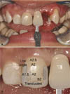
A complete maxillary arch impression was made with a silicone impression material (Exafine, GC, Tokyo, Japan). A customized abutment (Direct Casting Abutment; Dentium) was fabricated. Shade information was transferred to the dental laboratory after taking clinical photos with a shade guide (VitaLumin shade guide; VidentInc, Brea, CA, USA) and a detailed shade map (Fig. 5A and Fig. 5B). A metal ceramic fixed dental prosthesis was cemented with implant cement (Premier Implant Cement; Premier Products Co., Plymouth Meeting, PA, USA) (Fig. 6A).
The patient was followed for oral hygiene instruction and no complications were found after 6 months (Fig. 6B). However, due to insufficient long term clinical research on color stability for resin infiltration treatment, continuous post-treatment monitoring should be followed. It is also important to monitor the maintenance of proper surface gloss, oral hygiene and health of peri-implant soft and hard tissue.
DISCUSSION
The resin infiltration technique is primarily indicated for incipient enamel caries for its relatively thin penetrable surface layer.1,3 It is reported that the masking effect seems to be related to the depth and cariogenic activity of the lesion.1,8 As the infiltrant cannot penetrate the remineralized surface, a cariogenically inactive lesion may acquire little or no masking effect with the resin infiltration technique.1,8 Lesions deeper than the infiltration capacity of resin infiltrants might also demonstrate insufficient esthetic improvement.8 The diagnostic methods of white spot lesions such as clinical exams, photographic exams, optical nonfluorescent methods, and optical fluorescent methods might not precisely measure the depth of the lesion. Moreover, quantitative correlation between shade improvement and lesion depth has not been reported, yet. For esthetic improvement of a white spot lesion with varying depth, resin infiltration might be an effective treatment modality to discriminate relatively deep or inactive lesion, enabling minimal tooth reduction. Further studies would be needed to integrate various diagnostic methods with treatment options for minimally invasive treatment of white spot. As the long term color stability of resin infiltrants has not been reported to the author's knowledge, a careful recall check of color stability should be followed.

 XML Download