

 PDF
PDF ePub
ePub Citation
 Print
Print


INTRODUCTION
Fixed partial denture generally consists of two parts, the enduring substructure or coping and the esthetic veneer overlaid on the coping to match the neighboring tooth color. Different types of dental alloys have been used as substructures for metal-ceramic restorations. Because of their high fracture resistance and high reliability, metal-ceramic restorations have shown exceptionally high success rates even for a long-term treatment.1-3 However, it is more difficult to create a metal-based restoration with a natural tooth color and translucency when compared to an all-ceramic prosthesis because a metal substructure does not allow light transmission through a restoration.4,5 In contrast, various core ceramics have been shown to have varied degree of translucency, ranging from a very translucent material to an opaque core ceramic.4-6 The varied translucency of these core ceramics has made it uncomplicated to fabricate a restoration that can match its neighboring teeth in terms of color and translucency.
The translucency of ceramic materials depends on several factors such as relative refractive index, wavelengths of the light sources, numbers and sizes of porosity and inclusion etc.7 Contrast ratio is an optical parameter that is used to represent the degree of translucency of a material.7,8 Several studies have used this parameter to compare the light transmission capability or the masking ability of tooth-colored dental restorative materials.4-6,9,10 For determining the contrast ratio, the ratio of light reflectance of a material over black and white backgrounds (Yb/Yw) are measured using a spectrophotometer.7,8 While the contrast ratio for a completely opaque material is 1, a lower contrast ratio represents a more translucent substance. For tooth enamel and dentin, the contrast ratio was approximately 0.55 at 1 mm thickness.11
As previously mentioned, color match between a dental restoration and the adjacent natural teeth depends partly on the degree of translucency of dental restorative material. However, the ability to conceal the discolored abutment tooth or a metal post and core is also a subject of interest for all-ceramic restorations. The masking or covering ability of materials can be defined as a measure of the capability of a coating to hide a colored background and the contrast ratio of 0.98 of a covering layer is proposed for the perfect masking ability in industrial production.8 If the color of an underlying structure can be observed, it would result in the color difference between the covering material and the target color. The color difference in CIELAB units is given by the following equation;12
When ΔL*, Δa* and Δb* are the differences in lightness, chroma in red-green axis (a*), and chroma in blue-yellow axis (b*) of the measured colors of two objects.
In dentistry, the color difference or ΔE*ab is used to evaluate the color match between a dental restoration and the adjacent natural teeth. The perceptible thresholds (ΔE*ab ≈ 2.6-3.7) were set as guidelines for color matching determination according to the results from few in vivo studies.13,14 The perceptible thresholds obtained from in vitro studies were lower (ΔE*ab ≈ 0.4-1) because of their better viewing and measuring conditions.15,16 However, the color differences were reported even for the matched natural teeth.17 For a perfect match between natural upper central incisors, the reported ΔE*ab values ranged from 0.1 to 1.6. For a perfect match between an upper natural central incisor and a contralateral all-ceramic crown, ΔE*ab values varied from 0.2 to 2.9 with an average of 1.6.
Zirconium dioxide or zirconia has presently received considerable attention from dental practitioners because of its high fracture resistance and excellent biocompatibility. Most zirconia-based core materials obtained from different manufacturers are yttria-stabilized tetragonal zirconia polycrystals or Y-TZP. Even though they are all polycrystalline materials with comparable compositions, they could have slightly different microstructures.18 As a result, Y-TZPs could have different degree of opacity because of an increase or decrease in light scattering caused from the microstructural variations.19 For example, an increase in the light scattering inside the bulk material results from an increase in porosities or inclusions or the discontinuity of refractive indices at the grain boundary.19 In an esthetic viewpoint, a ceramic material with limited light transmission is not desirable because it does not imitate the optical characteristics of a natural tooth. On the contrary, a high opacity ceramic material is required when a restoration is made on an abutment such as a discolored tooth or a metal post and core. For zirconia-based core materials, they appear to be opaque materials and they could be used to mask the dark colors of an underlying substructure.3,20 However, there is limited information about the translucency of zirconia core materials. The objective of this study was to determine the effect of color of an underlying substructure on the overall color of zirconia all-ceramic crowns.
MATERIALS AND METHODS
The protocol for this study was approved by the Mahidol University Institutional Review Board (MU-IRB 2008/031.0506). Twenty adult subjects, of good to excellent dental health, were recruited from the pool of subjects on the waiting list of the Faculty of Dentistry at Mahidol University in Bangkok, Thailand. All selected subjects had healthy periodontal tissues, were free of active periodontal disease and caries, and showed no evidence of bruxing. For these 20 adult subjects, 7 were men and 13 were women. Their ages ranged from 17 to 55 years. Eligible subjects had at least one posterior endodontically treated tooth opposed by natural dentition in the maxillary or mandibular arch. This endodontically treated tooth was used as an abutment for an all-ceramic crown. After obtaining an informed consent to participate in the study, a maximum of two crowns were placed per patient.
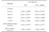
There were 7 premolars and 14 molars prepared for all-ceramic crowns. Color shade of a contralateral or an adjacent tooth was determined for use as an all-ceramic crown shade. All abutment teeth were prepared either for a metal cast post and core or a prefabricated fiber post with a composite core build-up as foundations for the all-ceramic crowns. The following minimal guidelines for the final tooth preparation were followed; axial tooth reduction of 1 mm, shoulder or deep chamfer design, occlusal reduction of 1.5-2 mm, and rounded inner angles/edges/transitions. The ceramic material used in this study was a zirconia-based core ceramic and a compatible glass-based veneering ceramic (ZENO®, Wieland Dental and Technik GmbH & Co, Germany, and IPS e. max Ceram, Ivoclar Vivadent AG, Liechtenstein). Twenty one crowns were made using a layering technique according to the manufacturer's instructions. These crowns were divided into three groups according to the remaining abutment tooth structure and the appropriate reconstruction of the core foundation as shown in Table 1.
After try-in and adjustment to produce proper anatomical contours and proper occlusion, each crown was glazed before insertion. The all-ceramic crowns were cemented using resin cement which had simply one shade (Multilink, Ivoclar Vivadent AG, Liechtenstein). Only one shade resin cement was used in order to limit the influence of cement shade on the color of all-ceramic crowns. Examinations of the overall prosthesis, the marginal area, the adjacent gingival tissues, and the occlusion were evaluated before cementation. The thicknesses of the core and veneering materials were measured before cementation at the middle 1/3 of the buccal, lingual and occlusal surfaces of each ceramic crown. Color measurements of all crowns were made using a shade measuring device (ShadeEye NCC®, Shofu Inc., Kyoto, Japan) before and after cementation using the CIE-Lab parameters. L* represents the lightness of a material, +a* represents color in the red axis and -a* indicates color in the green axis. The blue and yellow axes were designated by -b* and +b*, respectively. This ShadeEye NCC® intraoral shade measuring device has been used in some previous studies with an acceptable performance.21,22 The color of all-ceramic crowns was measured using a pulsed xenon Lamp as an optical light source and a vertical light receiving system. The instrument was calibrated against a standard calibration according to a manufacturer's recommendation before each color measurement. The contact plastic tip, having a diameter of 3 mm, was positioned at the middle 1/3 of the buccal surface of each crown, the tip made an intimate contact with the crown surface during the measurement. After each measurement, L*, a* and b* value was obtained and used for calculation of color differences between before try-in, before and after cementation of all-ceramic crowns. A repeated measure ANOVA was used for a statistical analysis of a color change between before try-in, before and after cementation of all-ceramic crowns at α=.05.
In order to obtain the optical properties of a zirconia-based material used in this study, 24 zirconia core ceramic (ZENO®, Wieland Dental and Technik GmbH & Co, Germany) were prepared in a laboratory. These rectangular core specimens (15 mm × 15 mm) with four different thicknesses (0.4, 0.6, 0.8 and 1.0 mm) were prepared considering a shrinkage during the sintering process. After sintering, ZirLiner and dentine shade A3 (IPS e. max Ceram, IvoclarVivadent AG, Liechtenstein) was applied onto the specimens and fired in a furnace (Programat P100, IvoclarVivadent AG, Liechtenstein) according to the manufacturer's firing instructions. A lithia-disilicate-based core ceramic (15 mm × 15 mm × 0.8 mm, Empress 2, Ivoclar Vivadent AG, Liechtenstein) and a base metal alloy (Wiron 99, Bego, Germany) were also prepared as controls. For a metal-ceramic system, six rectangular specimens, with a dimension of 15 mm × 15 mm × 0.3 mm were casted using a lost wax technique. For veneering procedures, lithia-disilicate-based ceramic and metal samples were veneered with dentin porcelains (IPS Eris, Ivoclar Vivadent AG, Liechtenstein and Vita VMK95, Vita Zahnfabrik, Germany).
After veneering, all specimens were ground to a final thickness of 1.5 ± 0.1 mm using a milling machine (Schick Dentalgerate S master 3, Vacalon, USA) and a diamond grinding disc with a grit size of 80 µm (S327010, Bredent GmbH & Co. KG, Senden, Germany). All specimens were glazed by applying a thin layer of the glaze paste onto the grinding surface and fired according to the recommended schedule. After firing, the thickness of each specimen was measured four times and the mean thickness was calculated prior to color measurement.
The contrast ratios of all specimens were measured before and after veneering, respectively, using a spectrocolorimeter (ColorFlex, Model 45/0, Hunter Associates Laboratory, Inc., Reston, VA, USA). All specimens were measured using the 45°/0° geometry with CIE illuminant D65 and 2 degree observer function. Calibration of the machine was made using a black glass and a white tile as recommended by the manufacturer. Each specimen was placed at the specimen port with the measuring window of 13 mm in diameter. The spectral reflectance data was obtained in the range of 400 - 700 nm at 10 nm intervals. Three measurements were made for each specimen and the mean contrast ratio was calculated. The mean contrast ratios before and after veneering of each group were calculated and statistical analysis of the data was performed using a mixed/split-plot design ANOVA (SPANOVA) test at α=.05. The Tukey's multiple comparison test was used to determine the rank of each group.
RESULTS
For all-ceramic crowns, the mean thicknesses of core ceramic at the buccal surface were 0.6, and 0.7 ± 0.1 mm for premolar and molar crowns, respectively. The mean thicknesses at the occlusal surface of the core ceramic were 0.7 ± 0.1 for premolar, and 0.8 ± 0.2 mm for molar crowns. The total thicknesses of all-ceramic crowns after veneering were 1.8 ± 0.3 mm for premolars, and 2.0 ± 0.3 mm for molars at the buccal surface.
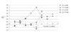
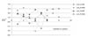
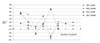
ΔL*, Δa* and Δb* values of all-ceramic crowns cemented on a metal cast post and core are shown in Fig. 1, Fig. 2 and Fig. 3. L*, a*, and b* values did not show a significant change when the values obtained before try-in, before and after cementation were compared. However, L* value of all-ceramic crowns with a metal cast post and core appeared to be decreased (-ΔL1*) and a* tended to be increased (+Δa1*) when compared between before try-in and before cementation. In contrast, b* was not affected much by the color of a background substructure. For the metal cores in these groups, the height of the metal part was approximately 2-3 mm covered the remaining tooth structure. L* and b* values of molar crowns with prefabricated post and composite core build-up also did not show a significant change when compared the values obtained before try-in, before and after cementation. But a* was significantly increased when compared between before try-in and before cementation (+Δa1*).
The color differences or ΔE*ab between the colors of an all-ceramic crown obtained before try-in and before cementation (ΔE*1), and between before try-in and after cementation (ΔE*2) were determined using Equation 1. The mean color differences of all-ceramic crowns are shown in Table 2.
For in vitro ceramic specimens, the mean contrast ratios before and after veneering of all-ceramic and metal-ceramic materials are summarized in Table 3. The contrast ratio of zirconia core specimen was significantly increased from 0.71 to 0.86 as their thickness was increased from 0.4 to 0.8 mm, and no significant difference was found at the thickness of 0.8 and 1.0 mm. After veneering to a final thickness of 1.5 mm, their contrast ratios was increased to 0.92 - 0.95. Compared with a translucent lithia disilicate-based all-ceramic system, zirconia core with a thickness of 0.4 mm had a level of contrast ratio similar to this system after veneering. The metal-ceramic system was used as a control group for a completely opaque system, and its contrast ratio was 1.00.
DISCUSSION
The aesthetic values of all-ceramic restorations are essentially based on their translucency. Even the zirconia-based restorations have significantly higher fracture resistance than other all-ceramic systems, but their limited light transmission as reported from few previous studies is their critical disadvantage.6,20 The contrast ratio of 1.00 was reported for one commercially available zirconia-based core ceramic, whereas the results from another study have shown that a zirconia-based restoration might not be as opaque as expected. An excellent match between a veneered zirconia crown and the adjacent natural anterior tooth has been reported.17 However, the information about the opacity of the zirconia core materials is still limited even though there are many zirconia core systems that are currently available in the market.
Light transmission of zirconia-based restorations is limited by its composition and microstructure. Different levels of light transmission were allowed for dissimilar polycrystalline zirconia-based specimens because of their microstructural dissimilarities.20 These microstructural dissimilarities such as grain sizes, the amount of porosity or inclusions, and the extent and orientation of the grain boundary, are the key factors which responded for the changes in light reflected and transmitted through zirconia materials.19,20 The variation in grain sizes and porosity of Y-TZP used in dentistry has been reported in few previous studies.18,23
For posterior zirconia-based all-ceramic crowns used in this study, the changes of L*, a* and b* values were detected after try-in and cemented on the abutment teeth with either metal post and core or prefabricated post and composite core build-up, even though these changes were not statistically significant. The color differences (ΔE*1 and ΔE*2) values were determined from the changes of L*, a* and b* values. Regarding to ΔE*1, these values indicated that the color of a background substructure could affect the overall color of posterior zirconia-based all-ceramic crowns. The cement layer would have minor influence on the overall color because ΔE*2 was comparable to ΔE*1. However, the mean ΔE*1 and ΔE*2 values (1.2-3.1) did not exceed the clinically acceptable limit (ΔE*ab< 3.7). This result implied that the color modification observed on all-ceramic crowns in this study was instrumentally detectable, but it would still be clinically acceptable. In a previous study conducted in both in vivo and in vitro environments, the contrast ratio could be related to the masking ability of ceramic veneers placed on the discolored abutment.10 Even the results from that study showed that the masking ability of 1 mm thick veneer was insufficient to conceal the discolored teeth but the threshold contrast ratio was determined to indicate the value above which the restoration could mask the discolored background. In order to produce ΔE*ab value of less than 3.7, the contrast ratio of a material should be at least 0.93-0.94 as indicated by the results from that previous study.10 In this study, the contrast ratio of zirconia all-ceramic crowns should be at least 0.945 at the core thickness of 0.6-0.8 mm according to the results from the in vitro investigation (Table 3). Therefore, the masking ability of the zirconia crowns was acceptable at these clinically relevant thicknesses. For a thin zirconia coping (0.4 mm) that had comparable opacity as that of a lithia-disilicate-based ceramic, it would be advisable to use a tooth-colored core materials to prevent the traceable dark shadow of a restoration.
As mentioned earlier that the changes of L*, a* and b* values were detected after try-in and cemented on the abutment teeth with either metal post and core or prefabricated post and composite core build-up, but the changing patterns were not similar. The optical properties of metal and nonmetallic material are different because of the differences in their atomic structures.24 For metallic materials, the incident light is absorbed superficially and then reflected from the surface. Therefore, metals are opaque and highly reflective and the metallic color could be effectively reflected through overlying translucent materials. For nonmetallic substance, the occurrence of refraction, and transmission of light at the interface and inside the bulk material is unavoidable. As a result of refraction phenomenon, the amount of light scattering plays an important role on the optical properties of nonmetallic materials.
For non-zirconia-based all-ceramic crowns, the effects of the core and cement shades were investigated in several in vitro studies.25-33 The results from few studies indicated that the core shade and color of the luting agents had minor influence on the overall color of all-ceramic restorations, especially when the ceramic thickness was more than 1.5 mm.25-27 On the contrary, the effect of the core shades and cement layer on the overall color of all-ceramic materials was significant in other studies as represented by the high ΔE*ab values that exceeded the acceptable limits.28-33 However, a similar suggestion has been drawn from these studies that the thickness of non-zirconia-based all-ceramic materials is a vital factor for this effect because they are translucent materials. With the thickness less than 1.5 mm, the background shade could be partly detectable through the all-ceramic materials. When the thickness of a ceramic is more than 1.5 mm, the final color of an all-ceramic crown would not be significantly affected by the color of a background substructure or cement. The effects of the core and cement shades were also investigated in anterior zirconia-based all-ceramic crowns.33 The perceptible color difference caused from the substrate and cement shades was observed in that study.
Because the limited numbers of zirconia crown were observed in this study, the results obtained from this study were the preliminary information for only one type of a zirconia restorative material. Another limitation of this study would be a geometric limitation of color measurement. To compensate the discrepancy between the measuring window and specimen size, the covering opaque backgrounds were used during the contrast ratio measurement and it could minimize the edge-loss effect. Future researches in this topic are required to obtain more information that can be used in choosing and designing of materials for all-ceramic fixed partial dentures.
CONCLUSION
No significant differences were observed between the L*, a* and b* values obtained before try-in, before and after cementation of posterior zirconia crowns cemented either on a metal cast post and core or a prefabricated post and composite core. However, the color of a background substructure could affect the overall color of premolar and molar zirconia restorations with clinically recommended core thickness based on the changes of ΔE*ab in this study.


 XML Download