

 PDF
PDF ePub
ePub Citation
 Print
Print


INTRODUCTION
Fabrication of a natural looking restoration is one of the challenges in esthetic dentistry because shade matching with natural teeth is a difficult task due to the complicated optical properties of teeth.1 An esthetic restoration should reproduce morphologic, optical, and biologic characteristics of teeth under varied clinical conditions. Switches of ambient light sources and condition cause perceived color shifts of restorations and shade guides.2,3
All-ceramic restorations can be made to match natural teeth in terms of color, surface texture, and translucency4; therefore, they address the demand for esthetic restorations.5,6 Optical properties of zirconia have introduced new opportunities for achieving superior esthetics.1 Based on a clinical evaluation of shade matching maintenance of an all-ceramic system, 97 to 100% of restorations were rated alfa.7 However, one of the clinical problems for all-ceramics is that the allowed thickness for a restoration is limited, which is generally regarded as 1.5 mm.4
Shade matching is one of the most pivotal esthetic tasks. Although shade matching is usually performed by visual methods, instrumental color taking enhances the validity of visual shade matching.8,9 Shade matching performance has been improved through the development of new shade guides and electronic color taking devices for dentistry.1 Electronic color taking devices showed excellent repeatability,10 and the use of spectrophotometer (SP) allowed accurate color evaluation of teeth and restorations.11 However, color parameters measured by instruments vary by the measurement protocols.12
Perceived color of an object is decided by reflected and transmitted visible light, and an object can only reflect and transmit the spectrum of light that shines on it. Since lightings show varied source-dependent spectral power distributions (SPDs), shade matching performance is highly influenced by the light sources.13 Therefore, the impact of illuminating lights on the color of dental substances is a significant clinical concern.3 Metameric colors are the color stimuli of identical tristimulus values calculated based on the reflectance values under a particular light source, but have different spectral reflectance values,14 and metamerism is probably the largest single cause of industrial shade matching problems.15 Since the SPDs of popular ambient light sources such as incandescent lamp, fluorescent lamp, and daylight differ, color of dental substances showed changes according to the illuminants used in SP or real light sources.3,16-18
It has been confirmed that the instrumental color values of teeth, restoratives, and shade guides vary by standard illuminant used in SP.19-24 It was also reported that color shift of all-ceramics by the switch of illuminants in SP was clinically perceptible.18 Perceptible color differences were observed in shade guide tabs due to the switch of illuminants in SP.8,23,24 As to the observer factor, shade matching performance was affected by the color temperature of illuminated lights; lower color temperature light decreased correct shade matching.25 Therefore, careful control of lighting conditions is essential to achieve an optically pleasing estoration.26
Although the daylight is regarded as an ideal light source, it cannot be easily standardized because of its variability by weather, time of the day, and season of the year. Therefore, the Commission Internationale de l'Eclairage (1) mathematically defined ambient lights. CIE standard illuminant D65 is defined to represent a phase of the daylight with a color temperature of 6,500°K, illuminant A is defined to represent an incandescent light (2,856°K), and illuminant F9 is defined to represent a fluorescent lamp light (4,150°K).27
If teeth and restorations are opaque, influences of the type of instrument, illuminating and measuring configuration, and the kind of illuminant or light source on the color determination should have been limited.28 However, color taking of translucent substances by SP results in deviated color values compared with the real color perceived by naked eyes.29 These deviations are mainly caused by edge-loss effect due to small measurement aperture of SP,12,30 thickness of translucent layer, and background conditions.31 These distortions in color values measured by SP would decrease when color is taken by a spectroradiometer (SR). SR does not show edge-loss effect, and the illuminating configuration is similar to that of ambient lighting condition; therefore, simulation of human color vision in this kind of instrument is higher than that in conventionally used SP. Light source-dependent color shifts of a shade guide were determined by SR.3 However, properties of light sources used in SR should be further specified,32-34 because the CIE illuminants are mathematically defined,27 whereas the SPDs of real light sources vary by the type, brand, and configuration of the source.
Visual thresholds for color differences are applied to correlate the instrumental color values with the clinical evaluation. Although the threshold for acceptability was reported to be 3.5 color difference (ΔE*ab) units and that for perceptibility was 1.8 ΔE*ab units based SP readings,35 2.6 ΔE*ab units was considered the clinically perceptible, while 5.5 ΔE*ab units was considered the clinically acceptable threshold based on SR readings.36 Human color vision is categorized into colorimetry, sensation, perception, and visualization.37 Since the instrumental color taking is in the colorimetry domain and the perceptible/acceptable thresholds are in the perception domain, correlating two domains needs careful interpretation.
Although there have been reports on the influence of illuminants on the SP-based color shifts of dental substances,16-18,38-40 limitations in SP color taking might have distorted the experimental results of those studies. Moreover, the illuminating configuration in the SP instrument is different from that in clinical condition. Therefore, the purpose of this study was to determine the influence of the switch of real light sources, simulating the CIE standard illuminants D65, A, and F9, on the SR-based color shift of clinically simulated ceramics. The null hypothesis assumed was that the shifts in color and three color coordinates (CIE L*, a*, and b*) would not be influenced by the switched light, shade of veneer ceramics, and brand of ceramics.
MATERIALS AND METHODS
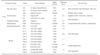
Specimens of seven core ceramics were fabricated, 11 mm in diameter, following the manufacturers' instructions. VITA Lumin A2 shade (VITA Zahnfabrik, Bad Sackingen, Germany) was selected. Thickness of the specimens was controlled with a polishing machine (AM Technology, Asan, Chungnam, Korea) to the manufacturers' recommended thickness required to mask a discolored abutment (Table 1). A sintering ceramic (VITA VM 7; VITA Zahnfabrik) was used as a reference core material.
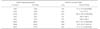
Veneer ceramics were prepared for each core material (Table 1 and Table 2), with the final thickness of layered specimen of 1.5 mm.4 Two shades corresponding to A2 and A3 shades (VITA Zahnfabrik) were selected. Thus, layered specimens were divided into A2- and A3-veneered groups. Seven specimens were made for each brand of the core and veneer ceramics. The number of specimens was determined based on previous color studies, in which generally five specimens were investigated.41-43 Detailed specimen preparation procedures have been reported previously.31
When the color of layered specimens was measured (Table 2), corresponding veneer specimen was laid over a core specimen. In this layering procedure, one veneer specimen for each material, representing the mean color value of seven specimens, was used. When layering, a drop of optical fluid (refraction fluid, 1.5 index; Cargille Lab, Cedar Grove, NJ, USA) was applied between the veneer and core specimens for an optical connection.31
Color of the layered specimens were taken according to the CIE L*a*b* color scale over a white tile (CIE L* = 94.4, a* = -0.1, and b* = 0.6) under each of three light sources. A spectroradiometer (PR-670 SpectraScan; Photo Research, Chatsworth, CA, USA), equipped with a lens (MS-75 MacroSpectar Lens; Photo Research), was fixed vertically over the upper part of a light-tight box (Color Sense II; Sungjin Hitech, Gunpo, Kyunggi-do, Korea) with a vertical distance of 355 mm from the specimen.3 A measurement spot size of 5.25 mm in diameter was selected by setting the automated aperture opening to 1 degree, which was prescribed by the manufacture. Two lamps that simulate the illuminant D65 (GretagMacbeth F20T12/65 6500 K lamp; X-Rite, Grand Rapids, MI, USA), one lamp that simulates the illuminant A (JD 100W/M2; Iwasaki Electric, Tokyo, Japan), and one lamp that simulates the illuminant F9 (F20T12/CW; Osram, Sylvania, Mississauga, Ontario, Canada) were installed on the inner top surface of the lighttight box by the manufacturer to illuminate the inside of the light-tight box with a similar light intensity regardless of the light source.3
Spectral reflectance values were obtained from 380 to 780 nm with 2 nm intervals (Spectrawin 2.0; Photo Research), which were converted to the CIE L*, a*, and b* values. Chroma was calculated as C*ab = (a*2 + b*2)1/2, and color shift was calculated as ΔE*ab = [(ΔL*)2 + (Δa*)2 + (Δb*)2]1/2.27
Vectorial shifts of lightness and chroma, and those of CIE a* and b* from the values under D65 simulator to those under A, or F9 simulators were determined. Amounts of shifts in color, lightness (CIE L*), CIE a* and b*, and also chroma, by the switch of lights were calculated. Influence of the kind of switched light (A or F9), shade of veneer ceramics (A2 or A3), and brand of core ceramics (n=8) on the shifts in color and color coordinates was evaluated with a three-way analysis of variance (ANOVA, α=.05). Brand was used as a factor instead of type of ceramics because ceramics in the same type could not be regarded as acting the same pattern by the switch of lights.
RESULTS
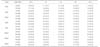
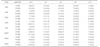
Amounts of shifts by the switch of lights are listed in Table 3 and Table 4. The range of shifts in color by the switch from D65 to A was 5.9 to 7.7 (mean ± standard deviation: 6.7 ± 0.6), that of lightness (the value under A simulator minus that under D65) was -1.3 to 1.6 (0.1 ± 0.8), that of CIE a* was 5.6 to 7.6 (6.5 ± 0.6), that of CIE b* was -0.1 to 1.3 (0.7 ± 0.4), and that of chroma was 1.1 to 2.6 (1.9 ± 0.4). The range of shifts in color by the switch to F9 was 7.7 to 10.2 (9.2 ± 0.8), that of lightness was 5.9 to 7.0 (6.4 ± 0.4), that of CIE a* was -0.9 to 0.1 (-0.4 ± 0.2), that of CIE b* was 4.9 to 7.8 (6.5 ± 0.9), and that of chroma was 4.9 to 7.7 (6.5 ± 0.9).
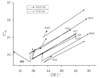
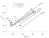
Vectorial shifts of lightness and chroma are presented in Fig. 1 and Fig. 2. In Fig. 1 to Fig. 4, 2M2 (A2) and 2M3 (A3) indicate the corresponding shifts of Vitapan 3D-Master shade guide (VITA Zahnfabrik) tabs under the same light switching conditions reported in a previous study.3 The ranges of lightness and chroma for the A2-veneered ceramics under D65 were 81.4 to 83.4 and 18.2 to 24.0, respectively, which shifted to 81.8 to 84.6 and 19.7 to 25.5 under A, and to 87.4 to 89.9 and 23.4 to 30.4 under F9. Those for the A3-veneered ceramics showed similar shifts.
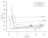
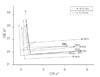
Vectorial shifts of CIE a* and b* are presented in Fig. 3 and Fig. 4. The ranges of CIE a* and b* for the A2-veneered ceramics under D65 were 0.3 to 1.7 and 18.2 to 24.0, respectively, which shifted to 5.9 to 7.7 and 18.8 to 24.4 under A, and to -0.1 to 1.2 and 23.1 to 30.4 under F9. Those for the A3-veneered ceramics were showed similar shifts.
Based on a three-way ANOVA, the shifts in color and three color coordinates were influenced by the kind of switched light, shade of veneer, and brand of ceramics (P<.05).
DISCUSSION
The null hypothesis of the present study was rejected because all the color values were influenced by three factors. Regarding the shifts of color coordinates by the switch of lights, CIE L* values under F9 were higher than those under D65 (Fig. 1 and Fig. 2), which might be caused by the difference in the light intensities of two simulators. However, it was confirmed that all the three simulators irradiated similar light intensities.3 Therefore, these shifts seem to reflect the light-switch induced lightness changes, which might be partially caused by fluorescent emission or other optical phenomena. As to the shifts in CIE a* and b* (Fig. 3 and Fig. 4), these shifts clearly reflected the SPDs of the switched lights. Fluorescent light tends to accentuate blue color, whereas incandescent light accentuates yellow-red range.13 In the present study, when light was switched from D65 to A, red and yellow hue increased (Fig. 3 and Fig. 4). When switched from D65 to F9, yellow hue and small amount of green hue increased (increased CIE b* and decreased a*).
With dental ceramics, acceptability thresholds in color parameters were determined.35 As results, the acceptability threshold was ΔL' = 2.4, ΔC' = 3.2, and ΔH' = 3.2. These parameters are used in the CIEDE 2000 color difference formula,44 and indicate the differences in the CIE L*a*b* lightness, chroma, and hue. Therefore, the thresholds for ΔL' and ΔC' were compared with lightness and chroma shifts of the present study. Lightness shifts by the switch from D65 to A (range: -1.3 to 1.6) were in the acceptable range (ΔL' < 2.4), while those from D65 to F9 (5.9 to 7.0) were not acceptable (Table 3 and Table 4). Chroma shifts by the switch from D65 to A (1.1 to 2.6) were in the acceptable range, while those from D65 to F9 (4.9 to 7.7) were not acceptable. Therefore, the shifts in color, lightness, and chroma by the switch to F9 could be regarded as visually higher compared with those by the switch to A. As to the threshold ΔE*ab values, the thresholds based on SRreadings36 were referenced in the present study. Although experimental methods were not the same, when the visually acceptable threshold(ΔE*ab < 5.5) are applied, color shifts in all specimens by both A and F9 switches were unacceptable (ΔE*ab = 5.9 to 7.7, and 7.7 to 10.2, respectively).
Influence of illuminant-dependent color shifts of shade guide tabs based on SP readings was determined, and the color differences between the values relative to the illuminants A and D65 were in the range of 0.9 to 2.7 ΔE*ab units.20 In the present study with ceramics, the corresponding values were in the range of 5.9 to 7.7 (Table 3 and Table 4), which were higher than those of the shade guide tabs. The shifts in color, lightness, and chroma of simulated all-ceramic specimens relative to three standard illuminants of SP were compared.18 As results, the range of color shifts was in the range of 1.5 to 3.6 ΔE*ab units by the switch from D65 to A and that from D65 to F2 switch was 1.3 to 3.0. Lightness shifts (ΔL*) were 0.6 to 1.2 by A switch and 0.5 to 0.9 by F2 switch. Chroma shifts (ΔC*ab) were 0.5 to 1.4 by A switch and 1.2 to 2.3 by F2 switch. Comparing with the results of the present study, the amounts of SP-based shifts were smaller than those measured by SR in the present study. Plausible causes for these discrepancies might be in the differences 1) of the measurement geometries of SP and SR, 2) in the illuminants and real light sources although the SPDs of the F2 and F9 simulators are similar, and 3) in the illuminating configuration. We think that the amounts of shift measured by SR of the present study are more clinically relevant than those determined by SP. Anyway, the color shifts by the switch of real light sources in ceramic materials are higher than those previously reported based on SP readings.
Color shifts of a shade guide due to the switch of three light sources were determined by SR.3 As results, the range of color shifts by the switch from D65 simulator to A simulator was 4.0 to 9.1 ΔE*ab units, and that from D65 to F9 switch was 3.2 to 8.5 ΔE*ab units. Comparing with the ceramics of the present study, color shifts in the corresponding shade guide tabs showed a similar trend, but were not the same (2M2 and 2M3 in Fig. 1 to Fig. 4). Based on these, it was confirmed that the shifts in color and color coordinates in clinically simulated ceramics are not the same to those of the corresponding shade guide tabs; therefore, matched color with a shade guide under a particular light source could be mismatched under a different light source.
Core and veneer specimens were optically connected by an optical fluid instead of firing together, which is a limitation of the present study. Besides, the shape and size of clinical restorations are different from columniform specimens used in the present study, which might have caused discrepancy. Further in vivo studies carried under clinical conditions should be performed.
CONCLUSION
Within the limitations of this study, perceptible color shifts of clinically simulated ceramics under different ambient light sources were confirmed by spectroradiometer readings. Color shifts under different light sources were in clinically unacceptable range (ΔE*ab > 5.5), which should be considered together with the inconsistencies in light-dependent color shifts among shade tabs, teeth, and restorations. Color matching and shade compatibility evaluation should be performed under optimal lighting conditions that simulate the light source, which is most relevant to the patient.
 XML Download