

 PDF
PDF ePub
ePub Citation
 Print
Print


INTRODUCTION
The adhesion mechanism of resin cements to dental tissues and especially to dentin, has been studied thoroughly in the last few decades. The establishment of effective micromechanical retention between the resin cement and the dentin tubuli takes place when the adhesive resin penetrates into the intratubular and intertubular dentin, forming resin tags and the hybrid layer.1 Micromechanical interlocking is the most important adhesion mechanism of resin to dentin. However, several factors such as acid-etching, moisture condition of the tooth, penetration depth of adhesive resin into dentin and dentin depth can affect the formation of hybrid layer and resin tags.2
The dentin bonding mechanism is essentially based on the infiltration of resin monomers into the porosities created by removal of mineral or inorganic material from the dental tissues. This exchange results in micro-mechanical interlocking in the porosities formed.3,4 Successful dentin bonding could be achieved through several routes. The socalled "etch-and-rinse" technique is the conventional three-step adhesion procedure. The tooth substrate is first etched with 30-40% phosphoric acid (H3PO4) and then rinsed off. Following acid etching, adhesive resin is applied on the conditioned tooth surface. For dentin, the bonding mechanism of etch-and-rinse adhesives primarily depends on micro-mechanical retention of resin with the exposed collagen fibrils. For enamel, total etch technique is the most effective and reliable method for long-term clinical success.5
In the "self-etch" approach, adhesives condition and prime dentin are applied at the same time, and no rinsing is required. In this procedure the clinical application time is shortened and technique sensitivity is significantly reduced. Self-etch adhesives can be categorized as "mild" and "strong". Strong self-etch adhesives with functional monomers have low pH (<1) and their bonding mechanism is reported to be similar to etch-and-rinse adhesives. 'Mild' self-etch adhesives (pH≈2) selectively demineralize the dentin surface and are reported to form a shallow hybrid layer. Adhesion is ensured by chemical interaction between residual hydroxyapatite and functional monomers.6
Several self-etching adhesive systems contain specific functional monomers which enhance the performance of adhesion. The functional monomers may help conditioning dental tissues, increase monomer penetration,7 and also improve the chemical adhesion to hard tissues of the tooth.8 10-methacryloyloxydecyl dihydrogen phosphate (10-MDP) is one of the most commonly used functional monomers;9 it is the hydrophilic phosphate monomer that increases resin diffusion and adhesion by causing acidic decalcification and binding to calcium ions or amino groups of tooth structure.10 It is reported to be one of the most successful materials in the market for chemical bonding.11,12 On the other hand, self-adhesive cements do not require conditioning the dentin or ceramic surfaces. Such cements have multifunctional phosphoric acid dimethacrylate modified monomers.2 However, their diffusion level into dentin and their hydrolytic stability are not optimal.12-17
Adhesion to deeper tooth substrates with contemporary strong adhesives is an interesting subject which is not studied thoroughly in the literature. The objectives of this study were to evaluate the adhesion of two different 10-MDP containing adhesive resin cements to deep dentin using either etch-and-rinse or two-step self-etch bonding techniques and analyze the failure types. The null hypothesis tested was that adhesion of 10-MDP containing resin cements would not show difference when used in combination with etch-and-rinse or two-step self-etch system.
MATERIALS AND METHODS
This study was approved by the Ethics Research Committee of Istanbul University Faculty of Medicine (Istanbul University, Istanbul, Turkey) (Approval no: 2013/317).
Intact human third molars without caries (N=180) were used for this study. The roots of the teeth were embedded in auto-polymerized acrylic resin (Takilon, SPD Salmoiraghi Produzione Dentaria S.r.l Mulazzano, Italy) and randomly divided into 6 groups (n=30 per group). Dental materials of 2.0 mm thickness from the occlusal surfaces of the teeth were removed by means of a slow-speed diamond saw (Isomet, Buehler Ltd. Lake Bluff, IL, USA) under water-cooling to expose flat deep dentin surfaces. Standardization of smear layer was achieved by grinding the dentin surfaces with 600 grit silicon carbide paper.
The adhesive systems were applied according to the manufacturers' instructions. Application procedures and product information is displayed in Table 1. A transparent polyethylene mold (diameter: 4 mm and height: 6 mm) was used to bond the resin cements onto dentin surfaces (Fig. 1A).
In Group 1, conventional adhesive resin cement (Panavia F 2.0, Kuraray, Tokyo, Japan) (PAN) was applied following etch-and-rinse and bonding (ED Primer, Kuraray). In Group 2, PAN resin cement was applied following bonding (ED Primer) without acid etching. In Group 3, self-adhesive resin cement (Clearfil SA, Kuraray) (CSA) was applied following etch-and-rinse and bonding (Clearfil SE, Kuraray). In Group 4, CSA resin cement was applied following bonding (Clearfil SE) without acid etching. In Group 5, only CSA resin cement was applied onto dentin surfaces neither with acid etching nor bonding. Lastly, in Group 6, only PAN resin cement was applied onto dentin surfaces neither with acid etching nor bonding. Light activation was performed for 20 seconds, using a quartz tungsten halogen curing device (Hilux 200, Benlioglu, Ankara Turkey) with irradiance of 600 mW/cm2. The specimens were stored for 24 hours in dark at room temperature prior to bond strength tests.
Bonding strength was evaluated using a mechanical shear test. Specimens were placed firmly in the universal testing machine (Shimadzu AG-IS, Shimadzu, Kyoto, Japan) and the shear force was applied until fracture of the specimen (Fig. 1B). The load was applied at the dentin/adhesive interface, as close to the surface of the tooth as possible. A crosshead speed of 0.5 mm/min was used for loading. Subsequently, specimens were evaluated under optical microscope (Leica M80, Leica Microsystems, Wetzlar, Germany) at ×40 magnification in order to investigate the mode of failure. The types of failures were categorized as; adhesive failure between tooth and resin cement (A), cohesive failure of resin cement (C) or mixed failure where resin cement was failed partially cohesively and partially adhesively on the specimen (M).
The specimens were sectioned longitudinally and interfaces were wet polished with silicone carbide papers of #600, #1000, #1200 grit in sequence. Following polishing, the interfaces were decalcified (37% H3PO4 for 10 seconds) and deproteinized (2% NaOCl solution for 1 minute) in order to evaluate the hybrid layer.
The hybrid layer and surface pattern were investigated for all experimental groups using scanning electron microscopy (SEM) (JSM 7000F, JEOL, Japan) by selecting one specimen with bonding strength closest to mean value of the group. The specimens were covered with a thin layer of platinum (Sputter-coated) for SEM observation. The whole fractured surface at the dentin side was observed under the SEM.
Statistical analysis was performed using SPSS 11.0 software for Windows (SPSS Inc., Chicago, IL, USA). The assumptions of normality and equal variances across groups were validated. Bond strength data (MPa) were submitted to analysis of variance (two-way ANOVA). Multiple comparisons were made with Tukey's post-hoc test (α=.05) with the shear bond strength as the dependent and adhesion protocols and the cement types as the independent factors. P values less than .05 were considered to be statistically significant in all tests.
RESULTS
Overall, PAN adhesive cement showed significantly higher mean bond strength (12.5 ± 2.3 - 14.1 ± 2.4 MPa) than CSA cement (9.3 ± 1.4 - 13.9 ± 1.9 MPa) (P<.001) (Table 2). Etching dentin with 35% H3PO4 increased bond strength significantly for both PAN (14.1 ± 2.4 MPa) and CSA (13.8 ± 1.9 MPa) compared to the application of two-step self-etch adhesive resin (12.5 ± 2.3 - 9.8 ± 1.6 MPa, respectively) (P<.05).
Application of bonding agent on dentin (9.8 ± 1.6 MPa) did not significantly increase the bond strength of CSA cement when compared to direct application (9.3 ± 1.4 MPa) (P>.05). Similarly, for the PAN adhesive cement the bond strength did not differ significantly between bonding(12.5 ± 2.3 MPa) and direct applications (12.8 ± 2.6 MPa) (P>.05).
Adhesive failures were more frequent in CSA cement groups when used in conjunction with two-step self-adhesive (68%) or no adhesive at all (66%) compared to other groups (37-43%) (Table 3). Generally, specimens with lower bond strengths failed to bond adhesively.
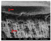
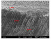
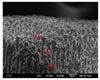
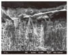
SEM observations demonstrated well-defined hybrid layer with visible resin tags in dentin in PAN cement group on 35% H3PO4 conditioned dentin and very minor detached areas were also evident at the interface (Fig. 2). The unconditioned dentin specimens presented thinner hybrid layer and less frequent distribution of resin tags (Fig. 3).
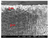
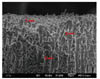
Hybrid layer quality was inferior in CSA compared to PAN cement in all conditions. On CSA specimens, resin tags were also visible on both H3PO4 conditioned and unconditioned dentin surfaces (Fig. 4, Fig. 5 and Fig. 6). Although resin tag formation was present, resin cement showed detached areas from dentin (Fig. 4). On unconditioned dentin surfaces a superficial interaction and a thin hybrid layer formation was apparent compared to PAN (Fig. 7).
DISCUSSION
Self-etch adhesive resins are easy to use and their shorter chair-time presents advantages. On the contrary, etch-and-rinse systems are more technique sensitive and require experience from clinicians.6,7 Since the use of self-etch adhesive system resulted in inferior bond results with both PAN and CSA compared to total etch system on dentin, the null hypothesis was rejected.
Total etch adhesive systems are reported to have better clinical results on the long term.18 Resin cement adhesion to dentin depends on surface energy of the dentin and the wettability of the resin cement on the dentin. H3PO4 application removes the smear layer and increases surface roughness and thereby, the wettability of the adhesive resin or the resin cement.19 It is also speculated that the water content of the dentin increases after acid etching which eventually helps the ionization of the acidic monomers in resin cements.20-22 This results in improved bonding penetration and more effective surface conditioning.20-22 Similarly, in the present study, with etch-and-rinse technique, better results were achieved compared to self-etch adhesive for both resin cements. Microscopy evaluations supported these findings. For the PAN resin cement, H3PO4 etching prior to bonding displayed higher mean bond strength. In the SEM observations, application of PAN to H3PO4 conditioned dentin surfaces resulted in visible hybrid layer, regular resin tags and no detached layers. Application of the same resin cement to unconditioned dentin after self-etching adhesive did not have major visual differences in SEM observations, but thickness of the hybrid layer was lower and resin tags were less frequent and more irregular.
CSA resin cement used in this study has self-adhesive properties. Self-adhesive resin cements etch and prime the dentin surface simultaneously without a need for acid application. However, the bond strength results achieved in the current study displayed that H3PO4 etching prior to the application of resin cement positively influences the bond strength of self-adhesive cement. It should also be noted that self-adhesive resin cements are reported to only interact with superficial dentin, without any presence of a hybrid layer or resin tags.23,24 This was observed apparent in the SEM images. On the unconditioned dentin, the hybrid layer was not present and the resin tags were irregular and less in number. In addition, detachment of resin tags was observed in some areas. However, H3PO4 etched dentin displayed more frequent resin tags and less detached layers. Similar results with self-adhesive resin cements were reported in previous studies.23,24 De Munck et al. reported low demineralization effect on RelyX Unicem, even on dentin surfaces without smear layer.23 Pisani-Proença et al. confirmed that the SEM observations of untreated dentin shows the need for further improvement for better infiltration and chemical binding of the self-adhesive cements. They also reported that H3PO4 etching of the dentin increases the bonding effectiveness of self-adhesive resin cements.25
It should also be stressed that both PAN and CSA resin cements demonstrated significantly higher mean bond strength when etch-and-rinse technique was used, being less effective for the latter. One reason for this could be attributed to the differences in the structure of the hybrid layers. SEM micrographs of PAN cement applied dentin displayed a thicker and attached hybrid layer on etched and rinsed dentin. On the other hand, CSA cement applied dentin showed thinner hybrid layer and detached local areas. Other reasons for this could be the variations in mechanical properties such tensile and flexural strength and Young's modulus.21-25 Although the statistically significant difference was stated based on the 2 MPa difference between self-etch and total etch systems, this difference may have less clinical meaning. Yet, clinical studies should verify the clinical performance of these two cement types applied on dentin with two adhesion modalities. Until then, it can be stated that etch-and-rinse may be the single most important factor in shear bond strength of resin cements to dentin evaluated in this study.
The functional monomer, 10-MDP, is present in both PAN and CSA resin cements. The stronger adhesion capabilities of these materials is due to this component; it is reported to be most promising monomer for chemical bonding to hydroxyapatite of enamel and dentin due to being stable against hydrolysis and forming strong ionic bonds with calcium.9 As a result of these positive qualities these resin cements may be preferred by clinicians. It should also be noted, however, this most promising material may have a decrease in adhesion when applied to deeper dentin. It is reported that shear bond strength decreases as the depth increases in dentin.22 This is observed being as a result of morphological differences; mainly increase in number of dentin tubules decrease in mineralized content. Also, vital deep dentin is highly hydrated and dentin fluid flows outward from dentin tubules. This movement of fluid may increase the pressure from direction of pulp and may hinder the adhesion. This wetness and movement may affect the optimal resin seal.23 Considering reports from previous studies and the results of this study where self-etching generally observed to be weaker than etch-and-rinse, the following can be suggested. In clinical situations where adhesion to deeper dentin is required such as large inlays/onlays or retreated crown or bridge abutment teeth were excessive tooth preparation was made, it may be appropriate to use total etch technique even with self-adhesive resin cements.
It should be noted that the results of the study represent possible early clinical failures. Adhesive joints are prone to degradation when they are thermocycled or long-term water stored. In deep dentin after aging, such cements show dramatic decrease in adhesion when only manufacturers' adhesion protocols were employed.17 The effect of aging conditions needs further investigations also considering etch-and-rinse bonding system.
CONCLUSION
Within the limitations of this study, it can be suggested that using etch-and-rinse technique for both conventional and self-adhesive 10-MDP containing resin cements result in greater success on the early clinical period. Clinicians should consider this especially in situations where adhesion is taking place mostly to dentin tissues, such as large inlay or onlay restorations and excessively prepared teeth for fixed dental prostheses.


 XML Download