

 PDF
PDF ePub
ePub Citation
 Print
Print


INTRODUCTION
Polymethyl methacrylate (PMMA) is primarily used for removable partial and complete denture fabrication as a base material. Reinforcements such as fibers, glass1 and polyethylene or activation and processing techniques such as injection-molding and microwave2 activation provided new benefits in polymer knowledge. However, this material still has some limitations about mechanical properties such as dimensional stability, color and flexural strength.
Eventually fracture of acrylic resin denture base is known to be the common clinical problem in prosthodontic practice. Causes of such fractures are known to be related to porosity, residual monomer, poor fit of denture base, occlusal imbalance, design and fabrication failures, material choices, stress after long clinical usage and accidents.3,4 Besides, in certain circumstances before complete fracture, a crack formation may be propagated bound to different types of stresses that denture base materials are subjected to. It is known that rough surfaces present suitable conditions for microorganism colonisation and/or biofilm formation,5,6 denture base cracks may become to be one of the best sites for microorganism propagation.
Biofilm is a microbial community that has dense and complex structure and may represent multiple organisms.7 They are often encapsulated within a matrix of exopolymeric material that consists of intricate networks of cells attached to biotic surfaces. They resist antimicrobials and immune cell challenge8 and are deeply embedded into cracks and porosities of dental materials as mentioned before. Metallic and non-metallic medical devices like catheters, implants, dental materials are suitable sites for colonization of various types of microorganisms.9 Corrosion at the interfacial surfaces of non-metallic materials usually starts with swelling after infiltration of little molecules or microorganisms. The chemical bonds are often subjected to corrosion with physiochemical process. Interfacial electrochemical process may be activated with the formation of biofilm on metallic and non-metallic surfaces resulting in an increased corrosion of colonized substratum. This development can detoriorate the materials with the presence of biofilm and is termed as biocorrosion.9
Three-dimensional structure of biofilm is known to provide a highly complex arrangement of microorganisms.10 Several studies regarding the developments and structures of biofilms on different dental materials including denture bases and their effects over oral health have been constituted.8-13 However the relationship between the biofilm related biocorrosion and crack and/or fracture formation still remains complicated even undefined.
The aim of this study was to investigate the destructive effects of biofilm formation and/or biocorrosive activity of 6 different oral microorganisms by evaluating the diffusion potential of the microorganisms, fracture propagation and scanning electron microscope (SEM) images (calculating biofilm covered surfaces) on three different denture base materials.
MATERIALS AND METHODS
Three different heat polymerized acrylic resins (Table 1) were used to prepare 50 × 15 × 4 mm (Type A; for three point bending (TPB) test, n=210), 8 × 8 × 1 mm (Type B; for SEM analysis, n=54) and 2 × 2 × 2 mm (Type C; for spectrophotometer analysis, n=180) samples (Table 2). Negative molds of the metal masters were obtained with a medium viscosity impression material. This technique was used to fabricate all types of the specimens. Wax patterns were invested in metal dental flasks. Acrylic resins were polymerized according to manufacturers' instructions. Flasks were left for 180 minutes cooling. Next, each specimen were deflasked and finished with 320, 400 and 600-grit silicone carbid papers. To simulate a crack line on the denture base, "V" type notch was carved in the middle of each specimen of impact test groups (A type) along with the 15 mm surface by using a milling machine and a milling tool as shown in the Fig. 1. The depth was 2 ± 0.2 mm. All type of specimens were ultrasonically cleaned for 20 minutes and immersed in distilled water for 48 hours at 37℃ before tests.
S. aureus, E. faecalis, P. aeruginosa, E. coli, S. mutans and C. albicans strains were inoculated in trypticase soy broth media and grown to stationary phase overnight. The samples were diluted 1:100 and each diluted bacterial culture (200 µl) was inoculated into each well in a fresh 96-well flat-bottom microtiter polystyrene plates, which also contain "C" type acrylic resin samples. Plates were incubated for 48 hours at 37℃ and visualized by staining with 0.5% crystal violet for five minutes after washing with water. The biofilm was quantified in duplicate, after adding 100 µL of 95% ethanol and the contents were transferred to new wells of microtiter plate. Optical density (OD) of stained adherent bacteria was determined with a micro ELISA auto reader at wavelength of 620 nm spectrophotometrically.14,15 These OD values were calculated as: "OD-control" and considered as an index of bacteria adhering to acrylic surfaces and forming biofilms.
Ethylene oxide was used to sterilize the specimens. Ten specimens of type "A", 3 specimens of type "B" and 10 specimens of type "C" denture base materials from 3 different brands (Table 1) were randomly inoculated into one of; Staphylococcus aureus (ATCC 6538), Streptococcus mutans (ATCC 35688), Enterococcus faecalis (ATCC 10541), Escherichia coli (ATCC 25922), Pseudomonas aeruginosa (ATCC 2327) or Candida albicans (ATCC 18804) culture as indicated in table 2 at 0.5 McFarland scale which corresponds to 108 cfu/mL for 168 hours at 37℃. A previously described method was modified and performed for biofilm formation.11 In brief, 1 mL aliquot of the bacterial and yeast cultures were introduced into 500 mL of brain heart infusion broth media in conical flasks and prepared denture base materials were inoculated into media. For the maintenance of bacterial and yeast density near the steady-state growth phase, 50% of the media were drained and replaced with the equal amount of a fresh sterile medium every two days and on day seven, the denture base materials were retrieved from the inoculated media for SEM examination. The control group was composed of 10 non-contaminated "A" type samples of each denture base material (30 total) and was kept in distilled water at 37℃ until the TPB test.
The contaminated samples of type "A", were rinsed with PBS and kept in running water for 15 minutes. The impact values of the specimens were measured under 2500 N maximum load (1 N preload) with 1 mm/min cross-head speed with TPB test by using an universal testing machine (Lloyd LRX, West Sussex, UK). The specimens were then supported on the jigs with a diameter of 3.2 mm with span length 50 mm. The "V notch" was placed face down on the jigs and the load was applied to the centre of the specimens. The data of the measurements were transferred to a personal computer, and the results were recorded.
After incubation, each specimen of type "B" was removed and kept in an ultrasonic cleaner for 1 minute and rinsed with PBS to remove non or weak adhered microorganisms. The samples were fixed in 2.5% glutaraldehyde solution for 1h at room temperature, then rinsed with PBS. Ethanol solutions with concentrations graded from 75% to 95% were used in 5 steps to dehydrate the specimens. Specimens were then dried and placed on stubs to coat with 20 A0 gold/palladium for SEM (JEOL 6400, JEOL Corp., Tokyo, Japan) analysis operating at 10 kV. Digital photographs as TIFF files at ×5,000 magnification were obtained from three different regions of each sample surface (Fig. 2). The images were transferred to a personal computer to calculate total area and area fractions of biofilms using Image J software.16 Since cleaning and dehydrating processes were performed accurately, the area aspects apart from denture surface were all accepted as biofilm surface of that microorganism. Area fraction of each image was recorded as the data of that sample.
Descriptive data were expressed as median, maximum, minimum and mean ± standard deviation. Statistical analysis was performed using "PASW 18.0 Statistics" and "STATISTICA-7" statistical software. When normality assumptions were satisfied Analysis of Variance (ANOVA) otherwise the equivalent non-parametric test: Kruskal-Wallis was used for group comparisons. When significant differences found between groups, we used Tukey and Dunnett's test (after ANOVA) and Dunn's test (after Kruskall Wallis) for multiple group comparisons. Results were considered statistically significant at α=0.05.
RESULTS
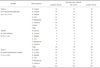
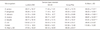
Table 3 shows the mean quantitative biofilm values and statistical significances according to microorganism and denture base material. There were significant differences among the adhesion potential of 6 different microorganisms and there were significant differences among their adhesion onto 3 different denture base materials. The highest median value within all the denture base materials was obtained with P. aeruginosa (0.095 ± 0.018) followed by S. aureus (0.085 ± 0.014), C. albicans (0.081 ± 0.017), S. mutans (0.079 ± 0.010), E. faecalis (0.070 ± 0.013) and E. coli (0.070 ± 0.012). E. faecalis and E. coli were found to be the least adherent microorganisms. The difference of adhesion between P. aeruginosa and S. aureus was not significant however there were significant differences between P. aeruginosa and other 4 microorganisms. The adhesion potential of the microorganisms over Lucitone denture base material was higher than the other materials. The difference was not significant compared with QC-20 but it was significant when compared with Ivocap Plus. P. aeruginosa exhibited the highest median value on Lucitone 550 denture base material surface (0.105 ± 0.020) while C. albicans exhibited the lowest value on Ivocap Plus (0.067 ± 0.021).
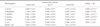
SEM images revealed the regions of typical biofilm formation of each selected microorganism on denture base materials (Fig. 2). Table 4 shows the mean biofilm covered regions (%) on denture base materials and statistical significances according to microorganism and denture base material. The type of denture base material did not alter the diffusion potential of the microorganisms significantly. The percentages of biofilm covered areas of denture base materials ranged from 52.57% to 70.96%. S. aureus and P. aeruginosa had significantly higher (≥68.66%) diffusion potential than the other tested microorganisms, but the difference among them was not significant. E. coli had the least diffusion potential and was significantly different from all the tested groups.
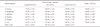
Table 5 shows the mean and standard deviation values of TPB test and statistical significances according to microorganism and denture base material. Biofilm formation of the tested microorganisms decreased the TPB test values compared to the control group and this was statistically significant. Lucitone 550 and Ivocup Plus denture base materials were significantly more resistant than QC-20 when mean TPB test values were consulted. The difference between Lucitone 550 and Ivocup Plus were not significant. E. coli produced the minimum destructive effect to the base materials when mean TPB test values of materials were evaluated, however this was not significantly different from E. faecalis (P=.699>.05), S. mutans and C. albicans.
DISCUSSION
This study investigated the effects of biofilm formation and/or biocorrosive activity of 6 different oral microorganisms on three different denture base materials. All tested microorganisms significantly decreased the TPB test values of the tested denture base materials.
After contamination with microorganisms, the TPB test values of denture base materials were decreased significantly compared with the control groups. It may be speculated that the structure of all the denture base materials were decomposed by the biofilm formation and/or biocorrosive activity of microorganisms.
Several studies have investigated the fracture resistance of denture materials.17-20 In this study Ivocap Plus appears to be the most resistant base material when the peak data of control group and mean values at TPB tests were evaluated. This was compatible with the results from the study of Hedzelek and Gajdus.21 However, when the percentages of TPB test value reduction were examined, Lucitone 550 was the most resistant material. The mean TPB test value of samples exhibited 18% resistance reduction at Lucitone 550 samples, 19% at Ivocap Plus and 24% at QC-20 denture base materials compared with control group after contamination processes of 6 different microorganisms. Evaluating the subgroups data of denture base materials and microorganisms of this study, the highest TPB test value reduction was observed in P. aeruginosa-QC group with 36%. Even E coli, known to have the least degenerative bioactivity,12 decreased the TPB test values of Lucitone 550 by 8%. Eventually considering all the mean TPB test values of three denture bases of our study it can be affirmed that microorganisms had biocorrosive activity and deteriorated at least 15% of the initial physical composition of tested denture base materials.
However, the difference of mean biofilm covered regions on denture base materials at SEM display were not statistically significant the mean quantitative biofilm values of microorganisms on Ivocap Plus was significantly different from Lucitone 550. This may be due to the dissimilar accumulation of microorganisms on the material surface (Fig. 2) that varies according to the production of extracellular polysaccharides which can only be demonstrated using advanced techniques like three-dimensional confocal scanning laser microscopy.13 Consequently, the two-dimensional SEM display may have inhibited quantitative estimation of this phenomenon.
Serrano-Granger et al.22 claimed that there was no relationship between the microorganism adhesion and acrylic resin type and/or composition. This was particularly compatible with our study; when biofilm covered regions were evaluated when we observed that microorganisms showed minimal adhesion to Ivocap Plus. This result was not statistically significant when compared with QC but significant compared with Lucitone 550. This may be attributed to the special fabrication technique of this material (injection molding) which may form samples with smoother surface and lesser microporosities. The other reason for minimal adhesion may be the higher residual monomer release after using injection molding technique.23 It is known that the PMMA monomer is toxic for living cells.23,24 During the experimental process, with the mentioned destructive effect, PMMA monomer may have inhibited the survival and/or adhesion of microorganisms tested on denture base materials. On the contrary, the higher adhesion to Lucitone 550 may be attributed to the reinforcing materials that poorly adhere to the polymer matrix forming microporosities that become suitable for microorganism accumulation.25
C. albicans was shown to have higher adhesion potential to denture base materials in most of the studies.10 Conversely, in our study C. albicans-Ivocap Plus group exhibited the least adhesion potential when all the subgroups of quantitative biofilm values were examined. Nevertheless, P. aeruginosa appeared to be significantly the most adherent microorganism. This result was compatible with the literature that P. aeruginosa and S. aureus have higher survival success over the plastic and/or metallic devices used in medicine.26 Additionally the mean biofilm covered regions on denture base materials at SEM display indicated the highest percentages for P. aeruginosa and S. aureus confirming the results of mean quantitative biofilm values of our study.
Within the limitations of this study it was shown that microorganisms diffused at least 52% of the denture base surface that cannot be neglected in dental practice. However it was a remarkable result that this rate did not exceed 71% (Table 4) which may be attributed to the environmental or local conditions of this study or the floral equilibrium of microorganisms in certain circumstance.


 XML Download