

 PDF
PDF ePub
ePub Citation
 Print
Print


INTRODUCTION
Recently, esthetic requirements in dental restorative treatment are on the rise. For this reason, recent attention-drawing material is zirconia which is a biocompatible material with stable structure. Flexural strength and fracture toughness of zirconia reaches up to 900-1200 MPa and 9-10 MPa·m1,2 respectively, and zirconia has been proven to be much more excellent in its strength than that of glass infiltrated full porcelain which is widely used in esthetic restoration.1,2 Material properties of zirconia like these show that zirconia can be substituted for metal and can be surely used even in long span fixed prosthesis.3,4
CAD/CAM system which was introduced to dentistry in 1980's has brought cost and time reduction in fabricating restoration by using computer for inputting, designing and cutting the form of restoration. In order to overcome disadvantage of previous method of framework fabrication in which post-sintered zirconia block was cut, pre-sintered zirconia block is being used recently. By doing so, fabrication time and consumption of bur have been reduced and thus cost and time was also reduced. This method, however, has disadvantage in that approximately 15-30% shrinkage occur in the procedure of sintering and hardening the cut block using CAM unit for the increased density of zirconia block.5 When using partially sintered block, therefore, it is more important to calculate shrinkage rate accurately than any other things in order to compensate shrinkage which inevitably occurs in sintering process, and also to gain precise product when using partially sintered block.
It is considered that internal and marginal fit plays a key role in longevity of prosthesis. In previous studies, many authors said that 120 µm or less marginal gap of prosthesis was clinically acceptable.6,7 According to a study on fit of zirconia framework which was used in all ceramic crown, marginal gap was reported to be 64-83 µm,8,9 and in another study on clinical fit of 3-unit fixed prosthesis, zirconia prosthesis (LAVA, 3M ESPE, Seefeld, Germany) for which CAD/CAM was used reportedly showed 80 µm of mean marginal gap.10 The reason why marginal fit is important is because increased marginal leakage may cause secondary caries, periodontitis, pulpitis etc. and also cause esthetic problem and eventually cause failure of prosthesis. Internal fit also is very important for longevity of prosthesis. It influences retention and support of prosthesis. There was a report from one previous study saying the thicker the cement became due to a large internal gap, the weaker the porcelain became.11 This effect is shown also in zirconia. Excessive thickness of cement increases radial crack12,13 and causes fracture of veneering porcelain.14 Fracture of veneering porcelain is considered to be one of the greatest reasons for removing zirconia restoration.15-18 As opposed to this, too small internal gap may cause unstable seating of prosthesis.19 Abduo et al.20 said that factors which influenced fit of prosthesis were fabrication system of zirconia, veneering, configuration, span length of zirconia etc.
Although the use of zirconia has increased and are being used more in multiple areas where the tooth are missing, studies on fit of zirconia fixed partial denture until the now have been limited to 3-unit fixed partial denture with nearly linear form; there has been few study on internal or marginal fit of 4 or more unit fixed partial denture with curved form.20 Particularly, there was no study in which both internal and marginal fit depending on the span length were simultaneously evaluated.
The purpose of this study, therefore, was to measure marginal and internal fit of single, 4-unit, 6-unit zirconia fixed partial denture core which had been fabricated using CAD/CAM system by using replica technique and to evaluate the effect that span length on fit while evaluating whether measured marginal gap was in clinically acceptable range or not.
MATERIALS AND METHODS
Experimental groups were divided into single, 4-unit and 6-unit groups, and preparation of each abutment teeth were done in order to make single crown of upper right central incisor, 4-unit fixed partial denture with abutment teeth of upper right and left lateral incisor where upper right and left central incisors were missing, and 6-unit fixed partial denture with abutment teeth of upper right and left canines where upper right, left central and lateral incisors were missing (Table 1). For tooth preparation, surveyor and diamond bur were used, and total taper of each abutment teeth was intended to be 6°. Preparation amount was 2 mm on incisal part and 1 mm on axial wall, and 1 mm width deep chamfer margin was formed.
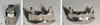
In order to prevent possible wear or fracture of master models from repetitive impression taking procedure, dentiformmodels (Nissin Dental Prod. Inc., Japan) where abutment preparation were done were duplicated and titanium master model weremade (Addtech Co., Seoul, Korea) (Fig. 1).
Impression of titanium model were taken with conventional method by using addition silicone impression material (Imprint II, 3M ESPE, St. Paul, MN, USA) and readymade tray, and total 30 improved stone (Fuji rock® EP, GC Corp, Tokyo, Japan) models including 10 models per each group were made (Fig. 2).
After scanning of the manufactured plaster model using CAD/CAM system, partially sintered zirconia blocks were cut and sintered, and finally zirconia cores were fabricated (Fig. 3). When fabricating cores, CAD/CAM system of Orapix (Seoul, Korea) and zirconia blocks were used.
Thickness of core and cement space were set as 0.6 mm and 40 µm respectively. Internal adjustment was not done so that fit of core that was fabricated by only CAD/CAM could be merely evaluated. After core fabrication was done, whether there were remnants inside, defects or distortions were checked and then cleaned with high pressure steaming.
After positioning fabricated zirconia cores on each models, jigs for positioning the zirconia coreon the model were made using Pattern resin® (GC Dental, Japan) so that zirconia could always be positioned at a same spot on the models. After filling fit checking material (Fit checker, GC Dental, Japan) in inner surface of core, it was placed on abutment tooth. After that, pre-made resin jig was positioned above the core. While maintaining adapted position by hand pressure, it was place in Universal testing machine (Shimadzu corporation, Kyoto, Japan) immediately. Universal testing machine was set to measurement mode, and its compressive force was limited to 40 N, and regular force (40 N) was maintained for 5 minutes until Fit checker was completely hardened.
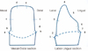
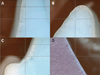
After 5 minutes, the core was removed from the model carefully. When doing so, the silicon film of Fit checker has to be fully attached to silicone core. Filling inner surface of silicone film of core with regular bodied addition silicone impression material (AquasilLV, Densply Caulk, USA) and hardening the filled material increase strength andalso enables gaining of stable film layer. Each 8 measuring points were established for labial and mesio-distal side respectively (Fig. 4), and cutting was done right in the center of the model labialy and mesio-distally (Fig. 5). By using measuring microscope (AXIO®, Carl Zeiss, Rochester, NY, USA) and I-Solution™ (IMT I-Solution Inc., Vancouver, BC, Canada), thickness of fit checker was measured at 16 measuring points of each abutment teeth (Fig. 6). Mean value was documented after 3 times measured by 2 experimenter for each measuring points.
Mean value and standard deviation value were calculated by adding measurement values of right and left abutment teeth of each group which was divided into bucco-lingual and mesio-distal.
Whether there was statistically significant difference between groups which were divided into bucco-lingual and mesio-distal or not was analyzed by multivariate analysis. After that, if there had been statistically significant difference, post-hoc test was done using Dunnett T3 test per each measuring points. In all analyses, statistical significance was accepted at 5% probability level.
RESULTS
Twenty seven specimens including nine single, nine 4-unit and nine 6-unit were used in fit measurement. There existed 1 specimen per each group of which measurement could not be done because fracture had occurred in the process of applying force after putting Fit checker inside the core and placing it on abutment.
Statistical analysis was performed by adding each measurement values of right and left abutment teeth of each group. By doing so, number of specimen of each group became nine single, and eighteen 4-unit and eighteen 6-unit. Statistical analysis was performed separately for bucco-lingual and mesio-distal.
Mean value and standard deviation value of gap at bucco-lingual measuring point were calculated (Table 2). As a result of multivariate analysis there was significant difference in at least one or more point. Which measuring point showed statistically significant difference was checked using ANOVA. As a result, statistically significant difference was shown at point 2, 4, 7 and 8. In order to investigate between which groups of point 2, 4, 7 and 8 showed the difference, post-hoc test was performed using Dunnett T3 test (Table 3).
Mean value and standard deviation value of gap at mesio-distal measuring point were calculated (Table 4). There was significant difference in at least one or more point in multivariate analysis. The result of ANOVA showed statistically significant differences at point 2, 4, 7 and 8 by span length factor. In order to investigate between which groups of point 2, 4, 7 and 8 showed the difference, post-hoc test was performed using Dunnett T3 test (Table 5).
As a result of measuring point analysis which showed statistically significant difference, measurement value increased from single, 4-unit and 6-unit in order in point 2, 4, 7 when bucco-lingual analysis was done. But in point 8, the gap of 4-unit group was measured to be the smallest. In the result of mesio-distal analysis, measurement value increased from single, 6-unit and 4-unit in order in point b, f and g (4-unit group showed the largest value).
DISCUSSION
Zirconia is an esthetical and biocompatible material, and its physical property is so strong that it can be used for fixed prosthesis of posterior region. Because of its property like this, it has become one of the most noteworthy materials in esthetic prosthetics. In order for zirconia to be used as prosthetic restoration material that can last long and function stably, not only its physical property but also the fit of prosthesis made by it should meet clinical requirements.
In fabrication of zirconia prosthesis using CAD/CAM, completely sintered block was used initially. However, this method has disadvantages in time and cost although fit could be more excellent than when partially sintered block which is used in most fabrication system is used. On the other hand, if partially sintered block is used, it will be better off when it comes to time and cost factor. However, if cutting is not done by accurate calculation due to shrinkage in sintering process, level of fit will decrease significantly.
According to previous studies, 120 µm or smaller marginal gap is clinically accepted.6,7 In the studies on fit of single or 3-unit fixed partial denture zirconia prosthesis fabricated by CAD/CAM system, marginal gap showed approximately 64-83 µm as a result.8,9 These show that zirconia can be used for fixed partial denture restoration adequately. However, these were the studies which had been mostly proceeded on linear type fixed prosthesis which was shorter than 3-unit. Study on prosthesis of which span is longer than this has not been done.10 In this study, therefore, whether fit of zirconia prosthesis is influenced by the span length was investigated using anterior teeth model.
As methods for measuring marginal or inner fit, a method of direct inspection, a method of inspection after cutting, a method of evaluation by impression taking, a method of evaluation using explorer and so on were suggested. Although a method of inspection after cutting may be the most accurate way, there is a shortcoming in that more specimens have to be made for the increase of the number of measurement. Therefore, replica technique where measurement of fit of various parts could be done easily with only small number of specimens was used in this study. This method had been considered to have low accuracy in the past,21 but it became known to be reliable compared to other methods for measuring fit by Rahme et al.22 and Laurent et al.23 study. Specimens for measurement were made using Replica technique, and thickness of fit checker was measured at 16 measuring points which were designated per each abutment teeth, and 2 experimenters recorded mean value after 3 times of measurements so that error could be minimized.
As a result of measurement, overall fit was acceptable (small gap) in marginal and axial wall area, but connection part between margin and axial wall and incisal part showed relatively large gap. Because it was considered that whether it was either right abutment tooth or left abutment tooth could not be a variable for analysis, mean value and standard deviation were calculated by adding measurement values of right and left abutment teeth together for statistical analysis in 4-unit and 6-unit group. In the case of this study, multivariate analysis was used because it was considered that gap of inner surface and margin could not be independent to each other although independent variable was a span length.
As a result of statistical analysis of bucco-lingual (1-8) and mesio-distal (a-h) measuring points, there were statistically significant differences at point 2, 4, 7, 8 and b, d, e, f, g among the groups. From this result, the fact that span length influenced fit of zirconia core was confirmed. As a result of analysis of each measuring points which showed statistically significant difference, measurement value increased from single, 4-unit to 6-unit in order at 2, 4, 7 point, but measurement value of 4-unit group came out smallest at 8 point. As opposed to this, 4-unit group showed the largest value at b, f, g point as a result of mesio-distal analysis. The value increased from single, 6-unit and 4-unit in order. This is considered to be because of small number of specimens that were used in the experiment and measurement error as well. Also, there were more points which showed significant difference inmesio-distal part than inbucco-lingual part. It is guessed that the reason for this is because more error occurred in the process of shrinkage at mesio-distal part since the form of zirconia core was not bucco-lingually but mesio-distally long. Although there was not enough number of specimen for more accurate multivariate analysis in statistical analysis process, Kruskal-Wallis test was performed to compensate it, and same result as that of multivariate analysis was gained.
It can be said that the meaning of this study lays in the fact that evaluation of effect of span length on fit in fabrication of prosthesis. However, the number of specimen was not enough for securing statistical significance which is the weak point of this study. Also, if fit of prosthesis had been measured after veneering, the study would have been closer to clinical setting. In some specimens, measurement was difficult because boundary of silicone film and impression material was blurred. For more accurate measurement, this method needs improvement. Also, in case of fixed partial denture, fit is not something that is independent to one another. Therefore, it is considered that comparative evaluation on volume will be more necessary than to just observe some part after cutting it. Also, if analysis such as micro CT24 and so on is additionally used, appearance of transformation or distortion in shrinkage process will be able to be evaluated. The further studies to investigate the difference of fit among the different manufacturers with increased number of specimens will be necessary. And improvement of fit through continuous analysis and evaluation will advance fit of zirconia.
CONCLUSION
Change of the span length influenced marginal fit and internal fit to some degree. In single or 4-unit fixed partial denture group, mean value of marginal fit was within clinically acceptable range. In 6-unit group, however, some margins showed values that were out of clinically acceptable range. Therefore, it was analyzed that the increase of the span length could possibly decrease fit between zirconia core and abutment tooth when fixed partial denture was 6-unit or longer.


 XML Download