

 PDF
PDF ePub
ePub Citation
 Print
Print


INTRODUCTION
Midfacial defects are classified as midline midfacial defects, that include the nose and/or upper lip; and lateral defects, which include the orbital contents and cheek. Combinations of these two categories are also common. Severe disfigurement and functional impairment are seen with acquired midfacial defects. Facial prosthesis is generally considered over surgical reconstruction to restore function and appearance in patients with facial defects that resulted from cancer resection.1,2 The health of the patient might not permit to undergo another surgery. An intraoral prosthesis such as an obturator is often needed to restore mastication, speech and improves patient appearance.3 Fabrication of an extra oral facial prosthesis is a challenging task as it tests the creative ability of the prosthodontist. The choice of material for extraoral prosthesis depends on the need to engage desirable undercuts, mobility of tissue bed, defect size and the weight of the prosthesis. Retention of the prosthesis may be a difficulty due to its size and weight. It may be retained by means of medical grade adhesives, resilient attachments, clips and osseointegrated implants. It can also be connected to obturator by magnets.4 This clinical report highlights the rehabilitation of a lateral midfacial defect with a two piece prosthesis that included an intraoral obturator and extra oral facial prosthesis with the use of magnets.
CASE REPORT
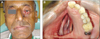
A 55-year-old man reported to the Department of Maxillofacial Prosthodontics for a definitive prosthetic rehabilitation with a surgical defect subsequent to radical surgery involving subtotal maxillectomy of left side performed six months before (Figs. 1A and 1B). The patient was wearing interim obturator prosthesis. Clinical examinations revealed complete healing of the surgical wound. The palatal defect was in continuation with the orbital defect. The margins of the defects and tissues within were healthy and normal.
Radiological examinations revealed loss of floor of left orbit and left half of the maxilla along with the teeth. The range of mandibular movements was found to be normal. Speech intelligibility and deglutition were severely affected since the patient's tongue could not make effective functional contacts due to lack of anatomic boundaries during speech and deglutition.
Hence prosthetic rehabilitation was planned with magnet retained intraoral-extra oral combination prosthesis. Fabrication of such prosthesis was planned in two sections to close the intraoral defect and create a partition between the oral cavity and nasal cavity for facilitating speech and deglutition. Firstly the extra oral prosthesis was fabricated, followed by fabrication of intraoral obturator prosthesis.
Fabrication of extraoral prosthesis
A facial moulage impression was made with irreversible hydrocolloid (Algitex, DPI, Mumbai, India) to record the facial defect along with surrounding normal structures (Fig. 2). Opened gauze squares were placed on the alginate for mechanical retention for the rigid plaster backing (0.25 inch), which is necessary for removal of the impression without distortion. When the plaster sets the impression is removed and a definitive cast was poured with Type II gypsum (Kalstone; Kalabhai Karson, Mumbai, India).
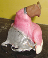
The facial prosthesis was sculpted with base plate wax and a magnet (cobalt-samarium, Ambica Corporation, New Delhi, India) was attached to it on the defect side. The wax pattern with the magnet was evaluated by positioning it on the patient's face. Investing of the wax pattern was done in Type II gypsum to form a mold for packing the silicone. After complete dewaxing, a silicone adhesive (2-butanone, Voco, Germany) was applied on the magnet. The prosthesis was packed with silicone (Maxillofacial silicone system, Factor II, Lakeside, AZ, USA) and colored using intrinsic stains and flocks (Silicone Coloring Kit; Factor II, Lakeside, AZ, USA) selected to match the patient's skin color. The silicone was kept for 24 hours at room temperature. After deflasking and finishing, extrinsic stains were applied and the prosthesis was fitted with medical grade adhesive to flush the borders of the prosthesis with the skin (Fig. 3).
Fabrication of intraoral obturator prosthesis
A preliminary impression of remaining maxillary arch along with the palatal defect was made in irreversible hydrocolloid impression (Algitex, DPI, Mumbai, India) material and a diagnostic cast was retrieved out of it. After adapting a layer of wax spacer an auto polymerizing resin tray was fabricated. Surveying of the diagnostic cast was done and necessary mouth preparation steps were undertaken.
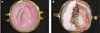
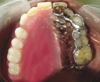
Definitive impression was made using a medium viscosity poly (vinyl siloxane) impression material (Reprosil; Dentsply, Konstanz, Germany) and poured in Type III gypsum to get the master cast. A wax pattern for framework was fabricated and casting was contemplated. The framework was polished, finished and tried in patient's mouth. A sheet of baseplate wax was adapted and contoured over the framework and jaw relations were recorded. Arrangement of teeth and wax contouring were completed. After try-in the waxed-up obturator was made hollow and processed in heat-polymerizing acrylic resin. Hollowing of the bulb was done by lost salt technique while the part under the teeth was hollowed with putty viscosity poly (vinyl siloxane) impression material (Express XT VPS Impression Materials, 3M, New Delhi, India). After processing an aperture was made on the mesial aspect of the bulb and posterior to molar tooth to remove the salt and putty, thus making the obturator hollow (Figs. 4A and 4B). Cobalt samarium magnet was attached on the supero-lateral aspect of the bulb with autopolymerizing resin so that it can contact the magnet on the extraoral prosthesis for mutual retention (Fig. 5).
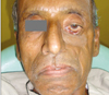
The prosthesis was delivered to the patient and final occlusal adjustments were completed. Movement of the prosthesis was minimized by maximal distribution of the occlusal force in centric and eccentric jaw positions. Premature occlusal contacts were eliminated and wide distribution of stabilizing components resulted in diminished stress created by horizontal forces. The patient experienced marked improvement in speech intelligibility and deglutition after the placement of the intraoral obturator (Figs. 6 and 7).
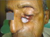
During the prosthesis insertion appointment the maxillary obturator was placed intraorally and the facial prosthesis was positioned extraorally against the obturator magnet. The patient attended recall visits every 4 to 6 months.
DISCUSSION
Orofacial defect results in communication between the oral and nasal cavity. It causes difficulty in deglutition, nasal regurgitation, loss of speech intelligibility and unesthetic appearance, leading to significant psychological problems. With acceptable cosmetic results, a reduced amount of retention can handicap the prosthesis. Unlike a conventional prosthesis, additional factors should be taken into consideration such as method of impression, materials to be used in laboratory procedures, prosthesis design, method of connection, direction of insertion and removal, esthetic factors, and maintenance protocol. With the understanding of the remaining anatomic structures, intraoral and extraoral prostheses that mutually retain one another can be constructed. Various methods of retention for facial prostheses have been described in the literature; they include eyeglasses, eye patches, extensions from the denture that engage desirable tissue undercuts, medical grade adhesives, magnets, and osseointegrated implants.5-7 The introduction of rare-earth permanent magnets made of alloys such as Neodymium-Ferro-Boron and samarium cobalt has resulted in magnets of very small dimensions. Tarnish and corrosion can be overcome by coating these magnets with nickel, gold and titanium.8 Although osseointegrated implants may provide the most reliable prosthesis retention but additional surgeries, expenses, inadequate available bone, and prior radiation to the area may contraindicate this type of treatment.9,10
Mutual retention of the intra- and extra- oral prosthesis can be successfully achieved through magnets. The force needed to separate magnets was designated as breakaway load expressed in terms of grams. The value of breakaway load is inversely proportional to the distance of separation and inclination between the magnets. These magnets are wholly biocompatible in the human body and exert no deleterious effects in human tissue. Inert magnetic field is harmless to human body and can be safely used for patients with cardiac pacemaker. There may be a gradual decrease in retentive force upon usage while some open field uncoated magnet exhibits significant cytotoxic effects which may be attributed to the release of corrosion by products from them. Therefore, magnets should be replaced as early as signs of corrosion develop.
The cast-metal framework improves retention, stability, support and bracing of the prosthesis and thus increases prosthesis longevity. Problem of two piece prosthesis is that the mobility of intraoral obturator can make facial prosthesis mobile especially during functions but the light weight of the prosthesis can alleviate this problem.11 Many methods and materials have been used to fabricate a hollow obturator such as salt, sugar and ice. A lightweight prosthesis which was fabricated in this case by lost salt technique and putty not only counteracted forces due to gravity but also enhanced speech intelligibility.12,13
CONCLUSION
The rehabilitation of a patient with a combined intraoral-extra oral defect has been presented. A two-piece prosthesis that included an intraoral obturator and extra oral facial prosthesis was fabricated. Magnets were incorporated into each unit to mutually retain each segment of the prosthesis. Marked improvement in appearance, speech intelligibility and deglutition were noted.

 XML Download