

 PDF
PDF ePub
ePub Citation
 Print
Print


INTRODUCTION
Dental implants have been used for treating single, partial or total edentulism with high success rates and predictability. At the conventional implant systems, the implants, an intraosseous part made of titanium, and a transmucosal prosthetic abutment, where the prosthesis is made, are screwed each other with gold or titanium screws.1
There are several connection designs between the implant and the abutment. An implant system, whose implant and abutment mating parts (conical and angulated) are overlapped, is known as morse taper and represents an alternative to internal or external hexagon connection designs.2 The use of morse taper connections between implant and abutment aims to enhance some properties such as better implant/abutment fit; hampered bacterial microleakage trough the gap; a decrease of perimplant bone loss; improved mechanical stability and avoided abutment loosening.2-4 In the morse taper connections, fixation and stability are not the function of screw once these properties are achieved by attrition resistance between conical parts of the abutment and implant4; and the good stability provided by morse taper systems seems to offer high flexure resistance at abutment/implant interface.3
Another important aspect for prosthetic rehabilitation is the space available for crown construction, mainly in the mandibular incisive region, where aesthetic requirement is greater. Some authors argued the use of single body implants, whose diameter is smaller, where the space is reduced, justifying that fracture susceptibility at the region of connection would be reduced in single body implants. However, single body implants require immediate rehabilitation.5
In an attempt to obtain a successful rehabilitation of these small spaces, a new abutment for morse taper implants was proposed in two sizes: a conventional abutment, whose diameter is 4.8 mm; and the reduced diameter abutment measuring 3.8 mm. Additionally, these abutments can be used for cemented or screwed prostheses, allowing change of planning after abutment placement. Thus, the hypothesis to be studied is that abutments of reduced diameter presents fracture resistance similar to conventional abutments. The aim of the present study was to evaluate fracture resistance of these implant-abutment connections under oblique compressive loads.
MATERIALS AND METHODS
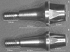
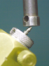
Twenty morse taper implants (3.5×11mm, Pross, Dabi Atlante, Ribeirão Preto, Brazil) were embedded in stainless steel cylinders of 21.3 mm in diameter and 25.6mm in height simulating 3 mm osseous resorption.6 Flex abutments (Pross, Dabi Atlante) measuring 3.8 mm (n = 10) and 4.8 mm (n = 10) in diameter (Fig. 1) were installed into the implants with 20 Ncm torque, as recommended by the manufacturer, measured by a digital torquemeter (TQ 680, Instrutherm Measure Instruments, São Paulo, Brazil). Oblique compressive loading tests were performed in a universal testing machine (DL-2000, EMIC, São José dos Pinhais, Brazil). Tests were performed using 20 sets (10 implants/3.8 mm abutments and 10 implants/4.8 mm abutments) positioned at 45 degree angle (Fig. 2), using a load cell of 1,000 kgf and 1 mm/min speed. The loading point was at a distance of 11 mm from cylinder surface.3 The values of maximum deformation force and fracture resistance were noted. The data were analyzed by Student's t test using SPSS software (version 17.0, SPSS Inc., Chicago, IL, USA).
RESULTS
Maximum deformation force of 4.8 mm and 3.8 mm abutments are presented in Table 1. No fractures were noted after mechanical test. Statistical analysis demonstrated that the evaluated abutments were statistically similar (P=.230), as expected because the diameter where bending occur was the same.
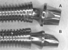
Fig. 3 represents implant/abutment sets after mechanical test. The apparent difference in abutment flexural dynamics was suggested as a result of higher head deformation in narrow abutments.
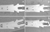
The Fig. 4 represents longitudinal section of implant/abutment sets before (A and B) and after (C and D) mechanical test.
DISCUSSION
The intimate contact between implant and abutment of conical connections aimed to reduce the incidence of mechanical problems reported in other connections, once loosening is prevented by an improvement of abutment stability.3 The frictional retention between the implant and abutment ensures a great anti-rotational property. Because oblique forces are dissipated along abutment/implant interface providing excellent prosthetic stability, and protection of fixation screw.7 Then, conical connections are indicated for rehabilitation of single crowns. A study that compared fracture resistance of internal hexagon and morse taper connections related that morse taper connection provides greater resistance to deformation and fracture than internal hexagon.8
Comparing the fracture resistance of internal hexagon and morse taper implant/prosthetic abutment connections, Coppedê et al. concluded that morse taper one-piece solid abutment provides greater resistance to deformation and fracture than internal hexagon.3
Thus, a morse taper abutment of small (3.8 mm) diameter is a new alternative to oral rehabilitation, providing a greater ease in the crown construction of upper lateral incisors and lower incisors because they have a smaller size.
In the present study, the maximum deformation force of 3.8 mm and 4.8 mm abutments was 95.25 kgf and 95.33 kgf, respectively. Considering that the incidence of maximum force applied to central incisor region varies from 13.2 to 23.1 kgf,9 these abutments presents mechanical properties enough for clinical use as indicated. Despite the maximum force reported to the first molar region was about 41.3 to 89.8 kgf,8 another tests of these narrow abutments must be performed before their use in this region. It must be considered that this is not a manufacturer indication for its use.
However, both the abutments presented permanent deformation at different positions after oblique compressive loading tests. The 4.8 mm abutment deformed in the body of the abutment while 3.8 mm, in the body and in the head. This difference can be attributed to the greater angle between the body/head of the 4.8 mm abutment, which caused greater flexion in this region and subsequent permanent deformation. On the other hand, the greater deformation in the head of 3.8 mm abutment can be caused by the reduced amount of metal at this site because of access hole to the abutment screw. Both abutments presented access holes to the screw because they can be used both for cemented or screwed prostheses. As identical screws are used for both abutments and their holes have similar size, the amount of metal in the head of the 3.8 mm abutment is smaller than of 4.8 mm, leading to easier head deformation, as shown in Fig. 3B. This head deformation would not happen in clinical situation because the head would be protected by a crown restoration. So, this is a limitation of this in vitro study and must be considered. Despite both abutments cause important deformation at the neck region of the implants, this occurrence was noted after forces much larger than those reported in the regions8 where the abutments are indicated. So, it could be suggested that this abutments/implant neck deformation would not happen clinically under normal masticatory forces.
Therefore, the results of the present study suggest that 3.8 mm abutments can be used clinically because they present satisfactory mechanical properties and strength compatible with the 4.8 mm abutments. Additionally, they represent a new alternative for rehabilitation with implants, once these abutments permit the choice for cemented or screwed crowns throughout the treatment.

 XML Download