Journal List > J Adv Prosthodont > v.3(2) > 1054044

Rastogi and Kamble: Comparative analysis of the clinical techniques used in evaluation of marginal accuracy of cast restoration using stereomicroscopy as gold standard

Abstract

PURPOSE

This study assessed the effect of preparation design on marginal adaptation and also compared the sensitivity and specificity of clinical evaluation techniques for marginal accuracy of cast restorations to stereomicroscopy.

MATERIALS AND METHODS

Three Ivorine molar teeth of different designs were prepared. (A)-A complete crown preparation with buccal shoulder and beveled finish line. (B)-A complete crown preparation with chamfer finish line. (C)-A three-quarter crown preparation with proximal boxes and beveled finish line. Twenty four castings were prepared with eight castings for each design respectively. Each casting underwent examination with an explorer, disclosing media, and a stereomicroscope. Stereomicroscopy at a value less than or equal to 30 microns was used as a gold standard to evaluate the significance of different designs on marginal adaptation. Chi-square tests of independence and Kruskal-Wallis were used to evaluate the effect of preparation design and compare the agreement between examination methods for detection of marginal gap size of greater than or equal to 30 microns (α=.05). Sensitivity and specificity for explorer and disclosing media as compared to stereomicroscope was calculated using statistical formula given by Park.

RESULTS

The preparation design did not significantly affect overall marginal adaptation. Examination by explorer and disclosing media at 30 µm revealed 39% and 10.06% sensitivity and 91% and 82% specificity respectively.

CONCLUSION

Preparation designs examined in this study did not significantly affect the marginal adaptation of the castings. Commonly used clinical evaluation techniques using explorer and disclosing media appeared to be inadequate for assessment of marginal accuracy.

INTRODUCTION

Several authors have emphasized that marginal accuracy and internal adaptations are critical factors for clinical success of cast restoration.1,2 It is desirable to have margins closed as much as possible to reduce width of cement line. Because of deficiencies inherent in the dental casting technique, a gap of varying width is likely to occur between a casting and a tooth. Open marginal configurations encourage micro leakage of bacteria and their by-products due to dissolution of the luting agents. This can cause severe effects on the health of pulpal tissues.1 The relationship between margin adaptation and periodontal health has been confirmed in experimental animals and humans.1
There is a continuing quest to determine the best way to minimize the width of the cement line within accepted technique constraints. Different finish-line designs have been advocated for several reasons. Preston3 and Shillingburg4 recommended the shoulder-bevel as the best type of finish line for the cast restoration. Rosner5 reasoned that a beveled finish line would fit better than a shoulder and trigonometric analysis to support his assertion. He recommended beveled margin parallel to the axial wall for smallest possible cement line. However due to the limited and contradictory theoretical, laboratory, and clinical evidence available, it is not clear which finish line design, if any, may offer the greatest advantage.6
There are several commonly used techniques to evaluate the marginal accuracy of cast restorations prior to clinical acceptance. These include use of dental explorer and elastomeric materials.7-9 Many disclosing agents, including chloroform and rouge, disclosing waxes, wax aerosols, and zinc oxide-eugenol have been used as aids in fitting crowns.10 McLean and von Fraunhofer11 used polyether elastomeric impression material to evaluate the potential cement film thickness beneath clinical restorations. They suggested a further application of this technique for the placement of cast restorations. This technique was further described by several authors using a light bodied-condensation reaction silicone impression material.7,8,12
Several studies have reported 30 µm to be clinically acceptable gap discrepancy.13 However, there is limited information on sensitivity and specificity of commonly practiced clinical evaluations (i.e. explorer and elastomeric disclosing material) in detecting a marginal discrepancy of this magnitude.
The indications for the use of die spacers in fabrication of cast restorations are well documented in the literature.6,14,15 In this in vitro study, die spacer was intentionally not used to create variations in casting fit for the purpose of the study. The aim of this study was to assess the effect of preparation design on marginal adaptation, as well as to assess sensitivity and specificity of clinical evaluation of cast restoration marginal accuracy when compared to stereomicroscopy.

MATERIALS AND METHODS

The method involved the evaluation of the marginal fit of cast restorations. Eight specimens of three different preparation designs were made, and castings were examined using an explorer, disclosing media, and a stereomicroscope for marginal adaptation.

1) Preparation of teeth

Three Ivorine maxillary first molar teeth (Columbia Dentoform Corp., Long Island City, NY, USA) were selected for study and mounted by securing roots with wax. The roots were then invested in dental stone (Kalstone, Kalabhai Dental Corp., Mumbai, India). These three ivorine teeth were then prepared with three different designs:
  • Design A: A complete crown preparation with a buccal shoulder (1 mm wide) and bevel as remaining finish line.

  • Design B: A complete crown preparation with a chamfer finish line.

  • Design C: A three-quarter crown preparation with proximal boxes and beveled finish line (Fig. 1).

Standardized tooth preparations were performed with the aid of paralleling device, the goniometric microscope (NRL-100-00, Rame-Hart, Mountain Lakes, NJ, USA). A cone angle of 6 degrees and a preparation height of 4 mm, (measured at the midfacial surface of the tooth), were used.

2) Fabrication of custom tray

Two layers of modeling wax (Maarc Modelling Wax, Shiva Products, Mumbai, India) were adapted over the prepared ivorine teeth for each design as a spacer. The custom tray was made with autopolymerizing acrylic resin (DPI-RR Cold Cure, Dental Products of India Ltd., Mumbai, India). The custom tray was bench cured for 24 hours. Eight custom trays were made for each preparation design, thus total of 24 custom trays were fabricated.

3) Impressions of teeth

Medium body addition silicone impression material (Aquasil Monophase, Dentsply International, USA) was mixed according to manufacturer's instructions and the custom tray was filled and seated on the prepared ivorine teeth with adequate finger pressure till the resistance of the stopper is felt. Once the impression material sets it is removed and inspected for any defects under 1.5×magnification. Thus, total of 24 impressions were made with eight in each of preparation design.

4) Die preparation

The type IV die stone (Pearl stone, Asian chemicals, Gujarat, India), with a water-powder ratio of 25 cc/100 gm was mechanically mixed using vacuum mixer, and poured into the impression using vibrator. After final set, dies were recovered. The dies were inspected for any discrepancies under 1.5×magnification. Thus a total of 24 dies were prepared with eight in each group.

5) Fabrication of wax pattern and investment

An even application of die lubricant (Die Lube Wax Sep., Dentecon, USA) was done on each die. Wax patterns were fabricated on the dies using type II blue inlay wax (Blue Wax, MDM. Corp., Delhi, India). Dip wax technique was used to form wax copings. The patterns were contoured parallel to the emergence profile and margins were manually sealed under 1.5×magnification.
Sprue wax of diameter 2.5 mm was attached to the occlusal surface of each pattern on the non functional cusps and was angled so that it was obtuse to the adjacent axial walls and occlusal surface. Each Wax pattern was invested immediately in phosphate-bonded investment (Moldavest Exact, Heraeus Kulzer., Germany) with the powder liquid ratio as 60 g of powder to 12 ml of liquid, after cleaning it with wax pattern cleaner (Wax Pattern Cleaner, J.F.Jelenko & Co., New Rochelle, NY, USA) this reduces the surface tension of wax and permits better wetting. The investment material was mechanically spatulated under vacuum spatulation time for 90 seconds. The wax patterns were carefully painted with the investment mixture by means of fine sable hair brush. The casting ring lined with cellulose acetate ring liner was then filled with the investment material under mechanical vibration and allowed to set on the bench for 1 hour.

6) Fabrication of castings

The rings were then, placed in an oven (Vulcan 3-550 PD Burnout Furnance, Dentsply Neytech., Burlington, NJ, USA). A standardized burn out and preheat procedure of 30 minutes at 23 - 270℃, 30 minutes at 270 - 580℃, and 30 minutes at 580 - 950℃, was followed. Casting was accomplished in an induction centrifugal casting machine (Ducatron serie 3, Ugin Dentaire, France) using non precious gold alloy with a composition of 80.07% copper, 7.80% Al, 3% Fe, 2.70% Zn, 1.70% Mn, 4.30% Ni. Castings were devested, cleaned, and air abraded with 50 microns aluminium oxide at 0.6 Mpa, (fine grit, Jelenko quartz abrasive; Armonk, NY, USA). Sprues were removed using silicon carbide disks, and the castings were finished externally using blue, green, and brown rubber wheels. Internal positive defects were removed using a ½ round bur under 1.5×magnification.
Castings were ultrasonically cleaned in distilled water and stored until further evaluation.

7) Measurement of marginal fit

Each casting underwent examination with an explorer, elastomeric disclosing media (Fitchecker II, GC Corp., Tokyo, Japan), and a stereomicroscope.
One examiner performed the clinical evaluations with an explorer and disclosing media. The examiner made a list of 20 sites without prior observation or examination of specimens to select 20 random sites for initial examination. These sites were then used to determine intraoperator reliability, which is established to be 95%. The reliability of stereomicroscope approximated 100%.
Twelve circumferential recordings were made of each casting, three on each buccal, lingual, mesial and distal surface, marked with a groove on original ivorine teeth and highlighted using a fine indelible marker, for a total of 288 examination sites.

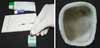
8) Evaluation of marginal adaptation using explorer

Margins were evaluated by examiner relying on his tactile sense and visual acuity with great concentration. Examiner evaluated the margins by feeling it with explorer (Trudent, New Delhi, India). Same explorer was used to evaluate all castings for the purpose of standardization. Explorer misfit was defined at any of the following: clinically unacceptable vertical marginal discrepancy, horizontal marginal discrepancy, under extended margins, and seating discrepancy examined at original magnification×1.5 (Fig. 2).

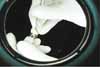
9) Evaluation of marginal adaptation using elastomeric disclosing media

Material (Fitchecker II, GC Corp., Tokyo, Japan) was manipulated according to manufacturer's instructions. Each experimental casting was filled with disclosing material then seated on the respective ivorine teeth with finger pressure appropriate to seat the casting on prepared tooth. An area was interpreted as "fit" if the seal of the casting is observed to be apparent by clearly identifiable thin translucent film. Areas where the film is very thin or thick indicate "misfit" (Fig. 3).

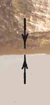
10) Evaluation of marginal adaptation using Stereomicroscope
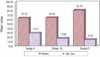
A stereomicroscope (Lawerence & Mayo pvt. Ltd., Mumbai, India) was used to examine these twelve circumferential sites at 30×magnification. The recordings were made and interpreted using software (Image J 1.40 g, Wayne Rasband, National Institute of health, USA). Stereomicroscopy at a value less than or equal to 30 microns measurements was used as a gold standard to evaluate the significance of different designs on marginal adaptation. Three sites for each buccal, lingual, mesial, and distal surface were given an overall evaluation of acceptable or unacceptable. Since in a clinical situation adequacy of marginal accuracy at each point is desired, these values were not averaged; instead a surface was ranked as fit where all 3 measurements were less than or equal to 30 microns using stereomicroscopy. If any of the 3 measurement points had a value of greater than 30 microns that surface was ranked as misfit (Fig. 4).
Chi-square tests of independence and Kruskal-Wallis were performed at a priority level of significance of α=.05 to determine the significance of each surface (buccal, lingual, mesial, and distal) and overall design with respect to marginal adaptation. Further Chi-square tests of independence were used to compare agreement between stereomicroscope, explorer, and disclosing media detection of marginal gaps less than or equal to 30 microns.
Sensitivity and specificity for explorer and disclosing media as compared to stereomicroscope was calculated using statistical formula (Sensitivity = TP/TP+FN, Where, TP = True positive,
FN = False negative; Specificity = TN/TN+FP, Where, TN = True negative, FP = False positive) given by Park.20

RESULTS

The three preparation designs did not significantly affect the overall marginal adaptation (P=.352) (Table 1). The mean marginal opening of three designs in Stereomicroscope was compared for statistical analysis using Kruskal-Wallis Anova test. The three designs showed no significant difference in the mean marginal opening (P=.1197) (Table 2, Fig. 5).
Analysis of different surfaces revealed no significant difference in marginal adaptation for the three designs examined (Table 3).
The percentage of 288 sites with inadequate marginal fit as detected by explorer, elastomeric disclosing media, and stereomicroscope (Table 4).
Explorer detected 33.33% of misfit sites (Fig. 6a).
Elastomeric disclosing media detected 10.06% of misfit sites (Fig. 6b).
Stereomicroscope detected 79.16% of misfit sites (Fig. 6c).
The overall agreement between the stereomicroscope and explorer was 50.69%, with a 19.09% correct acceptance rate (19.09% of the time the explorer ranked "fit" and the stereomicroscope showed a gap size of less than or equal to 30 µm) and 31.59% correct rejection rate (31.59% of the time the explorer ranked "misfit" and the stereomicroscope showed a gap size of greater than 30 µm) (Table 5).
The overall agreement between the stereomicroscope and elastomeric disclosing media was 29.16%, with a 19.09% correct acceptance rate (19.09% of the time the elastomeric disclosing media ranked "fit" and the stereomicroscope showed a gap size of less than or equal to 30 µm) and 10.06% correct rejection rate (10.06 of the time the elastomeric disclosing media ranked "misfit" and the stereomicroscope showed a gap size of greater than 30 µm) (Table 6).
The explorer at 30 µm had 39% sensitivity and 91% specificity and elastomeric disclosing media had 10% sensitivity and 82% specificity respectively (Table 7, 8).

DISCUSSION

This study was designed to evaluate the relationship of marginal accuracy of cast restorations to various tooth preparations. The three preparation designs evaluated in this study are most commonly used in the clinical practice. Previous studies could not come to an agreement as to which is the best finish line in terms of marginal adaptation. Some studies concluded that feather edge and bevel finish lines provide the best marginal seal16 while others suggested shoulder with oversized casting as best finish line in terms of marginal adaptation.17 Therefore this study was undertaken to determine the effect of these finish lines on the marginal adaptation of cast restorations. In the current study, it was determined that preparation designs assessed had no statistically significant effects on the marginal accuracy of cast restorations. This observation is consistent with that of the few recent studies.6,14
Christensen reported that when visible and invisible margins were evaluated with an explorer the barely acceptable range was 2 - 51 µm with a mean of 21 µm and 34 - 119 µm with a mean of 74 µm respectively.18 However the results of present study indicate that the most commonly used method i.e. explorer, for evaluating the fit of castings may be even less reliable than reported. When margins are evaluated with an explorer acceptance is more likely to be based on the size and character of overhangs and ledges than on the actual size of opening of the margins.
The advantages of using an elastomeric disclosing media to aid in clinical assessment of castings are well documented.7,13 In this in vitro study, it appears that these materials did not significantly aid in detection of marginal gaps as compared to the explorer. However the use of elastomeric disclosing media, in the assessment of the internal fit of castings remains valuable.7 In clinical practice, where assessment of an inter-proximal area may be more difficult with an explorer, the use of these materials may further assist in marginal discrepancy detection. So, an elastomeric disclosing media was used as one of the technique of clinical evaluation of marginal adaptation.
In current study, the sensitivity of explorer and elastomeric disclosing media as compared to stereomicroscope at 30 µm was 39% and 10% respectively. Higher values of sensitivity for explorer technique as compared to elastomeric disclosing media indicate that explorer technique is more reliable as compared to elastomeric disclosing media in detecting marginal gaps. However values of sensitivity for both the techniques evaluated is considerably low, indicating that these techniques may not be adequate to determine presence of marginal gaps of or less than 30 µm.
The specificity of the explorer and elastomeric disclosing media as compared to stereomicroscope at 30 µm was 91% and 82% respectively. Higher values of specificity for explorer technique as compared to elastomeric disclosing media indicate that explorer technique is more reliable as compared elastomeric disclosing media in detecting margins that are closely adapted to prepared tooth. Similar results were found in previous study done by Jahangiri.19 The limitations of this study were that the assessments of marginal accuracies were not performed intraorally and that the errors in fabrication and handling of dies and castings are assumed to be minimal. Within these limitations, it appears that clinical examination based on an explorer and the use of elastomeric materials may not provide satisfactory accuracy needed for detection of marginal gap sizes of less than or equal to 30 µm. A recent study has reported that clinical detection of marginal gap size with similar sensitivity and specificity as stereomicroscope occurs at greater than or equal to 124 µm.19
Although the stereomicroscope cannot be used to assess restorations intraorally, the future development of an intraoral apparatus may be of value. However, the use of a stereomicroscope as a supplement method for assessing castings on dies may provide a higher degree of marginal gap detection prior to examination of these castings intraorally. This instrument is easy to use and is not considered costly.
The software program used in this study was purpose of multiple measurements and storage of images, and future image analysis. This software program is not necessary for quality.

CONCLUSION

  1. The preparation designs examined in this study did not significantly affect the marginal adaptation and accuracy of the castings.

  2. Commonly used clinical evaluation techniques i.e. explorer and elastomeric disclosing media may be inadequate for assessments of marginal accuracy.

  3. Explorer technique proved to be better aid in detection of marginal accuracy as compared to elastomeric disclosing media.

  4. At 30 µm explorer revealed 39% sensitivity and 91% specificity and elastomeric disclosing media revealed 10.06% sensitivity and 82% specificity.

  5. For better evaluation of marginal accuracy of the cast restorations, the routine use of a stereomicroscope in the laboratory is indicated which provides a superior quality control prior to examination of restorations intraorally.

Above mentioned conclusions are within the limitations of this study. The assessments of marginal accuracies were not performed intraorally, so further research is required to obtain a better insight of the methods to assess the marginal adaptation of the cast restorations that can be used intraorally.

Figures and Tables

Fig. 1
Ivorine tooth preparations. Design A: complete crown preparation with a buccal shoulder and beveled finish line, Design B: complete crown preparation with a chamfer finish line, Design C: three-quarter crown preparation with proximal boxes and beveled finish line.
jap-3-69-g001
Fig. 2
Fit check using explorer.
jap-3-69-g002
Fig. 3
Fit check using elastomeric disclosing media.
jap-3-69-g003
Fig. 4
Marginal gap as seen under stereomicroscope.
jap-3-69-g004
Fig. 5
Comparison of mean marginal openings of three designs in stereomicroscope.
jap-3-69-g005
Fig. 6
Percentage of fit and misfit sites in explorer, elastomeric disclosing media, and stereomicroscope.
jap-3-69-g006
Table 1
Distribution of overall marginal adaptation of crowns by three designs using Stereo Microscope
jap-3-69-i001

Chi-square = 0.0000, df = 2, P=.352, NS

Table 2
Comparison of mean marginal openings of three designs in Stereomicroscope by Kruskal Wallis Anova test
jap-3-69-i002
Table 3
Marginal accuracy findings on buccal, lingual, mesial, distal surfaces of three preparation designs as detected by stereomicoscope
jap-3-69-i003
Table 4
Percentage of 288 sites with inadequate marginal fit as detected by explorer, elastomeric disclosing media, and stereomicroscope
jap-3-69-i004
Table 5
Results of stereomicroscopic evaluation of 288 marginal gap sites compared to explorer using stereomicroscope as gold standard
jap-3-69-i005
Table 6
Results of stereomicroscopic evaluation of 288 marginal gap sites compared to elastomeric disclosing media using stereomicroscope as gold standard
jap-3-69-i006
Table 7
Calculation of sensitivity for explorer and elastomeric disclosing media as compared to stereomicroscopy
jap-3-69-i007
Table 8
Calculation of specificity for explorer and elastomeric disclosing media as compared to stereomicroscopy
jap-3-69-i008

References

1. Felton DA, Kanoy BE, Bayne SC, Wirthman GP. Effect of in vivo crown margin discrepancies on periodontal health. J Prosthet Dent. 1991. 65:357–364.
2. Richter WA, Ueno H. Relationship of crown margin placement to gingival inflammation. J Prosthet Dent. 1973. 30:156–161.
3. Preston JD. Rational approach to tooth preparation for ceramo-metal restorations. Dent Clin North Am. 1977. 21:683–698.
4. Shillingburg HT Jr, Hobo S, Fisher DW. Preparation design and margin distortion in porcelain-fused-to-metal restorations. J Prosthet Dent. 1973. 29:276–284.
5. Rosner D. Function, placement, and reproduction of bevels for gold castings. J Prosthet Dent. 1963. 13:1160–1166.
6. Syu JZ, Byrne G, Laub LW, Land MF. Influence of finish-line geometry on the fit of crowns. Int J Prosthodont. 1993. 6:25–30.
7. Rissin L, Wetreich G. Utilization of elastomeric materials to evaluate the accuracy of cast restorations prior to cementation. J Prosthet Dent. 1983. 49:585–586.
8. Davis SH, Kelly JR, Campbell SD. Use of an elastomeric material to improve the occlusal seat and marginal seal of cast restorations. J Prosthet Dent. 1989. 62:288–291.
9. White SN, Sorensen JA, Kang SK. Improved marginal seating of cast restorations using a silicone disclosing medium. Int J Prosthodont. 1991. 4:323–326.
10. Walker PM. Remounting multiple casting prior to final cementation. J Prosthet Dent. 1981. 46:145–148.
11. McLean JW, von Fraunhofer JA. The estimation of cement film thickness by an in vivo technique. Br Dent J. 1971. 131:107–111.
12. Arakelian A Jr. A technique for seating castings. J Prosthet Dent. 1982. 48:357.
13. Eames WB, O'Neal SJ, Monteiro J, Miller C, Roan JD Jr, Cohen KS. Techniques to improve the seating of castings. J Am Dent Assoc. 1978. 96:432–437.
14. Belser UC, MacEntee MI, Richter WA. Fit of three porcelain-fused-to-metal marginal designs in vivo: a scanning electron microscope study. J Prosthet Dent. 1985. 53:24–29.
15. Campagni WV, Wright W, Martinoff JT. Effect of die spacer on the seating of complete cast gold crowns with grooves. J Prosthet Dent. 1986. 55:324–328.
16. Fusayama T. Technical procedure of precision casting. J Prosthet Dent. 1959. 9:1037–1048.
17. Pascoe DF. Analysis of the geometry of finishing lines for full crown restorations. J Prosthet Dent. 1978. 40:157–162.
18. Christensen GJ. Marginal fit of gold inlay castings. J Prosthet Dent. 1966. 16:297–305.
19. Jahangiri L, Wahlers C, Hittelman E, Matheson P. Assessment of sensitivity and specificity of clinical evaluation of cast restoration marginal accuracy compared to stereomicroscopy. J Prosthet Dent. 2005. 93:138–142.
20. Park K. Textbook of preventive and social medicine. 2005. 18th ed. Jabalpur: Banarsidas Bhanot publishers;117.
TOOLS
Similar articles