

 PDF
PDF ePub
ePub Citation
 Print
Print


INTRODUCTION
Ceramics have a long history in fixed prosthodontics for achieving optimal esthetics. Yttria-stabilized tetragonal zirconia (Y-TZP) is gaining use in dentistry due to its good mechanical properties and superior biocompatibility.1 It is currently used as core material in all ceramic dental restorations,2,3 implant superstructures,4 and orthodontic brackets.5 Its superior mechanical properties compared to other dental ceramics, such as higher strength and fracture toughness,2,6,7 which are due to the transformation toughening mechanism, are similar to that observed in quenched steel.
Color assessment is regarded as a complex psychophysiologic process subject to numerous variables. Dentin is considered to be the primary source of color for teeth, which is modified by the thickness and translucency of overlying enamel. The perceived color of natural teeth is a result of light reflected from the enamel surface, in addition to the effect of light scattering within enamel and dentin before it is ultimately reflected back.8 Clinically, it is important that ceramic restorations reproduce the translucency and color of the natural teeth. There are many factors affecting the match, such as translucency, opalescence, fluorescence, surface texture, and shape.9,10
Most all ceramic systems require the combination of 2 layers of ceramic material, such as a strong ceramic core and a weak veneering porcelain,11 with different opacity, shade, and thickness, to provide a natural appearance.12,13 All ceramic restorations without a metal substructure allow for greater light transmission within the restoration, thereby improving the color and translucency of the restoration; however, a perfect esthetic tooth-colored restoration cannot be ensured.14 If the majority of light passing through a ceramic restoration is diffusely transmitted and only part of it is scattered, the material will appear translucent.15 The amount of light that is absorbed, reflected, and transmitted depends on the quantity of crystals within the core matrix, their chemical nature, and the size of the particles compared to the incident light wavelength.16 Core translucency was also identified as a primary factor in controlling esthetics and a critical consideration in the selection of the materials.17 Some all ceramic core materials have high in vitro strength values.18,19 However, an increase in crystalline content to achieve greater strength generally results in greater opacity.17,20
Clinical perceptibility of color differences has been the subject of numerous investigations. The Commission Internationale de l'Eclairage (CIE) recommended calculating color difference (ΔE) based on CIELAB color parameters.21 The ΔE values are used to describe whether the changes in the overall shade are perceivable to the human observer. This magnitude of the color difference is based on the human perception of color; color differences greater than 1 ΔE unit are visually detectable by 50% of human observers.22 However, under uncontrolled clinical conditions, such small differences in color would be unnoticeable because average color differences below 3.7 have been rated as a match in the oral environment.23,24
Instrumental measurements can quantify color and allow communication to be more uniform and precise. In addition to the opacity and shade of the ceramic that determine the definitive shade of an esthetic restoration, other factors, including porcelain brand,8,25,26 batches,25 condensation techniques,25 firing temperatures,26 dentin thickness,27-29 and number of porcelain firings,28,30 can have an effect. Specific contributions of core and veneer thickness to the appearance of layered ceramics were determined,29 and it was concluded that there was a significant correlation between the thickness ratio of core and veneer ceramics and the color of the restoration. Even when adequate ceramic thickness exists, clinical shade matches are difficult to achieve31 because there is a wide range of translucency among the core materials of all ceramic systems at clinically relevant core thicknesses.16 The thickness and the combination of ceramic layers, such as the core, veneer, and other specialty ceramic materials, have been shown to control the appearance of all ceramic materials.15,23,32 Antonson and Anusavice32 found that it was important to determine the translucency of a layered core-veneering ceramic as a function of thickness. Moreover, Heffernan et al.10,16 investigated the influence of core and core-veneering ceramic thickness on overall translucency. Lee et al.33 showed that the layered color of varied all ceramic and veneer combinations was different depending on the type of all ceramic core material, even though the thickness of the layered specimen was set to 1.5 mm. These studies, however, did not address the particular contribution of each variable: the difference in core substructure, the changes in veneer thickness, or the number of firings.
The effect of multiple firings has been investigated in previous studies, and it has been reported that repeated firings did not noticeably affect the color of dental ceramics.28,30,34,35 However, O'Brien et al.25 found perceivable differences (ΔE = 1) between the color of ceramic specimens that were fired 3 and 6 times. Uludag et al.36 and Ozturk et al.37 reported perceptual color changes in L*a*b* color parameters as the number of firings increase. The purpose of this study was to evaluate the effects of various dentin ceramic thicknesses (0.5, 1, or 1.5 mm) and number of firings (3, 5, 7, or 9) on the color of zirconium oxide all ceramic system fabricated using CAD/CAM technology. The research hypothesis was that the color difference would be determined relative to the firing times and dentin ceramic thicknesses.
MATERIALS AND METHODS
The zirconia-based ceramic used in this study; the Lava™ All-ceramic system (3M ESPE Dental Products, St Paul MN, USA) utilizes CAD/CAM technology to produce a densely sintered and high strength zirconia framework with a 3% mol partially yttria-stabilized zirconia polycrystal content.
Thirty disc-shaped cores, 12 mm in diameter and 1 mm thick were fabricated from zirconia based all ceramic system using CAD/CAM technology. These cores were divided into three groups (n = 10) for veneering with dentin ceramic with thicknesses of 0.5, 1 and 1.5 mm. The cores were veneered with A1 (according to the VITA Shade Guide) dentin ceramic (LavaCeram, 3M ESPE, St Paul, MN, USA) with the use of the similar molds described by others25,28,32 which allowed standardization of the dentin thicknesses for each of group to be studied. Specimens were then fired in a dental ceramic furnace (Lava Furnace 200, 3M ESPE, St Paul, MN, USA). The thickness of each group of specimens was then measured with a digital caliper (Dial Caliper D, Aura Dental, Aura an der Saale, Germany) with an accuracy of 0.02 mm, and corrected with diamond rotary cutting instruments (ISO 173/016, Mani. Inc, Utsunomiya, Tochigi, Japan) until the desired thickness of dentin ceramic was achieved (0.5, 1, or 1.5 mm).
The color of each specimen was measured with a spectrophotometer (VITA Easyshade; VITA Zahnfabrik, Bad Säckingen, Germany). The spectrophotometer's CIE L*a*b* output is based on D65 illuminant and a 2-degree standard observer. Three measurements were made, and the average reading was calculated for each specimen. Instrument was recalibrated after measurement of each group (n = 10). The CIELAB measurements make it possible to evaluate the amount of perceptible color change in each specimen. The CIELAB color space is a uniform 3-dimensional color order system. Equal changes in any of the 3 coordinates can be perceived as visually similar. Total color differences were calculated with use of the following equation.38
ΔE*= [(ΔL*)2 + (Δa*)2 + (Δb*)2]1/2
The L* coordinate is a measure of the lightness-darkness of the specimen. The greater the L* is, the lighter the specimen. The a* coordinate is a measure of the chroma along the red-green axis. A positive a* relates to the amount of redness, and a negative a* relates to greenness of a specimen. The b* coordinate is a measure of the chroma along the yellow-blue axis; that is, a positive b* relates to the amount of yellowness, while a negative b* relates to the blueness of the specimen. ΔL*, Δa*, and Δb* are the differences in the CIE color-space parameters of the 2 colors.38
The results of the testing were analyzed with statistical software (SPSS, PC, Version 17.0; SPSS, Inc, Chicago, IL, USA). Repeated measurements of the data (number of firings and ceramic thicknesses) were analyzed with analysis of variance (ANOVA) for significant differences. Bonferroni post hoc test was used to perform pairwise comparisons (α=.05).
RESULTS
The analysis revealed that L*a*b* values of ceramic system (Table 1) were affected by number of firings [3, 5, 7 or 9] (P<.001) and dentin ceramic thickness [0.5 mm, 1 mm or 1.5 mm] (P<.001).
Significant interactions were present in L*, a* and b* values within each group of dentin ceramic thickness i.e. 0.5 mm, 1 mm or 1.5 mm. An increase in number of firings resulted in significant increase in L* values for both 0.5 mm and 1.5 mm thicknesses (P<.01, P=.013); however it decreased for 1 mm thickness (P<.01). The a* values increased for 1 mm and 1.5 mm thicknesses (P<.01), while it decreased for 0.5 mm specimens. The b* values increased significantly for all thicknesses-0.5 mm, 1 mm and 1.5 mm as the number of firings increased (P<.01, P<.01, P=.022).
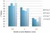
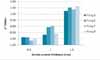
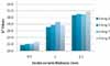
Significant interactions were also present in all color co-ordinates i.e. L*, a* and b* values among all three groups of dentin ceramic thicknesses and number of firings. As the dentin ceramic thickness increased (0.5 mm - 1 mm - 1.5 mm), significant reductions in L* values (P<.01) were recorded (Fig. 1). There were significant increases in both a* and b* values (P<.01) as the dentin ceramic thickness increased (Fig. 2, 3).
Mean color difference (ΔE) values were calculated for each group of thickness. The mean ΔE value increased as the thicknesses increased. Mean ΔE values were less than 3 when ceramic thickness increased from 0.5 mm to 1 mm (ΔE = 1.43) and from 1 mm to 1.5 mm (ΔE = 2.38). It was higher than 3 (ΔE = 3.37) when ceramic thickness increased from 0.5 mm to 1.5 mm.
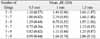
The ΔE values were calculated for each specimen within each group to determine mean color differences (ΔE), depending on repeated firings (Table 2). The ΔE value was smaller than 1 between firing 5 and 7, firing 5 and 9, and firing 7 and 9 for 0.5 mm thickness and between firing 3 and 9 for 1 mm thickness. The ΔE value was higher than 1 between all firings for 1.5 mm specimens.
DISCUSSION
This in vitro study measured the color changes of ceramic specimens prepared at different thicknesses and fired different numbers of times. The results of this study support the hypothesis that the color difference varies with respect to the firing times and dentin ceramic thicknesses. There were significant differences in color change within groups.
Most all ceramic systems consist of a ceramic core with a thickness of 0.5 to 1.0 mm and approximately 1.0 to 1.5 mm of space available for veneering ceramic.16 In the current study, the specimens had ceramic thicknesses of 0.5, 1, or 1.5 mm, with a core thickness of 1 mm. L* values, which reflect the brightness of the specimens, decreased for both systems as the total thickness of the specimens increased. Antonson and Anusavice32 studied the effect of change in the thickness of ceramics on the contrast ratio of dental core and veneering ceramic, and concluded that the contrast ratio was dependent on the type of the material tested. Heffernan et al.10,16 described the influence of core material thickness on its translucency and the influence of core plus ceramic veneer thickness on the overall translucency of specimens. Shokry et al.29 demonstrated that L* values decreased for leucite reinforced and spinell ceramics as the total thickness increased. The results of the present study are in agreement with the previous studies12,29,31,36,37 since the thickness of the layered ceramic influenced the final shade, partially due to the translucency, as the thicker ceramic disks were less translucent.
As the dentin ceramic thickness increased, significant reductions in L* values were recorded. There were significant increases in both a* and b* values as the dentin ceramic thickness increased. These changes in L*, a* and b* values are consistent with a study by Ozturk et al.37. An increase in the number of firings resulted in an appreciable increase in L* values that resulted in darker specimens for 0.5 mm and 1.5 mm thicknesses used in the present study. However it decreased for 1 mm thickness specimens. The a* color values of 1 mm and 1.5 mm thicknesses increased after repeated firings which resulted in redder specimens. However, the a* values decreased for 0.5 mm thickness specimens. The b* values increased appreciably by the number of firings in all specimens of 0.5, 1 and 1.5 mm thicknesses. This resulted in yellower specimens.
The mean ΔE values increased as the dentin ceramic thickness was increased. The ΔE value was smaller than 1 between firing 5 and 7, firing 5 and 9 and firing 7 and 9 for 0.5 mm thickness and between firing 3 and 9 for 1 mm thickness. The ΔE value was higher than 1 between all firings for 1.5 mm specimens. Color changes following repeated firings may also be attributed to the color stability of metal oxides during firing which can affect the resulting color of ceramic. Several studies have suggested that certain metal oxides are not color stable after they are subjected to firing temperatures, and color changes of surface colorants after firing have demonstrated pigment breakdown at firing temperatures.39-41
In the current study, mean color differences caused by various dentin thicknesses and repeated firings were below 3.7 ΔE units, which is rated as a match in the oral environment.23,24 Ceramic systems in the present study exhibited visual color changes during firing and demonstrated that changes in the thickness and repeated firings of ceramic have an effect on the final shade.
The results of this study suggest that dentin ceramic thickness and the number of firings of the all ceramic system tested significantly affect the final color of the all ceramic restorations. These are important factors for the definitive color of the restoration, and should be considered during shade selection and fabrication. The limitations of this study include the in vitro use of a spectrophotometer to evaluate shade differences of only 1 type of all ceramic material. Furthermore, the specimens were disc shaped rather than shaped like crowns. Further study of the clinical implications of the color and translucency of consistent layers for all ceramic restorations, such as core and veneer ceramics, luting cements, and abutment, on the final shade of restorations after repeated firings, should be performed.
CONCLUSION
Within the limitations of this in vitro study, the following conclusions were drawn:
The number of firings and dentin ceramic thickness have a definite effect on the final color of all ceramic system tested and these factors should be considered during shade selection and fabrication of the restoration.
As the ceramic thickness increased, significant reductions in L* values and significant increases in both a* and b* values were recorded. This resulted in darker, redder and yellower specimens.
The mean ΔE value increased as the dentin ceramic thicknesses increased for zirconium oxide based all-ceramic specimens tested. However, the mean ΔE values were less than 3.7ΔE units which is rated as a match in the oral environment.

 XML Download