

 PDF
PDF ePub
ePub Citation
 Print
Print


INTRODUCTION
Resilient lining materials are used on dental prostheses to absorb some of the energy produced by masticatory impact.1 A soft liner would distribute the functional and parafunctional stresses more evenly and thus have a dampening effect due to their elastic behavior, thus acting like a "shock absorber".2,3 Because of their ability to restore health to inflamed and distorted mucosal tissues, soft liners are used in the management of frail and chronically irritated tissues, thin and non resilient mucosal tissues, etc.4,5 Plasticized acrylic resins, silicones, vinyl resins, polyurethane and polyphosphazines have been tried as soft liners, of which the first two were selected for this study since they have a long term successful record of clinical application. One of the most common problems encountered with the soft liners is the failure of adhesion between the liner and the denture base.6-9 Such adhesive failure creates an environment for potential bacterial growth and accelerated breakdown of the soft liner.8 Since failure of soft liner often results from breakdown of bond between liner and denture base, measurement of bond strength is very important.10
Researchers have suggested various methods to improve bond strength, e.g. mechanical roughening by sandblasting or lasers, treatment with denture base monomer, etc.2,5,11 The effect of roughening the surface of denture base on the bond strength of soft liner is controversial, e.g. Craig et al.11 advocated a roughened surface to improve the adhesive bond whereas Amin et al.12 reported that roughening the acrylic resin base by sandblasting before applying a lining material had a weakening effect on the bond. Jacobsen reported that laser treatment of denture base before liner application resulted in reduction of bond strength.5 al-Athel et al. reported the effect of test methods on bond strengths of the liners.7 Tensile bond strength (TBS) of sandblasted surface specimens decreased when compared with the smooth surface while the shear bond strength increased, implying that the bond strength will depend on the test method used.7 In case of testing shear strength; the frictional force was increased when the surface was roughened, since more force was required to push the elevation of one surface through the other as compared to smooth surface.7 When the soft liner was applied to the untreated surface, the friction between the two surfaces was reduced, and therefore, the failure was seen at a lower load. Effect of liner thickness on tensile strength was studied by al-Athel et al.7 A significant difference was noted in the TBS of specimens with 4.5 mm and 3 mm liner thickness. A positive correlation was reported between the tensile strength values and rate of deformation of specimens.7 The tensile strength of lining material increased significantly with increasing rate of deformation to a limit of 40 mm/minute, beyond which the strength of the bond decreased. While information is available about mechanical surface roughening of denture base, there is paucity of information about the chemical treatment, particularly with denture base monomer. This study was undertaken to evaluate the effect of sandblasting and monomer pretreatment of denture base resin (Trevalon) on TBS of two long term resilient liners viz. Super-Soft, and Molloplast-B.
MATERIALS AND METHODS
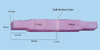
A heat activated poly (methyl methacrylate) (PMMA) resin (Trevalon, Dentsply India Pvt. Ltd., Gurgaon, India); and two resilient liners, Super-Soft (GC America Inc, IL, USA) and Molloplast-B (Detax GmbH & Co, Germany) were used. Super-Soft is a long term acrylic based resilient liner and chemically binds to acrylic denture base owing to similar chemistry whereas Molloplast-B is silicone based liner. A total of 60 PMMA resin specimens with cross sectional area of 10×10 mm were prepared from 120 blocks of acrylic resin (two acrylic resin blocks were required to form one specimen). The dimensions of specimen were such that they could be produced in conventional denture flasks and gripped easily in the Instron machine (Fig. 1), as described in a previous study.8 To standardize fabrication of specimens, six equal sized brass dies were prepared and utilized to fabricate a silicone rubber mold (MP Sai Enterprises, Mumbai, India). Wax blocks of equal size were prepared in mold, invested in dental stone in a denture-processing flask, followed by de-waxing, and packing of mold cavity with resin. After curing, blocks were retrieved from the flask and smoothed on 240 grit aluminum oxide paper.10 They were randomly divided into 2 groups, each group containing 60 blocks (to fabricate 30 specimens per group), which were followed by their surface treatment as follows. Ten specimens in each group acted as control, i.e. the surfaces were left untreated. Ten specimens from each group were treated by sandblasting; the nozzle measuring about 1.0 mm in diameter was held in light contact with each specimen, for 30 seconds.5 Aluminum oxide particles measuring about 250 µm was used as the sandblasting medium at a pressure of 0.62 MPa.5 Ten samples, for each liner to be tested, were swabbed with the monomer of the denture base resin (Trevalon) for 180 sec.2 To prepare one Super-Soft lined specimen, two acrylic resin blocks, separated by a brass spacer of dimension 10×10×3 mm (to standardize the thickness of liner), were invested in a dental flask.8 The spacer was removed once flasking was over.5 Super-Soft was mixed according to manufacturer's instructions, packed into the space created by brass spacer, and curing was done. After curing, the flask was brought to room temperature, samples were retrieved, and excess liner was removed with a sharp scalpel.2 Molloplast-B lined specimens were fabricated similarly, except that Primo (Detax GmbH & Co, Germany) adhesive was applied uniformly on surfaces to be bonded and allowed to dry for 60 minutes. The specimens were subjected to the tensile stress on an Instron universal testing machine (Instron-4467, Instron Corporation, UK) and deformed using a cross head speed of 5 mm per minute.7,13 The load at which failure occurred was recorded, together with the type of failure. Failure mode was recorded optically14 (Stereomicroscope SV8, Carl Zeiss, Oberkochen, Germany) although it can be recorded visually15 or by Scanning Electron Microscope.10 The maximum tensile stress before failure divided by cross sectional area of interface produced the tensile strength value for the specimen. Regarding modes of failure, adhesive failure refers to total failure occurred at the denture base soft liner interface; cohesive failure refers to tear within the liner, and mixed failure refers to both.7 Mean, standard deviation and coefficient of variation were determined for all groups. Results for each group (Super-Soft and Molloplast-B) were tested by one-way ANOVA to measure the variation between the three subgroups, i.e. control, sandblasting, and monomer; followed by Tukey HSD test. The surface treatment groups of the Super-Soft and Molloplast-B were compared using unpaired t-test.
RESULTS
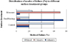
Means and standard deviations for Super-Soft are reported in Table 1; maximum mean TBS was observed with monomer treated group (4.025±0.616 MPa). These values are in agreement with those obtained by Kawano et al.,14 and Emmer et al.3 One-way ANOVA (Table 2) revealed significant differences among surface treatment groups of Super-Soft (P < .001). Maximum mean TBS of Molloplast-B was observed in monomer treated group (2.570±0.361 MPa) (Table 1). Oneway ANOVA (Table 2) revealed significant differences among various surface treatment groups for Molloplast-B (P < .001). Mean TBS of Super-Soft in various surface treatment groups (Table 1) was significantly higher (P < .05) than Molloplast-B. The modes of failure (observed together for both liners) were predominantly of mixed type in control category (60%). Adhesive modes were maximum (75%) in sandblast treated specimens, while cohesive failures were reported in 70% of monomer treated specimens (Fig. 2).
DISCUSSION
This study was planned to evaluate the effect of mechanical and chemical surface treatments on TBS between two resilient liners and PMMA resin. The tests developed by numerous investigators include peel test,5,7,15,16 tensile test,3,7,10,14-16 and shear test7,15; amongst these peel test closely simulates the intraoral forces encountered at the soft liner-denture base junction.14 However, during actual testing, forces will not be applied directly at the liner acrylic resin interface because direct gripping of the soft liner in the peel test may damage the integrity at the gripped region.14 In this study, tensile test was performed because it gives information on strength of bond in comparison to tensile strength of the materials and also because tensile properties are regarded as a general guide to the quality of rubbers.9,12 Fowler and Cantor et al. pointed out that, tensile failure was not caused by tensile forces alone as some shear forces developed because of the high Poisson's ratio of silicone lining materials.15 Khan et al. reported that soft denture liners should have a minimum of 0.44 MPa (4.5 kg/cm2) bond strength to be acceptable for clinical use.17
The surface treatment of denture base by monomer enhanced bond strength of both the liners. Super-Soft forms a strong bond with acrylic resin, even without a bonding agent as both have a similar composition.14 Molloplast-B, being a silicone based liner, requires an adhesive MMA,5 a solvent that dissolves the PMMA surface, and the bond strength of silicone liners will depend on tensile strength of the materials and the adhesive used.18 Therefore, using monomer and adhesive together prior to the resilient liner application may effectively increase the dissolution of the PMMA surface. It enables added fluid to penetrate between polymer chains and become entangled when the added monomer or solvent is evaporated.6 Sandblasting resulted in reduction of bond strength of both materials. Theoretically, sandblasting increases surface area and provides mechanical locks at bond site and should result in stronger bonds. According to Amin et al.,12 lower bond strengths were due to stresses that occurred at the interface of the PMMA and soft liner. Jacobsen et al.5 have considered the ability of soft lining material to penetrate into the irregularities of the PMMA. The penetration coefficient for liquids into a space is given by:
PC = γcos θ/2η
Where γ = surface tension, θ = contact angle, η = viscosity.
If this logic is applied to penetration of liners into the irregularities produced by sandblasting, increasing the viscosity of resilient liners for a given contact angle and surface tension reduces the penetration of the liner.5 This could explain the lower tensile strengths of sandblasted specimens observed in the study.
The bond failures were classified according to the criteria given by al-Athel et al.7 as adhesive, cohesive and mixed, and were considered together for both materials. Adhesive failures occurred when tensile strength of the soft liner was greater than its bond strength to PMMA14 and were mainly exhibited by sandblasted specimens. Cohesive failures occurred when tensile strength of the soft liner was less than bond strength and were mainly exhibited by monomer treated specimens. Control group mainly showed mixed type of failure indicating that the bond strength of the liner was nearly equal to the tensile strength of the liner. Results of this testing must be interpreted with caution as there are numerous factors which affect test results e.g. liner thickness, duration of application of monomer, strain rate, test methods, etc.2,7,19
This study may serve as a benchmark for the study of new resilient liners. Water sorption may also affect bond strength and further investigation is required to determine the influence of this factor.3,10 This study was entirely laboratory based and because the most appropriate testing environment is the mouth, long-term clinical studies of these materials are required.
CONCLUSION
Within the limitations of this in-vitro study, the following conclusions were drawn:
Monomer pretreatment of the resin surface produced higher bond strengths (P < .001), whereas sandblasting reduced the bond strength between the two liners and PMMA resin (P < .001).
Mean bond strength of Super-Soft was significantly (P < .05) higher than Molloplast-B in various surface treatment groups; although all subgroups tested had mean bond strength values greater than the minimum acceptable standard (0.44 MPa)17 for clinical application.
Mixed type of failures were more predominant in control specimens (60%), while adhesive modes were maximum (75%) in sandblasted specimens. In monomer treated specimens, 70% of failures were cohesive in nature.


 XML Download