

 PDF
PDF ePub
ePub Citation
 Print
Print


INTRODUCTION
Facial tissue loss or defects can be acquired, congenital, tumoral lesions or accidental. Facial deformity can cause functional and serious psychological problems that can affect an individual's social behavour.1,2 The field of maxillofacial prosthetics is concerned with the prosthetic reconstruction of missing/disfigured head and neck tissue. A prosthetic replacement of an exterior part is termed as epithesis, which is described as early as seventh century.3 Auricular reconstruction is challenging task in which a wide array of reconstructive options must be considered.4 Prosthetic replacement of missing facial tissues has several advantages over surgical reconstruction. The process is relatively inexpensive, allows for periodic evaluation and cleaning of the site. It is a short process and the maxillofacial clinician has complete control of color, shape and position of the prosthesis. Disadvantages include possible irritation of the tissue site, need for periodic remake and depending on adhesives or some other form of retention.5 The first well documented account of facial prosthetics is provided by Ambroise Pare who made varied contribution to standardize the indications for and materials used in facial prosthetics. Amongst the large number of materials that have been tried out in the history of Anaplastology as for example porcelain, natural rubber, gelatin and latex, two have established themselves: methacrylates and silicones.6 Long term success of a facial prosthesis depends mainly on retention.7 These prostheses are retained with different methods of retention such as medical adhesives, anatomical undercuts, and mechanical devices like spectacles, hair bands, magnets and implants.8 Since the introduction of percutaneous endosseous implants for use with bone conduction hearing aids in 1977, implants have acquired important role in the prosthetic rehabilitation of patients with craniofacial defects.9 Implants can vastly improve the retention and stability of a facial prosthesis. The co-ordinated participation of surgeon and maxillofacial prosthodontist is required in presurgical planning to determine the number, type and positioning of the implants in the defect.10
The aim of maxillofacial rehabilitation should provide a suitable prosthesis for patients with facial defects so that they are rehabilitated back to the society to face and accept the challenges of life. It encourages the best possible quality of life and upholds their self image during their traumatic psychological adjustment. This article describes an economical procedure for fabricating acrylic auricular prosthesis for a female patient who had bilaterally deformed ears resulting from trauma. Two stage impression technique was used and auricular prosthesis was fabricated in two parts, utilizing the external auditory canal for improving the retention of the prosthesis.
CASE REPORT
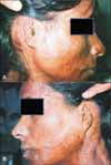
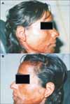
A 24 year, young female patient was referred to the Department of Prosthodontics, in Vidharbha Youth Welfare Society, Dental College and Hospital, Amravati (India) with bilateral auricular deformity. She was not willing to provide any information, including the reason for deformed ears. The disfigurement was suggestive of trauma making her quiet and avoiding interaction. Clinical examination revealed deformed helix, antihelix, concha, antihelical fold and lobules. Her face had scars throughout, as it appears after being burnt. (Fig. 1A and 1B) The skin surrounding the ears was wrinkled. Her hearing was normal, without any intrauricular problem as evaluated by speech recognition tests and signals to noise ratio by an otorhinolaryngoloist auricular prosthesis was planned for her in consultation with an otorhinolaryngologist. Patient education and counseling was done regarding the nature, function and limitation of the prosthesis.
She was draped, and her hair was protected by surgical cap covering the hairline. External auditory canal was blocked with gauze to prevent entry of impression material. Impressions of the auricular defect were made using irreversible hydrocolloid (Algitex, DPI, Mumbai, India) following standard procedures. Prebent L shaped paper clips were used for reinforcement and quick setting plaster of Paris (Kaldent, Kalahai Karson, Mumbai, India) was added for backing. Impressions were removed, boxed and poured and in type 3 gypsum material (Kalstone, Kalabhai Karson, Mumbai, India).11
Wax patterns of the ears were prepared on stone models using modelling wax (Deepti Dental Products, Ratnagiri India) by comparing with the appropriate size of the ear of another female of the same age group. They were checked on her face for proper orientation superoinferiorly and anteroposteriorly. Due importance was given to patients feedback regarding any modification in patterns. The wax patterns were not very retentive and stable on her face. After consulting an otorhinolaryngologist and performing an otoscopic examination of the ears, it was planned to utilize the external auditory canal for added retention of the prosthesis. She had the shape, curvature, extension of the canal and the anatomic undercuts of the existing deformed ears would be utilized for improving the retention and stability of the prosthesis.
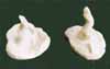
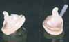
Patient was put at ease by educating her regarding the impression procedure, which was carried in assistance with an audiometrist. Lubricated cotton otodams of appropriate diameter were pushed in the canal beyond the second bend, with the tip of earlite to seal the canal and ensure safety of ear drum.12 Putty silicone impression material (Green Echo. Detax. GmbH & Co. K.G., Ettlingen, Germany) was manipulated and loaded in a plastic syringe (Cetylcide, Siemens' Erlangen, Germany) with tapered nozzle of approximately 5 mm diameter. Using proper bracing technique, the tip of the syringe was placed approximately 6 mm inside the canal opening. The material was gently expressed into the ear canal, allowing the material to flow back over the syringe tip. Once the material started to flow past the tip, the pressure of the material itself pushed the syringe out slowly, filling the surrounding concha and helix region. During the procedure her vagus/cough reflex, her gag and watering eyes reflexes were noted. She was asked to open and close her mouth along with jaw movements, like chewing motion etc.13,14 After curing the impression were gently removed from the canal by pulling her pinna up and back to break the seal (Fig. 2). The internal component was processed in heat polymerized clear acrylic resin (Trevalon, Dentsply, York, PA, USA). It was drilled in the centre. A polyethylene hollow tube of approximately 3 - 4 mm in diameter was cemented to maintain the opening of canal and prevent the occluding effect (Fig. 3).15
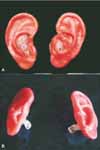
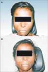
The internal component were attached to previous wax patterns and checked to verify the fit. Gentle clockwise rotational movement in open jaw position was done to place the prosthesis in place. Anticlockwise rotational movement for removal of the prosthesis was tried. After making necessary corrections, the patterns were sealed with the internal component15(Fig. 4) and invested in varsity flasks. Packing with heat polymerized Polymethyl-Methacrylate (PMMA) (Trevalon; Dentsply, York, PA, USA) was done in patient's presence for accurate color simulation. Main color characteristics were achieved at the packing stage. Combination of oil colors was used to obtain desired shade of skin which was incorporated in monomer. Monomer and polymer were mixed in proper proportion and the dough was packed in the mould. Trial closures were done until acrylic flash was reduced to minimum. Slow curing was done. After bench cooling, deflasking was done carefully. Surface imperfections were removed. The prosthesis was tinted with acrylic paint suspension (Fevicryl, Pidilite Industries, Mumbai, India) for final characterization (Fig. 5). Finished prosthesis was tried for retention and esthetics (Fig. 6A and 6B). Audiometry tests were carried to check her hearing. Patient was trained and instructed for proper placement and removal of the prosthesis. Proper care and maintenance of both tissue and prosthesis hygiene was emphasized. She was advised to remove the prosthesis at night, wipe the internal component with a dry soft cloth and keep it free of dirt and ear wax. A small brush and wax removing tool for cleaning the vent (used for cleaning hearing aids) was given to clear the wax. Hair oil, creams etc were not to be used with the prosthesis seated as they can clog the vent. Excessive exposure to sun should be avoided to prevent discoloration of prosthesis. During the recall visits she was assessed for her vagus and lymphatic reflexes. She was examined for swelling and soreness in her ears after wearing the prosthesis. She came for recall till 1 year, which was satisfactory.
DISCUSSION
Patients with auricular deformity or absence of auricle endures psychological affliction. Auricular defect can be repaired16 or reconstructed with autogenous tissue,17 but this may not be feasible for personal/medical reasons.18 A good alternative is to develop an auricular prosthesis with a suitable material Silicone is the material for choice for facial prosthesis because of its flexibility and life like appearance.19 In this case PMMA was used. Although this material has shortcomings of being inflexible and having esthetic limitations, it provided an economic rehabilitation to the patient, improving her quality of life and reintegrating her back to the society. Treatment was patient centered and patient directed.
The use of external auditory canal for achieving retention requires a sound knowledge of anatomy of ear and associated structures.13 The average length of the canal from tragus to tympanic membrane is 25 - 26 mm long and 6 - 7 mm in diameter. Size and shape of canal vary among individuals. The cartilaginous part of the canal is about 8 mm and the osseous part is 16 mm in length. The length of the impression extended about 17 - 18 mm in length, which was still far from the ear drum.15
Whenever feasible implant retained prosthesis should be given prime consideration, which has improved retention, stability, and comfort of the patient.1,20 Recent advances in techniques, including a new generation of Computed Tomography scanner and three dimensional (3D) systems facilitate the production of mirror image of auricular prosthesis with high level of accuracy, alleviating most of the limitations of conventional prosthesis21 Limitation to its use is high cost. Development in the field of tissue engineering has resulted in the formation of new tissue equivalents of bone and cartilage that will augment the outcome of prosthodontic rehabilitation in future.22 Evidenced based studies pertaining to the value of facial prosthetics have to be addressed to better understanding of the economical, functional and psychological burden of having a facial ablative procedure involving the orofacial, ocular, auricular and nasal tissues.5
CONCLUSION
Maxillofacial defects can be emotionally traumatizing considering the societal emphasis on physical appearance. An attempt to provide a cost effective and cosmetically acceptable bilateral auricular prosthesis for a female patient was made which was esthetically and functionally acceptable to her. Communication and education is the key for accepting the prosthesis. Successful use of prosthesis may depend on the patient's psychological acceptance of it. Patient's participation in the decision making process with realistic expectations is of vital significance.
 XML Download