

 PDF
PDF ePub
ePub Citation
 Print
Print


INTRODUCTION
Mandible fractures are belong to the most common fractures encountered in maxillofacial trauma.1 Because mandible is such a unique structure with hinge joint and masticatory muscles attached to the body of mandible, attention must be paid to avoid displacement during treatment.2 Displacement during fracture reduction leads to malocclusion. Some of the literature stated approximately 17% to 19% of cases will present with a transient malocclusion after an open procedure, 4% to 8% will require correction with an occlusal adjustment, and 0.5% to 3% require a secondary revision surgery.3,4 The treatment of the malocclusion includes occlusal adjustment, post-traumatic orthodontics, or corrective jaw surgery. During the treatment of the mandible fracture, dentists have to concern whether the occlusion of the patients has changed or not.
When fractured segments of the mandible are reduced in the displaced position, the three dimensional position of the condyle in the condylar fossa will be changed. Then, a question whether the temporomadibular joint could function with complete comfort in changed situation would arise. Dawson mentioned adapted centric posture in 1995.5 He stated that many temporomandibular joints function with complete comfort and apparent normalcy, even though they have undergone deformation caused by disease, trauma, or remodeling with adapted centric posture. In addition, he insisted that if the occlusion stays acceptably stable for up to three months and there are no other concerns, proceed with the restorative phase.5
The purpose of this article is to report post traumatic malocclusion and its prosthetic treatment. This clinical case report will briefly describe the patient who got open reduction of his fractured mandible in changed position, his changed occlusion, and its prosthetic procedures to establish stable occlusion in so-called adapted centric posture.
CASE REPORT
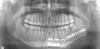
A 41 year-old-man was referred to the Department of Orthodontics at the Seoul National University Dental Hospital by the department of plastic surgery in July 2007, with difficulty in chewing after open reduction of left mandibular body fracture (Fig. 1). Without any treatment of his occlusal problem, he was transferred to the Department of Oral and Maxillofacial Surgery (OMFS) instantly, since he displayed the symptom of osteomyelitis due to the left mandibular first molar (#36) which was included in the fracture line. The mandible got saucerization along the fracture line with extraction of the associated tooth and reduced again with plates and screws in September (Fig. 2). After that, an iliac bone graft followed by plate removal was done in January 2008. Six month later, he was referred to the prosthodontic clinic for restoring missing tooth #36 and unoccluded premolars in the same segment (#34, 35) by the department of OMFS. The patient also complained that he had shifted his mandible to the right side when chewing since he had been injured, and he desired to improve esthetics with tooth whitening and closing the spaces between mandibulr anteriors.
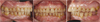
The patient demonstrated a slide to the right side from centric relation to maximal intercuspation avoiding premature conatact between teeth #27 and #38 (Tooth #37 was extracted when he was an elementary school student, after that the space for the tooth #37 has been closed with the tooth #38 completely.). Teeth #33, #34, and #35 looked like that they were located under the occlusal plane. In addition, diastemas were presented between teeth #31 and #41, and #41 and #42 (Fig. 3).
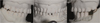
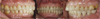
Three sets of study casts were made for record and treatment planning. A centric relation was recorded with bilateral manipulation using Aluwax (Aluwax Dental Products Co., Michigan, USA), and a maxillary cast was mounted on a semi-adjustable articulator using facebow transfer.6 The mandibualar cast was articulated with occlusal centric relation record (Fig. 4). Considering that the left segment of the mandible which contained tooth #38 had been displaced during the reduction procedure, the first premature contact point on the tooth #38 was eliminated on the articulated study cast. However, only the teeth #17 and #47 were occluded after occlusal adjustment. When the casts were adjusted to eliminate the contact between teeth #17 and #47, the mesial teeth #16 and #46 were occluded. The results were the same for the more mesial teeth as well. It was certain that both segments of the fractured mandible were reduced in the displaced position. From the all evidences and the patient's statement, it could be inferred that maxillomandibular fixation for his second open reduction in the department of OMFS using arch bar had been done in the shifted mandible position (Fig. 3). The articulated study casts continued to be adjusted until all the premolars and molars in the right side had contacts to the teeth of the opposite jaw. Avoiding working and nonworking interferences between teeth #27 and #38, it was necessary to adjust the tooth #38 which displayed severe mesial inclination. Next sets of articulated study casts were adjusted in the same way, and diagnostic wax-up was complete for unoccluded teeth #33, #34, #35 and diastema closure (Fig. 5). It satisfied the esthetic demand of the patient and the requirements for occlusal stability.7 Stable stops on all teeth were established and teeth #33, #34 guided working movement to left side in the diagnostic wax up model. A presentation of the treatment plan to the patient was made including risks, alternatives, and benefits of the each treatment options and we have chosen the following treatment: simple occlusal adjustment for right posteriors, porcelain fused to gold (PFG) crown and bridges for teeth #34, #35, X, #38, all ceramic crowns for teeth #31, #32, #33, #41, #42. For early period of treatment, home bleaching was done using Opalesence (Ultradent product, UT, USA), and #38 was endodontically treated prior to tooth preparation because of the large amount of tooth elimination for avoiding interference and gold space.
Occlusal adjustment was done just as it had been done in the articulated study models in twice carefully and adjusted occlusion functioned for one month.5 Tooth preparation for PFG was completed on teeth #34, #35, #38 and provisional crown and bridge were made from the diagnostic wax-up template. For another two month, it was assured that adapted centric posture had been stable.5 Tooth preparations for all ceramic crowns were made on teeth ##31, #32, #33, #41 and the teeth were provisionalized. The template of the diagnostic wax up model was used to determine necessary tooth reductions and adequacy of tooth preparation design. After final tooth preparation of the anteriors, final impression was made with poly vinyl siloxane impression material. A custom incisal guide table was made from Quicky resin (Nissin dental products Inc., Kyoto, Japan). Zirconia copings (LAVA, 3M ESPE, MN, USA) were tried in and 2R1.5 shade (VITA system 3D-master, Vita Zahnfabrik, Sãckingen, Germany) was selected at least one month after tooth whitening had been finished. All ceramic crowns #31, #32, #33, #41, #42 were fabricated and cemented with modified glass ionomor cement (Fuji-cem, GC, Tokyo, Japan). The patient satisfied with his lower anteriors in an esthetic point of view and anterior guidance was established in protrusive movement. Final impression was made for PFG crown and bridge on teeth #34, #35, #38 and interocclusal record was taken using pattern resin (Dura Lay, Reliace, IL, USA). Gold copings were tried in and PFG crown #34 and bridge #35, X, #38 were fabricated and cemented with Fuji-cem. A functional analysis of the occlusion confirmed that the goals of mutually protected occlusion have been achieved (Fig. 6).


 XML Download