

 PDF
PDF ePub
ePub Citation
 Print
Print


INTRODUCTION
Over the last two decades, the field of esthetic restorations has substantially expanded to satisfy patients' demands. Traditional treatment approaches (full coverage crown) would involve the removal of large amounts of sound tooth substances, which has adverse effects on pulp, gingiva, and crown biomechanics.1 Therefore, it is important to preserve much tooth structure in place. One of the most conservative treatment modalities is porcelain laminate veneers (PLVs). Since their introduction by Pincus in 1930, PLVs have been a popular dental treatment modality. The clinical survival rate of PLVs is high. Observation periods of PLVs reported in the literature range from 18 months to 15 years.2-4 One study reported that 98.4% of 186 PLVs placed over a 5-year period was rated as successful.2 Another study showed that the estimated survival probability of 182 PLVs over a period of 12 years is 94.4%.5 PLVs are indicated not only for treating tooth discoloration, but also for restoring fractured, worn dentition and malformed teeth. Swift et al. reported that PLVs provide suitable esthetics and reliable functional strength.6 With the conservative approach, esthetic and functional results may be achieved. To date, following the study of Magne and colleagues who demonstrated the efficacy of bonding porcelain to enamel (as strong as natural dentition), the use of PLVs may be expanded to more challenging cases.7
CASE REPORT
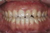
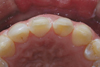
In this case, a 25-year-old male patient lost his maxillary left central incisor long before. From 2001, he had been treated with pre- and post-operative orthodontic therapy and gnathosurgery to improve his facial appearance. In this process, the maxillary left lateral incisor was moved on the position of maxillary left central incisor and the maxillary left canine was moved on the position of maxillary left lateral incisor. The patient wanted to improve esthetics of anterior dentition. First, the space between anterior dentitions was analyzed. Analysis showed asymmetry in anterior dentitions because of the absence of the maxillary left central incisor and size difference between maxillary central and lateral incisors. Analysis also showed lingual inclination of the left lateral incisor. The maxillary left lateral incisor was more yellowish than the maxillary right central incisor and incisal edge of the maxillary right central incisor was concave due to attrition (Fig. 1).
1. Treatment planning
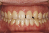
Porcelain laminate veneer was planned to improve esthetics of the anterior dentitions through transforming the shape of the teeth with porcelain laminate veneer. The treatment plan was to transform the maxillary left lateral incisor into a central incisor and the maxillary left canine into a lateral incisor. The restoration of the maxillary right central incisor was also included into the plan for esthetic improvement. The maxillary central incisors are mesio-distally larger than the lateral incisors for about 2 mm, and it was necessary to do minor tooth rearrangement by orthodontic treatment. After the tooth rearrangement, the space of the maxillary left lateral incisor became equal to that of the maxillary right central incisor. In addition, cross-bite of the maxillary left lateral incisor was resolved (Fig. 2).
2. Diagnostic wax-up
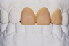
Diagnostic wax-up was performed including the maxillary right central incisor, the maxillary left lateral incisor and maxillary left canine on the study cast (Fig. 3). Mockup is critical for fabrication of the provisional restorations, enabling the patient and clinician to evaluate esthetics, and to preview the anticipated result.8
3. Intraoral preparations
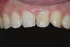
After duplicating diagnostic wax-up cast, putty index (Exafine putty type, GC, Tokyo, Japan) was made for tooth preparations. Selective preparations were performed with the guide of putty index using tapered diamond burs (Shofu, Kyoto, Japan). The preparation margins were placed at the equi-gingival level for esthetics. Preparation margins and surfaces were finished with a low speed white stone bur (Shofu, Kyoto, Japan) under water spray (Fig. 4, 5).
4. Provisional restorations
Provisional restorations were fabricated by Luxatemp® (DMG, Hamburg, Germany) using putty attained from the diagnostic wax-up cast. After 20 seconds of spot etching (Scotchbond etchant, 3M ESPE, MN, USA) for 20 seconds at the labial surfaces, provisional restorations were set using flowable light-cured composite resin (AELITEFLO™, Bisco, IL, USA).
5. Impressions
A week later, provisional restorations were removed and abutments were cleaned with a rubber cup. Before the impression was taken, retraction cord (UltraPak #00; Ultradent, UT, USA) was packed in the gingival sulcus to exposure the preparation margin. The final impression was taken with polyvinylsiloxane impression materials (Exafine putty type; GC Corporation, Tokyo, Japan / Honigum Light; DMG, Hamburg, Germany) light body polyvinylsiloxane (Honigum Light, DMG, Hamburg, Germany) using 1-step impression technique.
6. Placement and evaluation
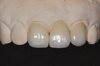
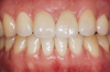
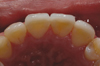
Porcelain laminate veneers were fabricated with IPS e.max Press system (Ivoclar-vivadent, Schaan, Liechtenstein). After IPS e.max Press copings were made, they were layered with glass ceramics (IPS e.max Ceram Powder; Ivoclar-vivadent, Schaan, Liechtenstein) and then finishing and glazing were done (Fig. 6). IPS e.max Press has high flexural strength (400 MPa) and esthetics.9 Also, the advantage of this method is needless of a refractory cast. Porcelain laminate veneers were cemented with the light-cured resin cement (Variolink II; Ivoclar-vivadent, Schaan, Liechtenstein). After a month from the delivery, as shown in Fig. 7 and 8, the gingiva was healthy and the interproximal area was fully filled with interdental papilla. Patient was satisfied with the harmonious shape and contour of the restorations which also showed good function.
CONCLUSION
This clinical report shows that porcelain laminate veneers can meet esthetic and functional desires of the patient with missing maxillary central incisor. Porcelain laminate veneer is a great choice to change tooth shape because of relatively minor tooth reduction, short treatment time and acceptable esthetics. In a case of changing a tooth shape with porcelain laminate veneers, pre-treatment evaluation, space analysis and diagnostic wax-up are important factors.
 XML Download