

 PDF
PDF ePub
ePub Citation
 Print
Print


INTRODUCTION
Patients with destroyed dentition have several characteristics such as loss of posterior occlusal support, severe occlusal wear of remained teeth, missing of multiple teeth, etc. The loss of posterior occlusal support may cause following problems: the attrition of anterior teeth, the reduction of vertical dimension (VD), and the alteration of normal occlusal plane. These problems will eventually cause the unesthetic facial appearance, the pathologic changes of the temporomandibular joint (TMJ) and masticatory muscles, and the inability of mastication.1 Patients with destroyed dentition require extensive restorative treatment.1 In such cases the restoration with implant-supported crowns and fixed partial dentures is preferred. However, in patients with financial, anatomical, and/or medical limitations alternative treatment modalities, such as removable partial denture (RPD), should be considered.2
The edentulous patients with compromised esthetic zone can be successfully treated with a rotational path RPD.3-9 Rotational path RPD has been often overlooked by the dental profession due to its complex prosthetic design and sensitive laboratory techniques.10 With better understanding of the principles of rotational path RPD, the dental clinician can deliver excellent esthetic outcome in compromised areas in which other treatment options may often be limited.
The rotational path design concept uses a rigid retainer portion of the framework as the retention component.11 The proximal plate provides retention through its intimate contact with the proximal tooth surface below the height of contour at a zero-degree tilt. These rigid retentive components have dual paths: the first path to engage into the undercuts and the second rotational path to fully seat the prosthesis.3,10,12 There are three basic types of rotational path RPDs: anterior-posterior (AP), posterior-anterior (PA), and lateral. Jacobson and Krol narrowed the rotational paths to two categories.11 Category I includes all prosthesis designs that first seat the rest that is associated with the rigid retainer. After the first rest is seated, the second segment rotates into place and conventional clasps are engaged.9 The rotational centers are located at the end of long rests. Category I includes all PA and AP paths of insertion that replace posterior teeth.10 Category II includes all lateral paths and AP paths that replace anterior teeth. The centers of rotation and the rigid retainers are located at the gingival extension of minor connectors.9,11 These prostheses use a dual path of insertion: at first RPD is inserted straightly from an incisal or occlusal direction to the rotational centers and then the conventional posterior or contralateral retainer is rotated into position. It is important that the initial straight path of insertion must be parallel with the cingulum rests to permit complete seating. The rotational path RPD offers the advantages of improved esthetics by eliminating anterior clasps, shortened treatment duration, cleanliness, and lower treatment cost over implants or fixed partial dentures.5,6,11
CASE STUDY
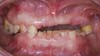
The patient was a 70 year-old female complaining of inability of mastication and unesthetic facial appearance. She had no specific medical history and dysfunctional habit. Typical features of destroyed dentition were observed in the clinical and radiographic examinations (Fig. 1). However, she had several problems: severe attrition of almost remained teeth, #14 horizontal root fracture, #13 periapical lesion and buccal fistula formation, insufficient interocclusal distance, loss of anterior guidance. In addition, severe ridge resorption resulted in asymmetric and unsupported upper lips. She had fear of surgery, financial limitations and severe ridge resorption of esthetic zone. For these reasons, we decided that not implant prosthesis but removable prosthesis was indicated for her. In this case, the extremely worn anterior dentition was planned to be restored with fixed prosthesis. All attrited teeth were restored with single porcelain-fused gold (PFG) crowns except four lower incisors, which were restored with 4-unit splinted PFG fixed dentures due to their very short crown lengths. The upper missing area was to be restored with rotational path RPD. Finally, the lower missing area was to be restored with conventional RPD. The rotational path RPD is the excellent treatment modality of restoring missing incisors when patients have contraindications for implants or when patients are financially limited.
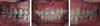
Preliminary impression was made and then recording base and wax rim were fabricated on the diagnostic casts for VD determination and centric relation (CR) registration. Anatomic landmarks, facial measurements and the resting positions of mandibular jaw were used to determine appropriate vertical dimension for the patient. As the result, it was decided that VD raising was necessary for esthetic and functional restoration. This increased vertical dimension was 4 mm from the tips of incisors. After that, CR registration was taken using bilateral manipulation1, and diagnostic wax up was performed to confirm the need of increasing VD for esthetic and functional rehabilitation. Prior to teeth preparation, composite resin was filled in the incisal and occlusal surfaces of all attrited teeth. This conservative correction was for conservation of destroyed tooth structure and ideal tooth preparation.1 Teeth were prepared using a omni-vac as the preparation guide. To compensate for short clinical crown lengths, axial surfaces were reduced as parallel as possible. The provisional restoration was fabricated based on the diagnostic wax up (Fig. 2). While the patient was wearing the provisional restorations for the 4 months, occlusal stability and TMJ were periodically checked. The patient was satisfied with the provisional restoration. Thus, it was decided to reproduce of temporary VD and CR state on the final restoration. It was performed using the recording base, waxrim and two pattern resin (Dura Lay, Reliance, Illinois, USA) coping crowns. This transfer procedure was described in the Fig. 3.
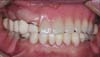
Final impression was made for PFG crowns. PFG crown copings were tried in the mouth to check for marginal fit. Before glazing, surveying was performed repeatedly for rotational path RPD. All PFG prostheses were cemented with resin modified glass ionomer cement (Fuji-cem, GC, Tokyo, Japan). To fabricate RPD framework, upper and lower master casts were made. Upper RPD was classified as Class III Mod. 1 RPD and was designed in lateral rotational path RPD with no clasp in the anterior region. Lateral or category II rotational path RPD in this report was surveyed in the next two steps (Fig. 4). The first surveying was performed at a zero-degree tilt to identify the mesial surface undercut of the anterior abutments (at least 0.010 inch) and buccal undercuts of the posterior abutments. The diagnostic cast was then tilled upward until the mesial undercuts on the anterior abutments were eliminated. The cast was again surveyed to determine if the anterior rest seats are accessible during the initial straight path of insertion. Lower RPD was conventional class I RPD with linguoplate major connector (Fig. 5). Both RPD frameworks were tried in the mouth and adapted with silicone material (Fit-checker, GC, Tokyo, Japan). RPD frameworks were very stable and showed very accurate fit and retention. For lower RPD, altered cast was made. A face-bow transfer was completed, and interocclusal registration was made with wax rim and bite material (O-bite, DMG, Hamburg, Germany). The casts were mounted in a semi-adjustable articulator. Denture teeth were set up to establish the stable and harmonious occlusion as such13: (1) Simultaneous bilateral contacts of opposing posterior teeth in the centric occlusion (CO) (2) unilateral balanced occlusion by denture teeth for left working side contacts (3) canine guidance by natural teeth for right working side contacts (4) contacts of opposing anterior teeth in the CO. After obtaining the patient's approval, RPDs were processed using pink denture resin (Rapidsimplified, Vertex, Zeist, Netherlands). The finished RPDs were placed in the mouth, and the following criteria were evaluated: adaptation of the clasps and rests, retention of the RPD, esthetics, and occlusion (Fig. 6). Finally the patient was instructed in placing the prosthesis and maintaining oral hygiene.
CONCLUSION
Treatment of patients with destroyed dentition is very difficult clinical procedure and challenging for dental profession. In this case, proper diagnosis and treatment plans are the necessity for not only esthetic and functional restorations but also the stability and adaptation of the neuromuscular system and TMJ. For compromised patients to implant dentistry, rotational path RPD is an alternative treatment option. It improves esthetics to replace missing teeth without placing a conventional clasp in the esthetic region. It is important that dentists evaluate each patient carefully and survey casts meticulously to ensure success.


 XML Download