

 PDF
PDF ePub
ePub Citation
 Print
Print


INTRODUCTION
Autopolymerizing resin has been conventionally used to make record bases.1,2 Recently, however, light-polymerized record bases have also been used to make resin-base full dentures where a metal frame is not used. Light-polymerized resin consists of a cross-linked urethan dimethacrylate matrix and low-level microfine silica, which shows advantages in maintaining the volume even after the fabrication and also it has a high elastic modulus.2
This resin material may contract due to polymerization but the level of such contraction varies by site. In particular, it would remarkably contract in the palatal portion of a maxillary full denture.3 A light-polymerized record base resin can also cause low fit due to contraction at the time of the polymerization in the maxillary palatal portion and shows an error during its fitting procedure.5 Such fitting error inhibits the sealing of the palatal posterior border.2,4 When a light-polymerization resin is used, the resin closest to the light source is polymerized first when exposed to the light and consequently the" resin uplift" phenomenon occurs which creates an empty space between the cast and the resin during the polymerization process.
In the maxillary edentulous cases the hard palate is relatively flat and inclined toward the residual alveolar ridge. The dented shape of the palatal portion in combination with the contraction of the resin causes the record base uplift in the palatal portion.5 Thus, it is considered that if polymerization and the resulting contraction of the light-polymerized resin toward the alveolar crest is distributed over the center of the palatal portion and the alveolar crest by dividing the light-polymerized resin into two pieces along the palatal portion when making the record base, the record base uplift phenomenon at the palatal portion due to the polymerization and the resulting contraction would be reduced. Although this clinical procedure was introduced by Oh and May6 in a case report published in 2008, they did not suggest proper guideline to apply the two-phase fabrication method.
The aims of this study were to suggest a method of fabrication of record base using a light-polymerized resin by the two-phase fabrication method for the improvement of the fit of the record base and to compare the degree of fit according to the separation site.
MATERIALS AND METHODS
A. Model fabrication
The edentulous cast of maxilla (Dentiform, Nissin Dental Prod., Inc., Kyoto, Japan) was obtained using light-body and regular-body silicone impression materials (Imprint II, GC Dental, Tokyo, Japan). The improved stone (Fuji Rock® EP, GC Corp., Tokyo, Japan) was poured to fabricate the cast. The labial/buccal undercut region of the residual ridge was blocked out on the edentulous maxilla model and was coated with a thin layer of petroleum jelly.
B. Guidelines for the fabrication of the record base
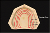
In the first case, a line was drawn along the end of the border (test group 1). In the second, third and fourth cases, lines were drawn along the palatal plane 5 mm (test group 2), 10 mm (test group 3) and 15 mm (test group 4) below the alveolar crest, respectively (Fig. 1). Each of these lines was a criterion for making two separate sections. For the control group the cast was made without separating the two sections.
Group 1: line connected to the alveolar crest along the end of the border. Group 2: line connected to 5 mm below the alveolar crest along the palatal line. Group 3: line connected to 10 mm below the alveolar crest along the palatal line. Group 4: line connected to 15 mm below the alveolar crest along the palatal line.
C. Fabrication of the record base specimen
Visible light cure resin (Eazipan LC, Vericom Co., Ltd., Anyangsi, Korea) was carefully attached to the cast. To prevent the formation of air bubbles, gentle force was applied to the cast using the fingers from the center to the edge of the cast. Excessive materials were removed using a surgical blade. The record base was divided into two U-shaped segments 5, 10, and 15 mm below the alveolar crest along the alveolar ridge. Care was taken to maintain the interval at 1.0 mm when dividing the case (Fig. 2). Light polymerization of the record base placed on the cast was performed under visible light at a wavelength of 475 nm and a wave strength of 90 mW/cm2 for 15 minutes (Solidlite® SHOFU Dental Co., Kyoto, Japan).
The gap between the two parts in the cast was filled again with light-polymerized resin, followed by light polymerization for an additional 15 minutes. The record base was removed from the cast and the edges of the record base were trimmed according to the predetermined border line (Fig. 1). Twelve specimens were made for each group.
D. Evaluation of the interior space of the record base of each group
After 24 hours the light-body silicone impression material (Aquasil XLV, DENTSPLY International, Inc., Milford, USA) was injected into the interior of the record base. It was then placed onto the cast and finger pressure was applied to stabilize it in a seated position, followed by immediate placement onto the universal test device (Shimadzu Corporation, Kyoto, Japan). The universal test device was set at the mode for measuring the compression strength; the compression strength was set to not exceed 40 N; and constant force (40 N) was applied for five minutes until the impression material hardened fully.
After five minutes the record base was removed and its edges were trimmed to match the edges of the record base. The residual silicon impression material inside the record base was removed. The weight of the impression material was weighed using the microscale that can measure weight up to the level of 0.1 mg (Ohaus Adventurer™, Itin Scale Co., Inc., Canada). Based on the density of the impression material (1.34 mg/ml), the volume of the impression material was calculated. This volume was that of the space between the record base and the master cast. The fit of the record base was indirectly measured by comparing these spaces.
RESULTS
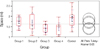
The mean inside volume of the control group was 1.88 (± 0.42) ml; of Group 1, 1.73 (± 0.23) ml; of Group 2, 1.68 (± 0.44) ml; of Group 3, 1.42 (± 0.20) ml; and of Group 4, 1.38 (± 0.30) ml. According to the one-way ANOVA and Tukey-Krammer HSD tests for the differences between the groups, the control group and Groups 3 and 4 showed significant differences, and Groups 3 and 4 showed significantly smaller inside gaps than the control group which was not made with the two-stage method (Fig. 3, Table 1).
DISCUSSION
Light-polymerizing resin has shown several advantages such as superior fit and strength, complete polymerization without leaving a residue, stable color and ease of manipulation and addition of other record base resins.7 Light-polymerizing resin however, can result in the contraction of the palatal portion during the polymerization. Particularly, the posterior portion of the palate as well as the distobuccal corner, is known to be the place where polymerization-induced contraction occurs most frequently during denture relining.8 Such contraction was reported to have also occurred with heat-polymerizing resin.9
Adequate closure of the posterior portion of the palate of a maxillary denture is necessary for the maintenance of a denture. An inaccurate denture can damage the final cast during the fabrication of the denture and may result in a wrong intermaxillary relation in patients. An inaccurate denture can also reduce the accuracy of the anterior record and the condylar head inclination of the articulator, which would make the record base unstable and pose difficulty in accurate oral examination.
Thus, the aim of this study was to present an alternate fabrication technique by applying the two-stage fabrication method and comparing the difference in the fit of the separation sites to improve the fit of the record base based on light-polymerizing resin.
In this study, significantly small inside gaps were observed in Groups 3 and 4 in which the record base was divided into two pieces along the palatal portion, 10 and 15 mm below the alveolar crest which indicated lower error during polymerization. This low error is believed to be attributable to the division of the light-polymerizing resin into two pieces along the palatal portion at a constant distance from the alveolar crest there by distributing the contraction force evenly and leaving stress on each of the pieces.
This study does not to suggest that the use of light-polymerizing resin is better than use of auto-polymerizing resin or that the record base should be made from light-polymerizing resin. A record base that has a metal frame can be ideal in clinical practice on account of the ease of maintenance of the denture, but when we make provisional denture or the patient has allergy to metal, light-polymerizing resin can be used to make record bases. It is believed that in this case the two-stage fabrication method can be applied easily to improve the fit. It was found that two-stage method (in which the cast is divided into two U-shaped pieces in regions 10 - 15 mm from the alveolar crest) was effective in improving the interior fit.
CONCLUSION
Although this study had some limitations, the error due to the polymerization-induced contraction of the posterior portion of the palate was reduced most significantly by the division of the record base into two pieces 10, 15 mm from the alveolar crest by polymerizing them, adding resin between them followed by repolymerization. It is thus recommended that the two-stage denture fabrication method in which the record base is divided at an appropriate region can be used in clinical practice as this method may show improvement in the fit of the light-polymerized record base.


 XML Download