

 PDF
PDF ePub
ePub Citation
 Print
Print


INTRODUCTION
A proper tooth preparation is essential for better aesthetics, acceptable prosthetic rehabilitation, fracture resistance, and healthy soft tissues.1 Dental burs are commonly used instruments during clinical and laboratory procedures in dental profession. They are manufactured in various shapes and sizes with different features to offer various utilization.123456
Diamond burs have better cutting efficiency and duration than other burs like carbide or stainless steel.78 Hardness of the surface particles, sterilization and/or disinfection procedures,9 storage conditions, corrosion, and multiple use10 are some of the determinants with potential to alter the cutting efficiency of the diamond burs.
Most of the clinicians may state that after multiple usage, the cutting/milling efficiency of burs evidently decrease.11112 Bae et al.4 concluded that cutting efficiency decreased as the number of cuts increased regardless of the type of burs. They claimed that this reduction is highest after the first use. These worn-out burs may require excessive pressure application during tooth preparation, which may cause undesired heat generation and waste of time.8121314 On the other hand, they cannot generate adequate roughness on tooth surfaces, which is essential for mechanical retention of cements like zinc-phosphate cement because of ineffective particle size.1516171819
The up-count limit to change the diamond bur during and/or after dental usage is not specified in the literature. In addition, the demand for efficient disposable dental instruments with low price, like single-use burs, are increasing nowadays.2021 Manufacturers claim that discarding a single-use diamond bur is more economical than sterilization. However, the cutting efficiency and durability of single-use diamond burs especially during full mouth preparations were not evaluated or specified in the literature either.121 Previous studies were based on investigating cutting efficiency,4512222324 performance,25 characteristics,262728 heat generation,293031 and abrasive properties32 of various burs only. The purpose of this study was to identify the changing frequency, by determining the alterations on the surface of the diamond burs when used on different surfaces. The null hypothesis of this study was that cutting efficiency of diamond burs will be unaffected by different surfaces.
Go to : 
MATERIALS AND METHODS
Twenty-six recently extracted human premolar teeth free of caries, restoration, and endodontic treatment were selected, cleaned and stored in tap water in room temperature. All teeth were evaluated by transillumination method for enamel cracks. Care was taken to select teeth with similar dimensions to diminish variability during experiments. The main group was named as Group T. Then, teeth were randomly grouped according to the number of preparations and were assigned to form 3 groups containing 1 (T1), 5 (T5), and 10 (T10) teeth, respectively. Each tooth was embedded vertically into an acrylic resin block (25 mm × 25 mm × 22 mm) 2 mm below the cementoenamel junction.
Sixteen disc shaped CoCr metal alloy (Remanium Star CL, Dentaurum Gmbh & Co. KG, Ispringen, Germany) samples (10 mm × 4 mm) were fabricated by direct metal laser sintering (DMLS) method (M2, Concept Laser; Hoffmann Innovation Group, Lichtenfels, Germany). This group was named as Group M. The samples were randomly grouped according to the number of preparations and were assigned to form 3 groups containing 1 (M1), 5 (M5) and 10 (M10) discs, respectively.
Sixteen disc shaped zirconia samples (10 mm × 4 mm) were fabricated from 3Y-TZP pre-sintered ceramic block (VITA In-Ceram YZ, Vita Zahnfabrik, Bad Säckingen, Germany) according to manufacturer's recommendations. This group was named as Group Z. The samples were randomly grouped according to the number of preparations and were assigned to form 3 groups containing 1 (Z1), 5 (Z5) and 10 (Z10) discs, respectively.
2 mm thickness of zirconia and metal samples were embedded into acrylic resin block separately. For the easy approach of the head of air-turbine handpiece to the embbaded samples, one side of the resin-block was modified. A custom modified milling device3334 was used to standardize the preparation protocol (Fig. 1).
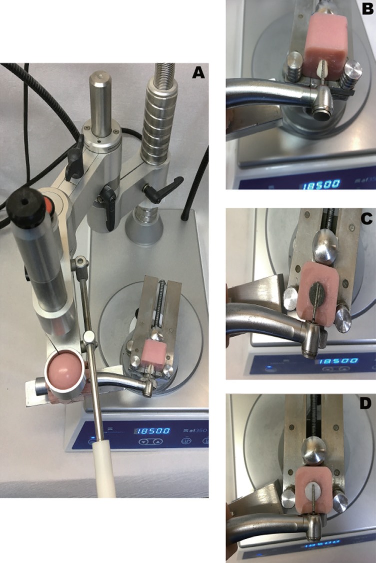
A high-speed air-turbine rotary handpiece (BA695LK; BA International, BA International Ltd., Northampton, England) with a coolant water spray of 25 mL/min was fixed onto the device, which operated at 300,000 rpm. Preparation cycle was composed of several cuts of a bur. In this study, a “cut” was defined as a groove, which was curved out using a bur guided with the custom modified milling device under constant pressure of 100 gr/10 sec. Figure 2 shows the cut design and direction of each sample. A long round tapered black band diamond bur (GZ Instrumente, G&Z Instrumente GmbH, ISO 806 314 200 544 018 12.0, Lustenau, Austria) was selected for this study. Each bur was coded as shown in Table 1. To mimic standard preparation, 5 vertical grooves were composed onto the 4 regions (buccal-palatinal/lingualmesial-distal) of the tooth.
 | Fig. 2(A) Preparation (five cuts) on tooth sample, (B) Preparation (one cut) on CoCr (DMLS) sample, (C) Preparation (one cut) on zirconia sample.
|
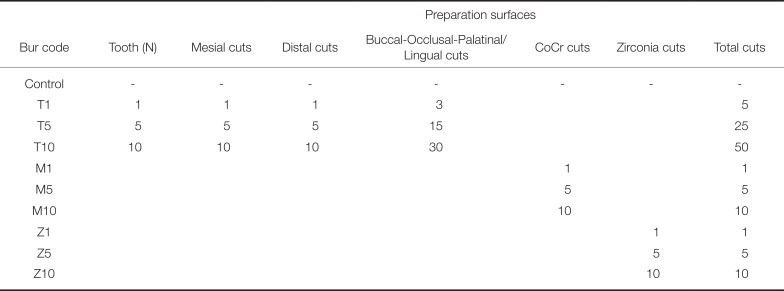
Table 1
The usage of the burs on the samples: preparation cycle
![]()
One bur was kept unused for control evaluations. For the tooth sample, 5 cuts were obtained in each study group for simulating tooth preparation with different enamel thicknesses on all surfaces (Fig. 2). This protocol was adapted to the other zirconia and metal samples as 1 – 10 cuts each time, as the surface characteristics and hardness of these materials are different from tooth. Substructure try-in phase was simulated.
After every preparation process, burs were cleaned with a toothbrush and placed into the ultrasonic cleaner (Bandelin Sonorex RK 102 P; Bandelin Electronic, Berlin, Germany) for 5 minutes. Then, surface roughness of each bur was measured using a profilometer (Time TR100; Phynix GmbH & Co., Köln, Germany), and six measurements were performed for every bur. Remaining 10 teeth were used to observe the cutting efficiency of each coded bur (Table 1) on the enamel surface of a tooth after multiple usage. First, teeth were separated 2 mm below from the cement-enamel junction using a high-speed instrument (Micracut 150; Metkon Endüstriyel San. Tic. A.Ş, Bursa, Turkey). Then the crown samples were separated into two in buccolingual direction from the midline.34 Each half of the teeth sample were prepared with the custom modified milling device under constant pressure of 100 gr/10 sec. By using same custom modified milling device, the burs were used (10 total) on a separate surface.
Scanning electron microscope (SEM, JEOL 6400, JEOL Corp., Tokyo, Japan) images were taken with 400× magnification at 20 Kv. SEM images were taken from the mentioned mesial surfaces and the appearance of mesial teeth surfaces were observed under 2.5× magnification using a stereomicroscope (Leica S6D; Leica Microsystems GmbH, Wetzlar, Germany). During the examination of the specimens under SEM and stereomicroscope, different Vickers hardness values of the specimens (Table 2) were taken into consideration.

Statistical software (SPSS v15.0; SPSS Inc., Chicago, IL, USA) was used for statistical analysis. One-way ANOVA was used to evaluate inter-group differences. The post hoc Tukey honest significant differences test was used for multiple comparisons of group differences (P = .05 for all tests).
Go to :
RESULTS
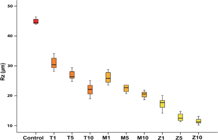
Mean roughness value of the Control group was found to be 44.83 ± 0.96 µm. Mean roughness values of the Groups T10 and M10 were decreased to 22.08 ± 2.14 µm and to 20.43 ± 1.14 µm, respectively (Fig. 3). When the average mean Rz value was measured for Group Z10, it was found that this value decreased drastically to 11.46 ± 1.0 µm. In comparison of all groups, the lowest mean Rz value was recorded for Group Z10 (Rz = 11.46 ± 1.0 µm) and highest mean Rz value was recorded for Group T1 (Rz = 30.85 ± 2.2 µm) (Fig. 3).
One-Way ANOVA revealed a statistically significant difference among all groups (Table 3). According to the Tukey honest significant differences test results, significant differences were observed within groups and P values are given in Table 4.
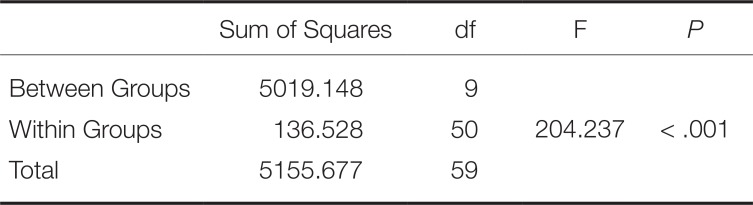
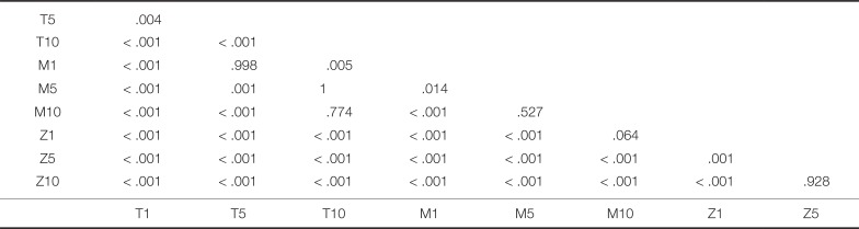
Table 3
One-way ANOVA results of mean Rz values of all groups
| Sum of Squares | df | F | P | |
|---|---|---|---|---|
| Between Groups | 5019.148 | 9 | ||
| Within Groups | 136.528 | 50 | 204.237 | < .001 |
| Total | 5155.677 | 59 |
![]()
Table 4
Results of Tukey's HSD, multiple comparison tests showing P values
![]()
For Group T, a statistically significant difference was found between Groups T1 and T10 (P < .001) and between Groups T5 and T10 (P < .001). However, no significant difference was observed between Groups T1 and T5 (P = .004). For Group M, a statistically significant difference was found between Groups M1 and M10 (P < .001). However, no significant difference was seen between Groups M1 and M5 (P = .014). For Group Z, a statistically significant difference was found within groups (P < .001). However, no significant difference was seen between Groups Z1 and Z10 (P = .928).
In comparison of Group T1 with the other groups, statistically significant difference was seen (P < .001). Also, statistically significant difference was seen among Groups T5, M5, and Z5 (P < .001). In addition, a statistically significant difference was observed among Groups T10, M10, and Z10 (P < .001). However, no significant difference was seen between T10 and M10 (P = .774).
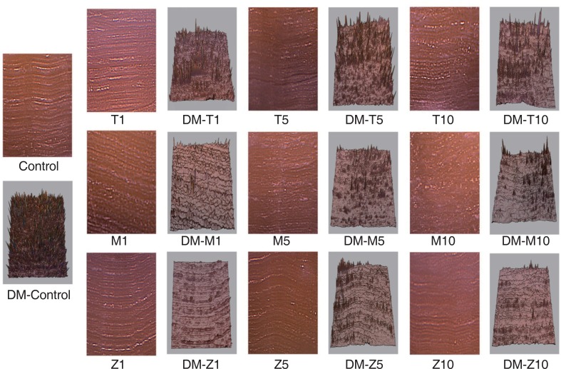
Compared with Groups T10, M10, and Z10, Groups T1, M1, and Z1 exhibited deeper grooves, pitting, waviness, and more irregularities on tooth surfaces (Fig. 4). In Group M10, grooves and ridges were shallower. Additionally, when compared to Groups T10 and M10, a regular morphology with more striation was observed instead of deep grooves and ridges in Group Z10. From Groups T10 and M10 to Z10, irregularities were decreased (Fig. 4, Fig. 5). Surface depth maps of machined teeth were created from the stereomicroscopic images with a software (Adobe Photoshop CS6, Adobe Systems, San Jose, CA, USA) to observe surface topography in detail (Fig. 5).

Go to :
DISCUSSION
The aim of the present study is to determine the changing frequency of diamond burs after preparations on three different surfaces. This study focused on observing the effects of reused diamond burs on tooth surfaces after prosthetic preparations or grinding process of CoCr and zirconia substructures. According to the findings of this study, surface roughness of diamond burs was decreased drastically after five preparations for all study groups (P < .001). Therefore, the null hypothesis was accepted and it was concluded that diamond burs need to be changed after five teeth preparations.
Three kinds of samples with different hardness values were used in this study. In literature, it was stated that Vickers hardness numbers of CoCr alloys that was manufactured by DMLS are lower than the conventionally produced ones.35 Therefore, wear of the diamond burs is expected to be less than the conventionally manufactured CoCr specimens. Conventionally produced CoCr specimens were not included in the study, as 3D additive manufacturing technology is getting popular nowadays. In this study, grinding procedures during try-in stages that are frequently encountered in the clinics were simulated. The results of the study presented that the highest wear value of the diamond burs was observed in Group Z. Group M and Group T followed the former. This difference is presumably because of the higher hardness value of zirconia than those of CoCr (DMLS) and tooth specimens. In addition, it was observed that the roughness of the diamond burs decreased after the preparation of all specimens. It was thought that the embedded diamond particles were separated from bur when the specimen cutting started and cutting efficiency was reduced due to the separation of the diamond particles from the matrix.56
According to the studies, dentists most apply 50 gr to 150 gr (corresponding to 0.5 – 1.5 N) mean cutting force on the tooth when using a high-speed rotary handpiece.12223242838 The cutting efficiency may vary with the force applied to the instrument. For this reason, constant force of 100 gr (1 Newton) was applied to the specimens in the study. The rpm of the air-turbine hand-piece might change during preparation.4222627 However, Ercoli et al.31 reported that there was no significant change in the rpm values of the different rotary cutting instruments even if the same air-turbine handpiece was used. For this reason, the same handpiece was used and torque values were not monitored.
Dentin surface topography has an important role on the cement retention. It was stated in the literature that preparation technique could affect the retention of the fixed prosthesis.15161734 In addition, McInnes18 and Al-Omari34 stated that surface roughness is important especially for the cementation of full crowns with non-adhesive zinc phosphate luting cement, as the luting is achieved from mechanical interlocking.1834 Also, the study of Solá-Ruiz et al.19 presented that in porcelain laminate preparation, oscillated instruments produced a rougher dentinal surface and this rough surface presented less microleakage. In our study, other factors like wettability and bond strength of adhesive cements were not included as the aim of this study was to investigate the cutting efficiency and changing frequency of the diamond burs. In this sense, for observing the changes in the topography of tooth surfaces, SEM and stereomicroscopic images were used.
In SEM examinations of machined tooth surfaces, when compared to the Control group, from T1 to T10, the cutting efficiency of the diamond bur was decreased and grooves and ridges on the tooth surfaces transformed to smoother areas. Similarly, these surface properties were also observed in tooth specimens that studied for Group M. When compared to Groups T and M, less shallow pits and grooves were observed in Group Z. This might be due to the highest Vickers hardness value of zirconia material. Although Group M have been manufactured by DMLS technique, it showed lower hardness value than the conventional type and enamel and also reduced cutting efficiency. Depth map presentations of the machined tooth surfaces also corroborate our findings on stereomicroscopic images.
The roughness on the prepared teeth surfaces revealed the negative shape of the burs used on 3 different specimens. This process allows us to observe surface roughness. Generally, Ra and Rz parameters are used in calculating surface roughness of materials.34 Ra averages all peaks and valleys of the roughness profile and then neutralizes the few outlying points so that the extreme points have no significant impact on the final results. However, Rz is calculated by measuring the vertical distance from the highest peak to the lowest valley and averaging these distances. Therefore, peaks have a much greater influence on the final value. Due to irregular surface of the burs, using the deepest and highest points in calculation of surface roughness gives us more reliable information.3334
In this study, comparison of control group revealed that mean roughness value (Rz) of Groups T5 and T10 was decreased by 40.2% and 51%. Also, decrease in Groups M5 and M10 by %50 and 54.5% and in Groups Z5 and Z10 by 71.5% and 74.5% were seen. This severe reduction in Rz values due to reuse of diamond burs exhibited that burs must be changed every five tooth preparations and the burs used in try-in process should not be used in tooth preparation. Also, it was stated in literature that reused diamond burs cause decrease in cutting efficiency and rates, increased chair time, and heat generation and potential heat damage to dental tissues. These are important factors that affect changing frequency of reused diamond burs and reuse protocols.530
In the present study, only one zirconia brand (VITA In-Ceram YZ) and one type of metal alloy brand was used. Other brand and types of metal alloy and zirconia specimens may show different machinability. In addition, the diamond burs from only one commercial brand and type were tested. Therefore, to obtain more information, it will be necessary to perform further studies using different brands and types of burs and materials.
Go to :
CONCLUSION
Within the limitations of this in vitro study it can be concluded that diamond burs exhibit wear with repeated use in clinic. The most important relevance of this wearing is the reduction in surface roughness of the bur and thus the reduction in cutting efficiency. For this reason, especially in prosthetic prepartions, diamond burs should be changed after every 5 preparations. Also, diamond burs that were used in try-in stages of metal and zirconia should not be used for tooth preparation.
Go to :
 XML Download