Abstract
PURPOSE
The aim of the present study was to record the metal-ceramic bond strength of a feldspathic dental porcelain and a Co-Cr alloy, using the Direct Metal Laser Sintering technique (DMLS) for the fabrication of metal substrates.
MATERIALS AND METHODS
Ten metal substrates were fabricated with powder of a dental Co-Cr alloy using DMLS technique (test group) in dimensions according to ISO 9693. Another ten substrates were fabricated with a casing dental Co-Cr alloy using classic casting technique (control group) for comparison. Another three substrates were fabricated using each technique to record the Modulus of Elasticity (E) of the used alloys. All substrates were examined to record external and internal porosity. Feldspathic porcelain was applied on the substrates. Specimens were tested using the three-point bending test. The failure mode was determined using optical and scanning electron microscopy. The statistical analysis was performed using t-test.
RESULTS
Substrates prepared using DMLS technique did not show internal porosity as compared to those produced using the casting technique. The E of control and test group was 222 ± 5.13 GPa and 227 ± 3 GPa, respectively. The bond strength was 51.87 ± 7.50 MPa for test group and 54.60 ± 6.20 MPa for control group. No statistically significant differences between the two groups were recorded. The mode of failure was mainly cohesive for all specimens.
Go to : 
Metal-ceramic works are still considered the preferable choice for fixed dental restorations, due to their high strength and superior aesthetic appearance. One of the main factors affecting the success of these restorations is the choice of proper dental alloy and its treatment for achieving a satisfactory metal-ceramic bond.
Nowadays, base metal alloys (Co-Cr and Ni-Cr) are mainly used for the fabrication of fixed metal-ceramic restorations. In particular, Co-Cr base alloys are a reliable alternative since Ni has been implicated in allergic reactions of patients.123456 The excellent mechanical properties such as high Modulus of Elasticity, and the high corrosion resistance of Co-Cr alloys, in combination with their significantly low cost, are factors that make these alloys the first choice for dental metal-ceramic applications.3789
Although the predominant fabricating technique of dental metal-ceramic substrates is the casting technique, over the recent years the evolution of digital technology has developed new techniques of construction, such as Stereolithography (SLA), Fused Deposition Modeling (FDM) and Selective Laser Sintering (SLS)1011 as well as Hot pressed technique.12 The SLS technique is applied mainly to metals, but can also be applied to polymers and ceramics. It constitutes the new challenge in manufacturing the fixed dental prosthetic restorations. The advantages of the SLS technique are the high productivity, the little or no loss of material, the minimal porosity, the improved mechanical properties, the shaping ability and the fit of the metallic dental substrates. The high surface roughness of the metal substrates produced by this technique is referred as the main drawback.91011121314
There are various classifications for SLS technologies, either according to the binding mechanism (Solid State Sintering, Chemically Induced Binding, Liquid Phase Sintering, Full Melting) or the sintered materials (polymers, metals, ceramics or compo-sites). However, in the dental materials the terminology is not yet clearly defined. So, the term SLS has been preferred for non-metallic materials (ceramic and polymers) while for metallic materials (alloys) the term used is Direct Metal Laser Sintering (DMLS) or Selective Laser Melting (SLM).11
The DMLS is an additive manufacturing process which was developed by EOS (GmbH, Munich, Germany), and has been available commercially since 1995 with the trade name “EOSINT M250 laser sintering machine”.15 For the rapid bonding of powder particles in the DMLS process liquid phase sintering and melting/solidification are the feasible mechanisms.16 In this process, the design of the prosthetic restorations is digitally held (3D-CAD file) and then the construction is accomplished by sintering the powder using powerful laser (usually a CO2 laser).911
The rapid development of the above-mentioned manufacturing techniques brought the need to evaluate the metal-ceramic bond strength of these restorations. Many in vitro methods have been applied for testing the metal-ceramic bond strength (shear, tensile, shear and tensile, flexure and torsion forces) among which the most commonly used are three and four-point flexural (bending) tests.1718 The three-point bending test is adopted by ISO 9693:1999, because of the simple construction of specimens, the quantification of the metal-ceramic bond strength and the use of simple mechanical testing devices.192021 In a three-point bending test, the upper portion of the specimen is under compression, the lower under tension, while the probe tips are in shear mode.19
Although the advantages of the DMLS technique are obvious in the construction of the metal substrate of dental metal-ceramic restorations, the knowledge concerning their bond strength is limited, so the present study aspires to contribute in this direction.
The aim of the present study was to record the metal-ceramic bond strength between a feldspathic dental porcelain and a Co-Cr alloy, using the Direct Metal Laser Sintering technique (DMLS) for the fabrication of metal substrates. To compare the results the metal-ceramic bond strength between the same feldspathic porcelain and a casting Co-Cr alloy will be used as control. Research hypothesis is that there will not be a statistically significant difference in the metal-ceramic bond strength between the two techniques used for the fabrication of the metal substrates.
Go to : 
Twenty metal substrates were constructed in dimensions according to specification ISO 9693 and were equally divided into two groups. The substrates of the first group (test group, n = 10) were constructed using the DMLS technique and the substrates of the second group using the casting technique (control group, n = 10). Additionally, another six substrates, three for each technique, were constructed to record the Modulus of Elasticity (E) of the used alloys.
To prepare the metal-ceramic specimens, thirteen plastic patterns were fabricated in slightly bigger dimensions than the ones proposed by the ISO 9693 and were positioned isometrically in a casting ring. The plastic patterns were invested (Giroinvest, Amann, Pforzheim, Germany) and casted (SmartCast, GalloniAsseg, Milano, Italy) using a Co-Cr alloy (Wirobond C, Bego, Bremen, Germany). The removal of the investment material was accomplished by sandblasting with 110 µm aluminum oxide (Al2O3) particles (Sandy2, Carlode Giorgi, Milano, Italy). All produced substrates were punctually adjusted to the ISO:9693 dimensions (thickness 0.5 ± 0.05 mm, width 3 ± 0.1 mm, length 25 ± 1 mm).
One of the substrates that was produced by the previously referred casting technique, was scanned by Computer Aided Design technology (CAD) and the data were sent to the special DMLS device, operating in 161.5-191.5 Jmm−3 laser energy conditions (EOS M270, EOS, Munich, Germany). Co-Cr powder was used for the fabrication of metal substrates (EOS-Cobalt-Chrome SP2, EOS, Munich, Germany). Another thirteen metal substrates were produced by this technique.
All substrates were subjected to macroscopically external inspection and then were examined for internal porosity, using an X-Ray unit (CMP 200, CPI, Cham, Switzer-land) operating under the following conditions: 80 kVp and 32 mAs.
After porosity testing, followed the ceramic mass positioning. A feldspathic dental porcelain (VMK-Master, Vita, Bad Sackingen, Germany) was applied in layers (bonding agent, opaque, dentin) at the center of one of the parallelepiped sides, according to the manufacturer's instructions, and was adjusted in dimensions, according to ISO 9693 (thickness 1.1 ± 0.1 mm, width 3 ± 0.1 mm, length 8 ± 0.1 mm).
To determine the Modulus of Elasticity (E) and to record the metal-ceramic bond, three-point bending test was used. Three of the thirteen metal substrates from each group were used to determine the E and the other ten to record the metal-ceramic bond strength, using a universal testing machine (Tensometer10, Monsanto, Akron, OH, USA).
The load was applied on the opposite side that the porcelain was applied, with crosshead speed of 1.5 mm/min, while the distance between the fulcrums was 20 mm.
The mode of failure of the fractured parts of the specimens was defined using an optical stereoscopic microscope (Eclipse ME 600, Nikon-Kogaku, Tokyo, Japan) under reflected light, at 5× magnification. Representative specimens were observed in a Scanning Electron Microscope (SEM) (Quanta 200, FEI, Hillsboro, OR, USA) and depicted areas were analyzed by X-Ray Electron Dispersive Spectroscopy (EDS) to study the elemental distribution in the deboned areas (mapping). In case that over fifty percent of the fracture surface was covered by ceramic material, (including bonding agent) the mode of failure was defined as cohesive, while in case that this percentage was lower than fifty percent the mode of failure was defined as adhesive.
Finally, the results of bond strength were statistically analyzed using t-test.
Go to : 
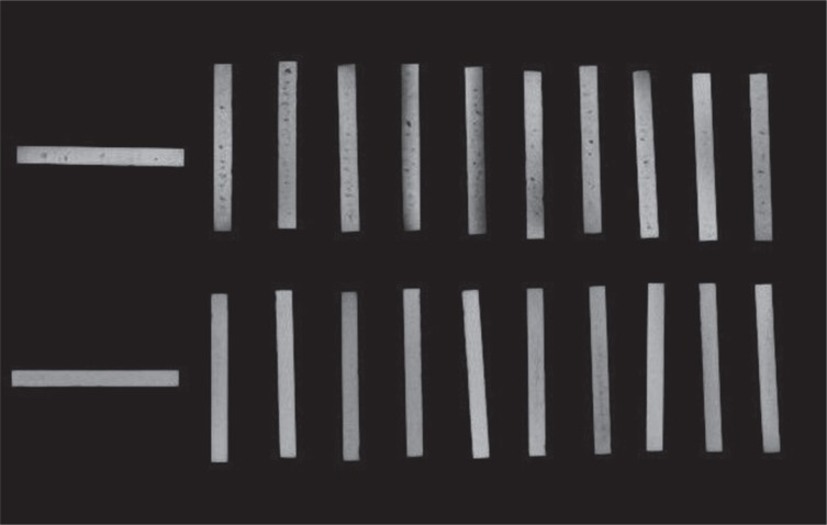
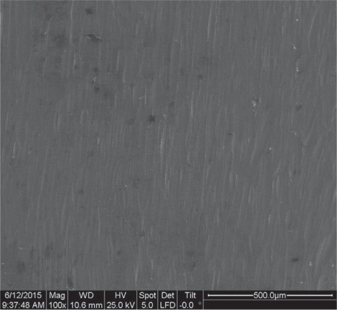
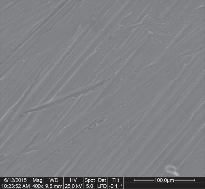
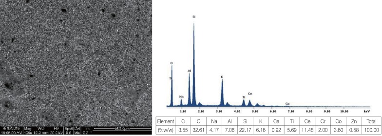
Radiographic inspection showed that metal substrates constructed by the casting technique showed dispersed microporosity within the accepted limits for in vitro research, although microscopically they did not reveal any external porosity (Fig. 1). In opposite, the metal substrates constructed by DMLS technique showed little or no microporosity, as it is shown in SEM micrographs (Fig. 2, Fig. 3).
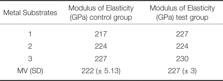
The E of the used Co-Cr alloys accrued from the stress-strain curve applying to the relative mathematic formula according to ISO 9693. The E was found varying within values available in the literature (Table 1).
No statistically significant differences were recorded in the strength of the metal-ceramic bond between the two applied techniques (P < 0.39). The values of the metal-ceramic bond strength are shown in Table 2.
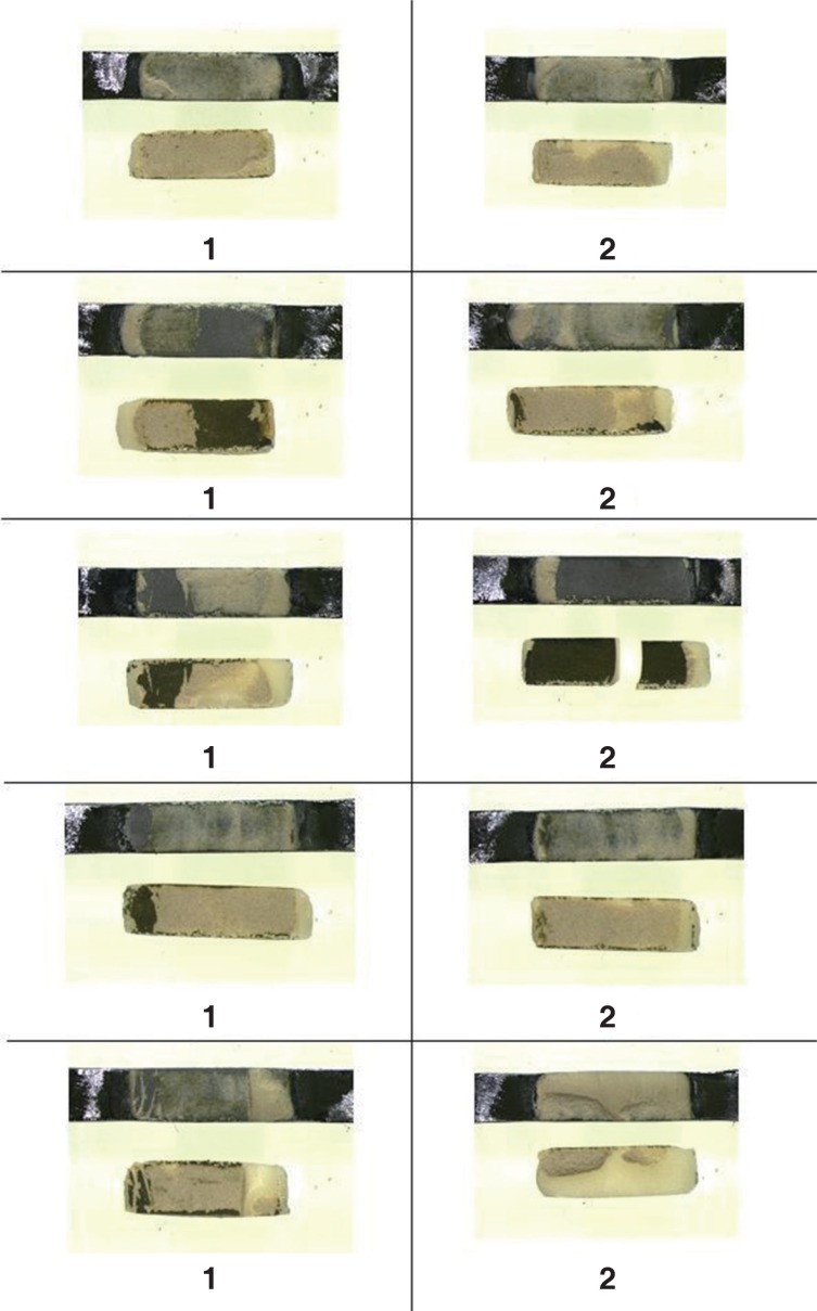
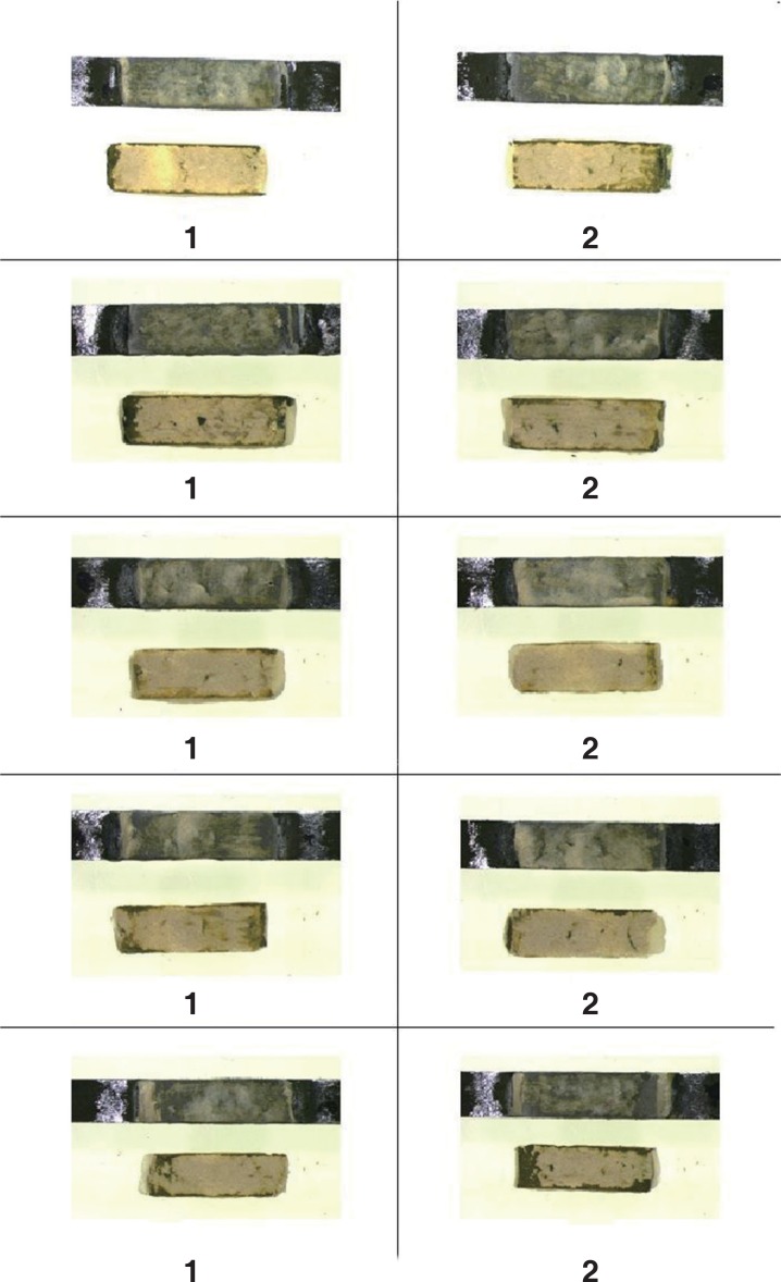
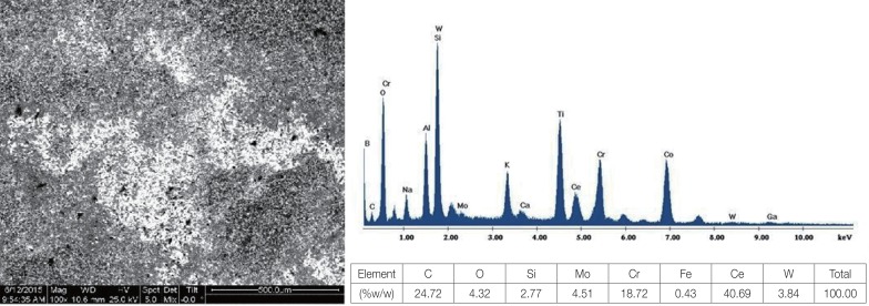
The mode of failure for both groups of specimens was cohesive. In stereoscopic microscope all the fractured specimens revealed the presence of elements, which represent the bonding agent and the ceramic mass (Fig. 4, Fig. 5). The EDS of the fractured specimens for both groups (except the increased amounts of Cr and relative amounts of Co and Mo) showed descending order of Ce, C, O and Si elements for the control group and O, Si, Ce and C elements for the test group, confirming the cohesive mode failure of both groups. The increased amounts of Ce represents residues of bonding agent on the metal substrate, while the increased amounts of Si represents residues of feldspathic porcelain (Fig. 6, Fig. 7).
Go to : 
The research hypothesis of the present study was verified because there was no statistically significant difference in the metal-ceramic bond strength between the two applied techniques.
Many researchers1314 studied a variety of parameters concerning the microstructure, the mechanical and electrochemical properties of fixed metal-ceramic substrates fabricated by Co-Cr dental alloys using the SLM technique. Al Jabbari et al.,14 recorded internal porosity only in substrates produced by casting but not in the ones produced by SLM technique, which is in accordance with the results of the present study. Takaichi et al.,13 observed dense sintering when the laser power was over the 400 Jmm−3, while internal porous structure observed when laser power was lower than 150 Jmm−3. These results are in accordance with the results of the present study where 161.5 – 191.5 Jmm−3 laser power had been used.
Moreover, many researches9222324252627 studied the metal-ceramic bond strength when the SLM technique was used. Some of them922 tested the metal-ceramic bond strength applying the shear test where others2324252627 using the three-bending test. All researches presented no statistically significant difference of the metal-ceramic bond strength independently of the test used. Absolute differences which were recorded among the previously mentioned studies and, waved between about 70 MPa the higher and 31 MPa the lower, can be attributed to the different Co-Cr dental alloys and feldspathic porcelains used. Also, superficial roughness of the metal substrates due to different porosity between the two applied techniques may play role in the overall metal-ceramic bond strength.
The results of the three-point bending test for the metal-ceramic bond are in agreement with the mean value of the previously referred studies (~55 MPa for casting technique and ~52 MPa for DMLS technique). Three-point bending test was applied according to ISO 9693, where the minimum acceptable failure value is 25 MPa. All experimental projects recorded values over this number.
Two studies926 examined the mode of failure in the fractured specimens using EDS. Xiang et al.,26 recorded increased ceramic elements on the metal substrates in both applied techniques, but ceramic amounts were higher when the SLM technique was used, which led to cohesive mode of failure. The results of Xian's study are in complete agreement with the present study. Conversely, Akova et al.,9 found a mixed mode of failure in the specimens fabricated with the casting technique, while the specimens fabricated by SLS technique waved between mixed and adhesive mode of failure. These results can be attributed to the different test (shear test) used for the metal-ceramic bond strength. Generally, there is confusion in literature about the definition of the metal-ceramic mode of failure. Some researches account the bonding agent within the ceramic material and others not. Also, the way of computing the remaining ceramic material on the metallic substrate for the definition of the mode of failure is controversial. In the present study, the 50 percent method was followed, as described in the Materials and Method section.
The strength of dental prosthetic restorations is an important factor for their survival in the dynamically changing biological environment of the human oral cavity (temperature change, change in pH, chemical changes) under the influence of alternative chewing forces. More specifically, the stresses developing in dental restorations during mastication are multiaxial. So, choosing both the appropriate materials and the suitable manufacturing technique for fabricating dental restorations can ensure their protection from failure.
Go to : 
Within the limitations of the present in vitro study, the authors can conclude that the metal substrates constructed by the DMLS technique showed little or no micro-porosity compared to the cast technique. The metal-ceramic bond strength of the specimens, in which the metal substrates were fabricated using the DMLS technique, cover the lower acceptable limit of 25 MPa, in accordance to ISO 9693 while no statistically significant difference in the metal-ceramic bond strength was recorded between specimens produced by cast and DMLS applied techniques. The failure mode for both techniques was cohesive.
Go to : 
References
1. Bezzon OL, de Mattos Mda G, Ribeiro RF, Rollo JM. Effect of beryllium on the castability and resistance of ceramometal bonds in nickel-chromium alloys. J Prosthet Dent. 1998; 80:570–574. PMID: 9813808.

2. Willis HH, Florig HK. Potential exposures and risks from beryllium-containing products. Risk Anal. 2002; 22:1019–1033. PMID: 12442995.

3. Al Jabbari YS. Physico-mechanical properties and prosthodontic applications of Co-Cr dental alloys: a review of the literature. J Adv Prosthodont. 2014; 6:138–145. PMID: 24843400.

4. Alkmina LB, Araújo AA, Silval P. Microstructural evidence of beryllium in commercial dental Ni-Cr alloys. Mat Res. 2014; 17:627–631.
5. Chen WC, Teng FY, Hung CC. Characterization of Ni-Cr alloys using different casting techniques and molds. Mater Sci Eng C Mater Biol Appl. 2014; 35:231–238. PMID: 24411373.

6. Henriques B, Soares D, Silva FS. Influence of preoxidation cycle on the bond strength of CoCrMoSi–porcelain dental composites. Mater Sci Engin: A. 2012; 32:2374–2380.

7. Molina A, Echeverri D, Parra M, Castro IJ, Garzon H, Valencia CH, Olave G. Metallographic characterization of overdentures bars manufactured by over-castting abutments for dental implants. Rev Fac Odontol Univ Antioq. 2013; 25:26–43.
8. Atluri KR, Vallabhaneni TT, Tadi DP, Vadapalli SB, Tripuraneni SC, Averneni P. Comparative evaluation of metal-ceramic bond strengths of nickel chromium and cobalt chromium alloys on repeated castings: An in vitro study. J Int Oral Health. 2014; 6:99–103.
9. Akova T, Ucar Y, Tukay A, Balkaya MC, Brantley WA. Comparison of the bond strength of laser-sintered and cast base metal dental alloys to porcelain. Dent Mater. 2008; 24:1400–1404. PMID: 18417202.

10. van Noort R. The future of dental devices is digital. Dent Mater. 2012; 28:3–12. PMID: 22119539.

11. Koutsoukis T, Zinelis S, Eliades G, Al-Wazzan K, Rifaiy MA, Al Jabbari YS. Selective laser melting technique of Co-Cr dental alloys: A review of structure and properties and comparative analysis with other available techniques. J Prosthodont. 2015; 24:303–312. PMID: 26129918.

12. Henriques B, Soares D, Silva FS. Microstructure, hardness, corrosion resistance and porcelain shear bond strength comparison between cast and hot pressed CoCrMo alloy for metal-ceramic dental restorations. J Mech Behav Biomed Mater. 2012; 12:83–92. PMID: 22659369.

13. Takaichi A, Suyalatu , Nakamoto T, Joko N, Nomura N, Tsutsumi Y, Migita S, Doi H, Kurosu S, Chiba A, Wakabayashi N, Igarashi Y, Hanawa T. Microstructures and mechanical properties of Co-29Cr-6Mo alloy fabricated by selective laser melting process for dental applications. J Mech Behav Biomed Mater. 2013; 21:67–76. PMID: 23500549.

14. Al Jabbari YS, Koutsoukis T, Barmpagadaki X, Zinelis S. Metallurgical and interfacial characterization of PFM Co-Cr dental alloys fabricated via casting, milling or selective laser melting. Dent Mater. 2014; 30:e79–e88. PMID: 24534375.

15. Khaing MW, Fuh JYH, Lu L. Direct metal laser sintering for rapid tooling: processing and characterisation of EOS parts. J Mater Proces Technol. 2001; 113:269–272.

16. Simchi A. Direct laser sintering of metal powders: Mechanism, kinetics and microstructural features. Mater Sci Engin: A. 2006; 428:148–158.

17. Akagi K, Okamoto Y, Matsuura T, Horibe T. Properties of test metal ceramic titanium alloys. J Prosthet Dent. 1992; 68:462–467. PMID: 1432762.

18. Ban S, Anusavice KJ. Influence of test method on failure stress of brittle dental materials. J Dent Res. 1990; 69:1791–1799. PMID: 2250083.

19. ISO 9693. Metal-ceramic dental restorative systems. Second edition. Geneva, Switzerland: International Organization for Standardization;1999.
20. Ritter JE. Critique of test methods for lifetime predictions. Dent Mater. 1995; 11:147–151. PMID: 8621037.

21. Pröbster L, Maiwald U, Weber H. Three-point bending strength of ceramics fused to cast titanium. Eur J Oral Sci. 1996; 104:313–319. PMID: 8831067.

22. Liu J, Chi S, Xu J, Wang Y, Zhan D. Effect of preparation methods on the metal-porcelain bond strength of Co-Cr alloys. Hua Xi Kou Qiang Yi Xue Za Zhi. 2014; 32:115–118. PMID: 24881202.
23. Bae EJ, Kim JH, Kim WC, Kim HY. Bond and fracture strength of metal-ceramic restorations formed by selective laser sintering. J Adv Prosthodont. 2014; 6:266–271. PMID: 25177469.

24. Bae EJ, Kim HY, Kim WC, Kim JH. In vitro evaluation of the bond strength between various ceramics and cobalt-chromium alloy fabricated by selective laser sintering. J Adv Prosthodont. 2015; 7:312–316. PMID: 26330978.
25. Liu J, Liu Y, Sun R, Zhan DS, Wang YY. Metal-ceramic bond strength of Co-Cr alloy processed by selective laser melting. Zhonghua Kou Qiang Yi Xue Za Zhi. 2013; 48:170–172. PMID: 23751533.
26. Xiang N, Xin XZ, Chen J, Wei B. Metal-ceramic bond strength of Co-Cr alloy fabricated by selective laser melting. J Dent. 2012; 40:453–457. PMID: 22402336.

27. Wu L, Zhu H, Gai X, Wang Y. Evaluation of the mechanical properties and porcelain bond strength of cobalt-chromium dental alloy fabricated by selective laser melting. J Prosthet Dent. 2014; 111:51–55. PMID: 24161258.

Go to : 


 PDF
PDF ePub
ePub Citation
 Print
Print



 XML Download