

 PDF
PDF ePub
ePub Citation
 Print
Print


INTRODUCTION
The replacement of missing teeth with osseointegrated dental implants has become an evidence-based treatment modality and a routine procedure in dentistry for more than four decades. Despite frequent occurrence of peri-implantitis and other complications, the survival rate for titanium implants is 90 – 95% over a period up to 20 years.1 Commercially pure titanium and titanium alloys are gold standard dental implant materials because of their biocompatibility, excellent mechanical properties, and long term clinical success.234
Despite its great advantages, titanium exhibits grayish discoloration on the peri-implant mucosa and jeopardizes aesthetic outcomes of restoration, especially if there is insufficient soft tissue to mask in the anterior segments.56 Although the prevalence is low (0.6%), titanium allergy can be detected in dental implant patients.7 Furthermore, titanium might induce hypersensitivity in susceptible patients and can play a critical role in implant failure.8 Some studies have also reported corrosive behavior that occurs after titanium comes in contact with saliva and fluoride.910 To compensate for the drawbacks of titanium, many researchers have tried to create tooth-colored biocompatible ceramic materials. Because of its aesthetic superiority, excellent biocompatibility and mechanical properties, ambitious efforts were made to introduce zirconia for applications in implant dentistry.411
Pure zirconia is a polymorphic crystal that can be found in three different crystalline phases depending on the temperature: monoclinic (room temperature until 1170℃), tetragonal (1170 – 2370℃), and cubic (2370℃ until melting point). The transformation from the tetragonal to the monoclinic phase is associated with a 3 – 4% localized volume expansion that induces compressive stresses in the compromised areas.12 The addition of stabilizing oxides like magnesia (MgO), yttria (Y2O3), and ceria (CeO2) prevents this phase transformation and maintains a metastable tetragonal phase at room temperature. 3 mol% Y2O3-stabilized tetragonal zirconia polycrystals (3Y-TZP) exhibits high strength and toughness as well as tetragonal phase stability at room temperature. Based on these, 3Y-TZP has been introduced as an alternative to titanium which shows superior mechanical properties compared to other ceramics.1314 Many studies have been performed to compare the osseointegration of standard titanium and zirconia implants and have reported no significant differences between 3Y-TZP and titanium implants.15161718 In vitro studies revealed comparable osteoblast adhesion, proliferation, and differentiation between differently treated Y-TZP disc surfaces and sandblasted/acid-etched titanium surfaces.1920 Several in vivo studies also proved that 3Y-TZP implants undergo osseointegration comparable with that of titanium implants.212223
Despite its excellent mechanical properties and biocompatibility, however, a major shortcoming of zirconia is its inherent accelerated aging and low temperature degradation (LTD). LTD is related to a lattice relaxation process induced by thermally activated oxygen vacancy diffusion.24 It consists of a spontaneous, slow transformation of the crystals from the tetragonal phase to the monoclinic phase at low temperatures (150 – 400℃). In a humid environment, this could decrease the strength of the materials and lead to catastrophic failures over time.25
Various approaches to eliminate or reduce LTD have included a ceria partially stabilized zirconia/alumina nanostructured composite (NANOZIR),2627 alumina-toughened zirconia (ATZ),282930 and 3Y-TZP co-doped with niobium oxide (Y,Nb)-TZP.2431323334 The resistance of (Y,Nb)-TZP to hydrothermal degradation is attributed primarily to t-ZrO2 phase stability as a result of Y-Nb ordering in the t-ZrO2 lattice31 as well as a reduction in the oxygen vacancy concentration in Y-TZP as a result of the substitution of Nb5+ for Zr4+.243135 In order to utilize this advantage of niobium in dental implant treatment, it is important to analyze the osteogenic potential of niobium oxide containing tetragonal zirconia polycrystals as proper osseointegration around the implant body is a major successful criteria for implant treatment.1234
Our previous study has shown that sandblasted (Y,Nb)-TZP discs have a similar osteogenic potential to that of anodized titanium.36 However, the correct combination of each composition to achieve optimal osseointegration is still challenging for the development of new materials. In this study, we synthesized new niobium oxide containing (Y,Nb)-TZP discs with specific ratios and denoted as YN4533 and YN4533/Al20. This study was performed to evaluate the osteogenic potential of new (Y,Nb)-TZP discs, YN4533 and YN4533/Al20, and compared with that of most widely used zirconia ceramic 3Y-TZP.
MATERIALS AND METHODS
Zirconia discs containing niobium oxide were synthesized according to specific ratios. The overall composition of YN4533 is 92.2 mol% ZrO2, 4.5 mol% Y2O3, and 3.3 mol% Nb2O5. YN4533/Al20 discs were prepared with the same concentration of YN4533 with an additional 20 vol% of Al2O3. YN4533 and YN4533/Al20 were test groups and 3Y-TZP used as a control. 3Y-TZP, YN4533, and YN4533/Al20 disc-shaped green compacts (15 mm diameter and 1 mm thickness) were prepared by cold isostatic pressing of the powder mixtures at 200 MPa followed by sintering for 2 hours at 1500℃ for 3Y-TZP, 1450℃ for YN4533, and 1600℃ for YN4533/Al20. The different sintering temperatures were used because the optimum sintering temperature for each material depends on the composition of the specimens to achieve maximum strength without deterioration and based on preliminary studies.2431 All zirconia discs were gradually polished and finished with diamond pastes to produce mirror-like surfaces. After polishing, half of the zirconia discs in each group were sandblasted with 50-µm alumina (Al2O3) at 2 bar pressure for 1 minute to create rough surfaces. Mirror-like smooth surface groups were denoted as 3Y-TZP-M, YN4533-M and YN4533/Al20-M while sandblasted rough surface groups were denoted as 3Y-TZP-R, YN4533-R and YN4533/Al20-R. The average surface roughness (Ra) and surface topography were analyzed using a 3-D confocal laser microscope (LSM 5 Pascal, Carl Zeiss, Germany). The Ra values represent the mean ± SD of three independent experiments. Surface morphologies of zirconia discs were observed via a field emission scanning electron microscope (FE-SEM; HITACHI S-4700, Tokyo, Japan).
Mouse pre-osteoblast MC3T3-E1 cells were purchased from ATCC (Manassas, VA, USA). The cells were cultured in α-minimal essential medium (α-MEM, Hyclon) supplemented with 10% fetal bovine serum (FBS) and 1% penicillin/ streptomycin and incubated in a humidified atmosphere of 95% air/ 5% CO2 at 37℃. The osteogenic media included 10 mM β-glycerophosphate, 50 µg/mL ascorbic acid in α-MEM with 10% FBS and 1% penicillin/streptomycin. A confocal laser scanning microscope (LSM700, Carl Zeiss, Germany) and ZEN2011 software were used to evaluate cell attachment and morphology. 24 hours after seeding onto the zirconia discs, cells that attached onto the discs were fixed with 4% formaldehyde. 4′,6-diamidino-2phenylindole (DAPI, Invitrogen) was used for detection of cell nuclei and Alexa Fluor 568 phalloidin (Invitrogen) was used for detection of the cytoskeleton.
Cell proliferation was examined by a PicoGreen assay using the Quant-iT PicoGreen assay kit (Invitrogen) 1, 4, and 7 days after seeding cells on the zirconia discs. Cells adhered to the zirconia discs were washed with PBS and lysed with TE buffer (10 mM Tris-HCl, 1 mM EDTA, pH 7.5) to allow for formation of DNA samples. Then, 100 µl of the DNA samples was mixed with 100 µl of PicoGreen reagent. Samples were loaded in triplicate and florescence intensity was measured on a microplate reader (FLUOstar Optima, BMG LABTECH, Ortenberg, Germany). Florescence intensity was converted into DNA concentration with a DNA standard curve per the manufacturer's instructions. To evaluate osteoblast differentiation, cells were seeded on the zirconia discs and cultured in osteogenic media, which includes 10 mM β-glycerophosphate and 50 µg/mL ascorbic acid in growth media. Cells were harvested at 5, 8, and 11 days and RNA was isolated using Trizol lysis reagent (TRIzol Reagent, Invitrogen). The Primescript RT reagent kit (Takara Bio, Shiga, Japan) was used for reverse transcription and then real-time PCR was performed using Takara SYBR premix Ex Taq (Takara Bio, Shiga, Japan) on an Applied Biosystems 7500 Real Time PCR system (Foster City, CA, USA). All samples were run in triplicate. The osteoblast differentiation marker genes were alkaline phosphatase (Alp) and osteocalcin (Oc). The results were normalized to glyceraldehyde-3-phosphate dehydrogenase (GAPDH) to account for variations in RNA quantitation. The marker genes were synthesized by Integrated DNA technology (IDT, Coralville, IA, USA). Alp activity was measured using an ALP kit (Sigma-Aldrich). Cells were seeded on the zirconia discs and cultured in osteogenic medium for 10 days. Cells were washed twice with PBS and stained as described by the manufacturer.
All quantitative data are presented as the mean ± SD and each experiment was performed at least three times. The data analysis was performed using two-way ANOVA-test and Tukey post hoc test. Differences were considered as being significant at P < .05.
RESULTS
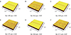
The average roughness values (Ra) and topographies of all zirconia discs under three-dimensional confocal laser scanning microscopy (3D-CLSM) are shown in Fig. 1. The Ra values of the mirror-like surface of 3Y-TZP, YN4533, and YN4533/Al20 were 0.09 ± 0.01 µm, 0.09 ± 0.01 µm, and 0.08 ± 0.02 µm, respectively. The surface roughness of the mirror-like surface discs was similar. To increase roughness, we sandblasted the zirconia discs with alumina particles. After sandblasting, the roughness of all zirconia discs increased significantly. As a result, the Ra values of the rough surfaces of 3YTZP, YN4533, and YN4533/Al20 were 0.62 ± 0.05 µm, 0.72 ± 0.04 µm, and 0.71 ± 0.07 µm, respectively. Although there was no significant difference between the rough surface discs, slightly higher Ra values were noted for modified zirconia discs.
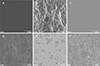
The surface morphologies of zirconia discs were analyzed by using scanning electron microscopy (SEM) (Fig. 2). Mirror-like zirconia surfaces showed a smooth and fine dotted pattern, which is assumed to be from the process of sintering. After sandblasting with alumina particles, all zirconia discs exhibited irregular rough patterns. The surface morphologies of 3Y-TZP and new (Y,Nb)-TZP discs did not differ significantly and were in good agreement with their Ra values (Fig. 1).
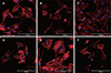
Fig. 3 shows MC3T3-E1 pre-osteoblast cells after 24 hours of culture on the mirror-like surface and the rough surface of zirconia discs. Cell attachment and morphology were analyzed with confocal laser scanning microscopy. The cells on the mirror-like surface of all zirconia discs showed regular size and morphology and a widely spread cytoskeleton. However, cells on the rough surface exhibited some morphologic irregularities, with a thin cytoskeleton and a less-stretched appearance on both 3Y-TZP and new (Y,Nb)-TZPs. All newly modified (Y,Nb)-TZP discs displayed good cell attachment similar to 3Y-TZP, and cell to cell contacts were observed on all zirconia discs regardless of surface roughness.
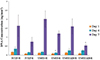
A PicoGreen assay was performed to examine cellular proliferation. Fig. 4 shows cellular proliferation on the zirconia discs for 1, 4, and 7 days. Cells proliferated well on all zirconia discs and the proliferation rate increased as time went on. Mirror-like surfaces had higher cell proliferation than rough surfaces, and this was highest at day 7. This indicates that MC3T3-E1 cells proliferate well on the smooth surface and match well with cell morphologies. Significant differences were found only between day 4s of 3Y-TZP mirror and YN4533 rough surface groups and days 7s of 3Y-TZP mirror and YN4533/Al20 rough surface groups. There was no statistically significant difference between cells grown on 3Y-TZP, YN4533, and YN4533/Al20, within the same surface roughness groups.
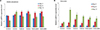
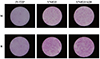
Quantitative real time polymerase chain reaction (RT-PCR) was performed to evaluate mRNA expression levels after 5, 8, and 11 days of culture. Fig. 5 (A and B) show the mRNA expression patterns of alkaline phosphatase (Alp) and osteocalcin (Oc), which are marker genes of osteoblast differentiation. Although the morphology of cells cultured on the rough surface appeared smaller and less stretched, cell differentiation between smooth and rough new (Y,Nb)-TZPs did not differ significantly. Osteoblast differentiation patterns of new (Y,Nb)-TZP discs were not influenced by the surface roughness, however rough 3Y-TZP discs showed more cellular differentiation than smooth 3Y-TZP discs. Significant differences were found when compared with the 3Y-TZP mirror surfaces. Both mirror and rough new (Y,Nb)-TZP discs showed significant Alp activities at all experiment days except day 5 of YN4533 mirror, while significant Oc levels were seen at all experiment days. Moreover, Alp gene expression level of both mirror and rough new (Y,Nb)-TZP discs showed significantly higher than that of both mirror and rough 3Y-TZP discs at experiment day 8, while osteocalcin level showed significantly higher at experiment day 5. Osteoblast differentiation patterns on new (Y,Nb)-TZP discs were similar to or slightly higher than that of 3Y-TZP. We performed Alp staining to confirm the differentiation capacity of modified zirconia. Cells were stained at differentiation day 10. As shown in Fig. 6, new (Y,Nb)-TZPs had a higher differentiation capacity than 3Y-TZP, regardless of surface roughness. Although there were no significant differences in bone marker gene expression, Alp staining showed better osteoblast differentiation on new (Y,Nb)-TZPs than 3Y-TZP.
DISCUSSION
Modified zirconia newly combined with yttrium, niobium, and aluminum oxides were developed in this study to overcome the drawbacks of 3Y-TZP. Several researchers have already shown that niobium has higher biocompatibility and osteoconductivity than titanium.373839 Other previous studies revealed that the LTD phenomenon in zirconia was substantially reduced by the addition of Nb2O5.3132334041 In order to utilize this advantage of niobium in dental implant treatment, we analyzed the osteogenic potential of niobium oxide containing tetragonal zirconia polycrystals and compared with that of most widely used zirconia ceramics 3Y-TZP.
There is ample evidence that the increased surface roughness of commercially pure titanium results in a higher percentage of bone-to-implant contact and removal torque values, or faster osseointegration. This principle is the same for zirconia surfaces. However, it is difficult to modify a dense, hard zirconia surface to achieve sufficient roughness and this may adversely affect its mechanical strength. The sandblasting technique is the most commonly used technique to increase surface roughness of zirconia.42 In this study, we sandblasted all zirconia discs with alumina particles (Al2O3). Albrektsson and Wennerberg43 classified implants into four different categories depending on their surface roughness (Ra): smooth (Ra < 0.5 µm), minimally rough (Ra between 0.5 and 1.0 µm), moderately rough (Ra between 1.0 and 2.0 µm), and rough (Ra > 2.0 µm). Most currently used titanium implants have a moderately rough surface to facilitate osseointegration.4344 Several studies revealed that zirconia, (3Y-TZP) and titanium implants have comparable biocompatibility and osseointegration.15161718 Although the average roughness values of zirconia discs used in this study (0.62 ± 0.05 µm, 0.72 ± 0.04 µm, and 0.71 ± 0.07 µm) were less than that of current titanium implants, they have a comparable osteogenic potential to titanium.
We found that MC3T3-E1 cells attach more weakly to rough surfaces than to smooth ones, and this was consistent with the cell morphologies on these two surfaces (Fig. 2 and Fig. 3). Cellular proliferation was predominant on the mirror-like surfaces and there was no significant difference between 3Y-TZP and new (Y,Nb)-TZP discs, YN4533 and YN4533/Al20, regardless of surface roughness (Fig. 4). Cell proliferation rates increased as time went on and highest at day 7 for all zirconia discs. Significant differences were found only when compared with the 3Y-TZP mirror surfaces, between day 4s of 3Y-TZP mirror and YN4533 rough surface and day 7s of 3Y-TZP mirror and YN4533/Al2O rough surface groups. These results are in agreement with a previous study that showed that cells on polished surfaces proliferated more rapidly than those on the rough surfaces,36 but was not consistent with another study that stated that cell proliferation was significantly greater on rough zirconia surfaces than on smooth surfaces.45 The sample discs used in this study were minimally rough, while samples from Taniguchi's study45 were moderately rough. When performing zirconia surface roughing, it is important to achieve the minimum effective roughness without jeopardizing the mechanical properties. In our study, cell morphology and cellular proliferation were associated with and influenced by the surface roughness of zirconia discs.
Cell differentiation of 3Y-TZP increased with surface roughness. However, although the differentiation patterns of all new (Y,Nb)-TZP discs were increased, their osteogenic responses were not influenced by surface roughness. Statistically significant differences were found when compared with 3Y-TZP mirror discs. Moreover, Alp gene expression level of both mirror and rough new (Y,Nb)-TZP discs showed significantly higher than that of both mirror and rough 3Y-TZP discs at experiment day 8, while osteocalcin level showed significantly higher at experiment day 5 (Fig. 5). This indicates that new (Y,Nb)-TZPs have comparable osteogenic potential to 3Y-TZP discs. On the basic of the available data from systematic reviews, osseointegration of 3Y-TZP implants might be comparable to that of titanium implants, however, they are prone to low temperature degradation.15161718 Our tested bioceramics, new (Y,Nb)-TZP, has the potential to solve this problem. Bosshardt18 stated that yttria-stabilized zirconia can be toughened by adding alumina and our study revealed that addition of 20 vol% Al2O3 into YN4533 does not affect its osteogenic potential. It was important to note that although osteocalcin levels of new (Y,Nb)-TZPs increased as time went on, alkaline phosphatase activities decreased at day 11. In addition to RT-PCR, we also performed Alp staining to confirm the osteogenic potential of modified zirconia (Fig. 6). Alp staining showed that the osteogenic potential of all zirconia discs increased with surface roughness. Alp staining also revealed that new (Y,Nb)-TZP discs have superior osteogenic potential compared to 3Y-TZP, and these are biomaterials that have been widely used and already proven for use in medical and dental restorations.4647 This also indicates that niobium may improve the biocompatibility of zirconia.
The results of this study indicate that niobium oxidecombined zirconia has significant potential for use as an implant biomaterial. Niobium oxide, contained in modified zirconia discs, has shown excellent biocompatibility and osteogenic potential.3739 Besides, oxygen ions in niobium oxide may stabilize the tetragonal structure, resulting in enhanced crack resistance and biaxial strength in addition to resistance to low temperature degradation. This study revealed that new niobium oxide containing (Y,Nb)-TZP discs, YN4533 and YN4533/Al20 have comparable or better osteogenic response than 3Y-TZP when considering alternative titanium bioceramics implants. However, further studies might be necessary to confirm good osteogenic potential, proper peri-implant soft tissue integration, and the mechanical strength of new (Y,Nb)-TZP bioceramics (YN4533 and YN4533/Al20) in the form of implant fixtures.
 XML Download