Journal List > J Adv Prosthodont > v.10(2) > 1053962

Woo, Cho, Lee, Lee, Cho, and Lee: Precision of the milled full-arch framework fabricated using pre-sintered soft alloy: A pilot study

Abstract

PURPOSE

This study aimed to evaluate the marginal discrepancy of full-arch frameworks in implant-supported prostheses fabricated using pre-sintered soft alloy (PSA).

MATERIALS AND METHODS

Full-arch metal frameworks were fabricated on the edentulous implant model using casting alloy (CA), fully-sintered hard alloy (FHA), and PSA (n = 4 in each group). To evaluate the misfit of the framework to the abutments, the absolute marginal discrepancy (AMD) values of the frameworks were measured in cross-sectional images that had been drawn as part of the triple-scan protocol. The AMD values were compared among the tested alloy groups using the Kruskal-Wallis test, with a post hoc Mann-Whitney U test (α=.05).

RESULTS

The FHA and PSA groups showed lower marginal discrepancies than the CA group (P<.001). However, the FHA group did not differ significantly from the PSA group.

CONCLUSION

Soft alloy milling is comparable to hard alloy milling, and it is more precise than casting in terms of the marginal fit of implant-supported, full-arch prostheses.

INTRODUCTION

The passive fit of a framework is an important prerequisite for the long-term success of implant-supported dental prosthesis.1 The production of a passively-fitting full-arch framework for several implants is challenging because the implants do not tolerate movement due to the absent of a periodontal ligament.23 The misfit of the prosthesis to the abutment may cause biological or technical failures, such as soft tissue irritation, crestal bone loss, screw loosening, and fracture.45
One-piece casting using a casting alloy (CA) is considered the technique of choice for fabricating full-arch frameworks for implant prostheses because the method produces a stable and homogenous structure.678 When the framework does not fit passively, it can be segmented and reconnected by soldering or laser welding. However, adjustments are time-consuming and disrupt the homogeneity of the framework, and may result in new errors of adaptation.9 With the development of computerized technology, milling techniques can be used as an alternative for making the framework.101112 The milling process is less technique-sensitive and there is a decreased need for subsequent adjustment. However, when a fully sintered hard alloy (FHA) is used, the technique can be associated with rapid abrasions inflicted onto the milling bur, as well as long milling times.11 To overcome these limitations of FHA, pre-sintered soft alloys (PSAs) have recently been developed.13 Soft-block milling and subsequent sintering associated with PSAs facilitate the fabrication of milled prostheses. The fit of single crown made from a PSA has been reported to be clinically acceptable.1415
Prosthesis misfits have been evaluated using various methods. Mechanical sectioning allows direct measurement of misfits, involving cross sectioning of the final restoration on the abutment and observation of the fit discrepancy using a microscope.16 The replica technique uses silicone replicas of the space between the intaglio of the restoration and outer abutment surface to measure the fit discrepancy.17 Microcomputed tomography can be used to visualize and measure the misfit of the restoration by making multiple projections of the restoration and reconstructing the projections with dedicated software.18 As a mathematical method, the mean internal gap can be calculated by applying the weight technique that uses the surface area of the abutment, and the weight and density of the silicone material.1920 Prosthesis misfit can also be assessed by analyzing the resulting stress distribution induced to an underlying solid model when fitting the prosthesis on the abutments.21 Currently, the computer-aided digital approach allows for three-dimensional visualization of the space between the restoration and abutment, and facilitates various geometric analyses.22 This method includes digital scanning of objects, superimposition of images, and discrepancy measurement in specialized software.23
The present study aimed to evaluate the marginal discrepancy of the full-arch framework in implant-supported prostheses fabricated using PSA. The marginal discrepancy was measured using 3-dimensional analyses and compared between CA and FHA. The null hypothesis was that there was no difference in the marginal discrepancy between the fabrication systems.

MATERIALS AND METHODS

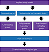
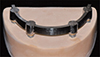
The overall study procedure is described in Figure 1. The edentulous study model with 4 implants (USII, Osstem, Seoul, Korea) placed in the canine and 2nd premolar areas was prepared. Four prosthetic implant abutments (Freefrom ST, Osstem, Seoul, Korea) were connected to each implant. In this study model, 12 implant-supported bar frameworks were fabricated using the different systems (CA, FHA, and PSA, n = 4 in each group) (Fig. 2). In the CA group, wax framework patterns were made and subsequently cast using a non-precious metal alloy (4-all, Ivoclar Vivadent, Schaan, Liechtenstein). In the FHA and PSA groups, the study model was scanned using a desktop scanner (Ceramill Map 400, Amann Girrbach, Koblach, Austria) and the bar framework was designed using dental software (Ceramill Mind, Amann Girrbach). In the FHA group, the metal frameworks were processed using solid metal blocks (M3, Medipion, Daejeon, Korea), whereas in the PSA group, they were processed using soft metal blocks (Ceramill Sintron, Amann Girrbach). Subsequently, the frameworks were sintered in an argon gas atmosphere at 1300℃ in a sintering furnace (Ceramill Argotherm, Amann Girrbach). All the fabrication procedures for each group were performed according to the manufacturer's instructions.
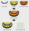
Marginal discrepancy was evaluated using the triple-scan protocol (Fig. 3).24 The first scan was of the study model alone, the second was of the fabricated frameworks alone, and the third was of the framework placed on the abutments with the fixation at the right premolar part. The framework was fixed by one terminal abutment to expose the discrepancies of the opposite terminal abutment. All digitization was completed using a 3D scanner (Breuckmann SmartScan, AICON 3D Systems GmbH, Braunschweig, Germany) with a homogenous measuring-point distance of 5 µm, according to the manufacturer's instructions. The scan datasets were exported in surface tessellation language format to an image analysis software (Geomagic DesignX, 3D Systems, Rock Hill, SC, USA), where the first and second scan images were superimposed to the third scan image using an area-designated best-fit algorithm. After the superimposition process, the third scan image was deleted. The cross-sectional images were obtained at the left premolar abutment bucco-lingually and mesio-distally. Absolute marginal discrepancy (AMD) values, the distance between the most external points of the crown margin and the abutment finish line, were measured at the margins of the buccal, lingual, mesial, and distal regions. The triple-scan protocol and misfit measurements were performed by one trained investigator.
The AMD values were compared among the groups using the Kruskal-Wallis test, with a post hoc Mann-Whitney U test and Bonferroni correction. The significance level was set at .05.

RESULTS

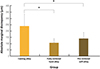
Table 1 lists the AMD values at each measurement point in the three groups. The FHA group showed the lowest mean discrepancy, followed by the PSA and CA groups (Fig. 4). The FHA and PSA groups showed no significant difference; however, the CA group was significantly different from the other groups (P<.001). The standard deviation of the data from the FHA and PSA groups was lower than that of the CA group.

DISCUSSION

This study was designed to investigate the marginal fit of the full-arch framework fabricated using the soft-metal milling method. The results from the PSA group were comparable to those from the FHA group and both groups presented a smaller discrepancy in marginal fit than the CA group; thus, the null hypothesis was rejected. The results of this study corresponded well with those found in an earlier marginal fit study, in which the milling method showed higher framework accuracy than the casting method.2526 Meanwhile, Part et al.14 reported that single restorations made using soft alloy had a higher fit accuracy than those made using hard alloy. This finding conflicts with the results of the present investigation, perhaps because the size of the tested frameworks differed among the studies.18 Therefore, it may be that the effect of PSA use differs according to the framework size.
The triple-scan protocol was utilized in this study to measure the AMD. Digital measurement tools visualize restoration misfits 3-dimensionally, enabling further geometric analyses, such as color-coded discrepancy distribution and volume calculation of specific regions.2327 For this reason, further experiments using various 3-dimensional analyses are recommended. Clinical studies on the fit accuracy of the full-arch frameworks fabricated using PSA are also needed to confirm the results of the present study.

CONCLUSION

With the limitations of this in vitro pilot study, the full-arch frameworks fabricated using soft-alloy milling exhibited a marginal accuracy that was equivalent to those fabricated using hard-alloy milling. Soft-alloy milling is an alternative to hard alloy milling and casting.

Figures and Tables

Fig. 1

Workflow of study.

jap-10-128-g001
Fig. 2

Study model with implant-supported bar framework.

jap-10-128-g002
Fig. 3

Triple-scan technique of the marginal fit analysis. The framework image (a) and model image (b) are aligned with the framework/model image (c) to superimpose the images (d). The cross-sectional image (f) is obtained by virtual sectioning of the combined model (e).

jap-10-128-g003
Fig. 4

Overall absolute marginal discrepancy in the three groups. *Significant difference.

jap-10-128-g004
Table 1

Mean and standard deviation of absolute marginal discrepancy at the various measurement points

jap-10-128-i001
Buccal Lingual Mesial Distal
Casting alloy 366.3 ± 67.7 137.7 ± 34.2 239.5 ± 20.5 216.2 ± 35.3
Fully-sintered hard alloy 144.4 ± 21.0 114.3 ± 14.9 60.1 ± 3.3 107.3 ± 19.6
Pre-sintered soft alloy 105.8 ± 26.7 189.6 ± 57.9 112.2 ± 10.9 155.5 ± 42.6

Notes

This research was supported by Basic Science Research Program through the National Research Foundation of Korea (NRF) funded by the Ministry of Science, Information and Communication Technologies (ICT) & Future Planning (NRF-2014R1A1A1006073).

References

1. Holmes JR, Bayne SC, Holland GA, Sulik WD. Considerations in measurement of marginal fit. J Prosthet Dent. 1989; 62:405–408.
crossref
2. Henry PJ. An alternative method for the production of accurate casts and occlusal records in osseointegrated implant rehabilitation. J Prosthet Dent. 1987; 58:694–697.
crossref
3. Aparicio C. A new method for achieving passive fit of an interim restoration supported by Brånemark implants: a technical note. Int J Oral Maxillofac Implants. 1995; 10:614–618.
4. Contrepois M, Soenen A, Bartala M, Laviole O. Marginal adaptation of ceramic crowns: a systematic review. J Prosthet Dent. 2013; 110:447–454.
crossref
5. Pimenta MA, Frasca LC, Lopes R, Rivaldo E. Evaluation of marginal and internal fit of ceramic and metallic crown copings using x-ray microtomography (micro-CT) technology. J Prosthet Dent. 2015; 114:223–228.
crossref
6. Ziebert GJ, Hurtado A, Glapa C, Schiffleger BE. Accuracy of one-piece castings, preceramic and postceramic soldering. J Prosthet Dent. 1986; 55:312–317.
crossref
7. Sarfati E, Harter JC. Comparative accuracy of fixed partial dentures made as one-piece castings or joined by solder. Int J Prosthodont. 1992; 5:377–383.
8. Rinke S, Rasing H, Gersdorff N, Buergers R, Roediger M. Implant-supported overdentures with different bar designs: A retrospective evaluation after 5–19 years of clinical function. J Adv Prosthodont. 2015; 7:338–343.
crossref
9. Eisenmann E, Mokabberi A, Walter MH, Freesmeyer WB. Improving the fit of implant-supported superstructures using the spark erosion technique. Int J Oral Maxillofac Implants. 2004; 19:810–818.
10. Ortorp A, Jemt T, Bäck T, Jälevik T. Comparisons of precision of fit between cast and CNC-milled titanium implant frameworks for the edentulous mandible. Int J Prosthodont. 2003; 16:194–200.
11. Paniz G, Stellini E, Meneghello R, Cerardi A, Gobbato EA, Bressan E. The precision of fit of cast and milled full-arch implant-supported restorations. Int J Oral Maxillofac Implants. 2013; 28:687–693.
crossref
12. Agustín-Panadero R, Peñarrocha-Oltra D, Gomar-Vercher S, Ferreiroa A, Peñarrocha-Diago M. Implant-supported overdenture manufactured using CAD/CAM techniques to achieve horizontal path insertion between the primary and secondary structure: A clinical case report. J Adv Prosthodont. 2015; 7:264–270.
crossref
13. Lee DH, Lee BJ, Kim SH, Lee KB. Shear bond strength of porcelain to a new millable alloy and a conventional castable alloy. J Prosthet Dent. 2015; 113:329–335.
crossref
14. Park JK, Kim HY, Kim WC, Kim JH. Evaluation of the fit of metal ceramic restorations fabricated with a pre-sintered soft alloy. J Prosthet Dent. 2016; 116:909–915.
crossref
15. Kim EH, Lee DH, Kwon SM, Kwon TY. A microcomputed tomography evaluation of the marginal fit of cobalt-chromium alloy copings fabricated by new manufacturing techniques and alloy systems. J Prosthet Dent. 2017; 117:393–399.
crossref
16. Kelvin Khng KY, Ettinger RL, Armstrong SR, Lindquist T, Gratton DG, Qian F. In vitro evaluation of the marginal integrity of CAD/CAM interim crowns. J Prosthet Dent. 2016; 115:617–623.
17. Shamseddine L, Mortada R, Rifai K, Chidiac JJ. Fit of pressed crowns fabricated from two CAD-CAM wax pattern process plans: A comparative in vitro study. J Prosthet Dent. 2017; 118:49–54.
18. Alfaro DP, Ruse ND, Carvalho RM, Wyatt CC. Assessment of the internal fit of lithium disilicate crowns using micro-CT. J Prosthodont. 2015; 24:381–386.
crossref
19. Nakamura T, Dei N, Kojima T, Wakabayashi K. Marginal and internal fit of Cerec 3 CAD/CAM all-ceramic crowns. Int J Prosthodont. 2003; 16:244–248.
20. Lee KB, Park CW, Kim KH, Kwon TY. Marginal and internal fit of all-ceramic crowns fabricated with two different CAD/CAM systems. Dent Mater J. 2008; 27:422–426.
crossref
21. De Santis R, Gloria A, Russo T, D'Amora U, Varriale A, Veltri M, Balleri P, Mollica F, Riccitiello F, Ambrosio L. Reverse engineering of mandible and prosthetic framework: Effect of titanium implants in conjunction with titanium milled full arch bridge prostheses on the biomechanics of the mandible. J Biomech. 2014; 47:3825–3829.
crossref
22. Bae SY, Park JY, Jeong ID, Kim HY, Kim JH, Kim WC. Three-dimensional analysis of marginal and internal fit of copings fabricated with polyetherketoneketone (PEKK) and zirconia. J Prosthodont Res. 2017; 61:106–112.
crossref
23. Lee DH. Digital approach to assessing the 3-dimensional misfit of fixed dental prostheses. J Prosthet Dent. 2016; 116:836–839.
crossref
24. Holst S, Karl M, Wichmann M, Matta RE. A new triple-scan protocol for 3D fit assessment of dental restorations. Quintessence Int. 2011; 42:651–657.
25. Real-Voltas F, Romano-Cardozo E, Figueras-Alvarez O, Brufau-de Barbera M, Cabratosa-Termes J. Comparison of the marginal fit of cobalt-chromium metal-ceramic crowns fabricated by CAD/CAM techniques and conventional methods at three production stages. Int J Prosthodont. 2017; 30:304–305.
crossref
26. Zhou Y, Li Y, Ma X, Huang Y, Wang J. Role of span length in the adaptation of implant-supported cobalt chromium frameworks fabricated by three techniques. J Adv Prosthodont. 2017; 9:124–129.
crossref
27. Park JM, Hämmerle CHF, Benic GI. Digital technique for in vivo assessment of internal and marginal fit of fixed dental prostheses. J Prosthet Dent. 2017; 118:452–454.
TOOLS
ORCID iDs

Sung-Am Cho
https://orcid.org/0000-0002-8315-7833

Kyu-Bok Lee
https://orcid.org/0000-0002-1838-7229

Jin-Hyun Cho
https://orcid.org/0000-0002-2453-9372

Du-Hyeong Lee
https://orcid.org/0000-0003-2803-7457

Similar articles