

 PDF
PDF ePub
ePub Citation
 Print
Print


INTRODUCTION
Poly methyl methacrylate (PMMA) has been used as a denture base material because of its desirable characteristics such as ease of processing, chemical stability, durability, moderate cost, light weight, color matching, and stability.1 Despite of these favorable properties, the acrylic denture base material has not fulfilled all the intended mechanical properties. Hence, there is a need for improvement of the mechanical properties of poly methyl methacrylate-based prostheses, and different methods have been introduced to improve its mechanical properties such as chemical modification by addition of rubber graft copolymers like butadiene styrene and reinforcement of poly methyl methacrylate by the addition of fibers, fillers, and materials like ultra-high modulus polyethylene.23456789101112 Glass fiber,5 carbon fibers, aramid fiber, and nylon fibers67 are some of the fibers used to strengthen acrylic resins.
Incorporation of metal wires is another approach for reinforcement of PMMA.89 Poor adhesion between the resin matrix and the wire is a problem with this technique. Therefore, metallic fillers have been considered by different researchers for the improvement of physical and mechanical properties of acrylic resins. Sehajpal and Sood11 evaluated the effect of the addition of various amounts of powdered silver, copper, and aluminum on some physical properties of PMMA. They concluded that metal fillers can increase thermal conductivity and compressive strength of poly methyl methacrylate.10 Asar et al.12 added various amounts of metal oxides including Al2O3, TiO2, and ZrO2 to heat-cured acrylic resin and found that modification of acrylic resin with certain amounts of metal oxides led to a significant increase in mechanical properties and a decrease in water sorption and solubility. They concluded that metal oxides may be useful to prevent denture fractures resulting from fluids of the oral environment.12
The introduction of nano-scale materials offers new aspects for improvement of the mechanical properties of polymers. Interest in nanostructures has been driven by their high surface area to volume ratio, which increases the interfacial interaction and the specific characteristics of them including new biological, physical, and chemical properties.13 Recently nanotubes have been considered for reinforcement of PMMA denture base resins. Carbon nanotubes,1415 zirconia nanotubes,16 titania nanotubes, and halloysite nanotubes17 are some nanotubes which are employed for reinforcement of denture base resins. Wang et al.15 reinforced poly methyl methacrylate denture base material using 0.5 wt%, 1 wt%, and 2 wt% of Multiwalled carbon nanotubes. Their results indicated improved flexural strength by addition of 0.5% and 1% MWCNTs but not for the 2% MWCNTs reinforced group due to the poor dispersion of CNTs fillers throughout the matrix. Yu et al.16 modified denture base PMMA with ZrO2 nanotubes and ZrO2 nanoparticles and concluded that ZrO2 nanotubes had a better reinforcement effect than ZrO2 nanoparticles. Abdallah17 evaluated the mechanical properties of PMMA resin reinforced with halloysite nanotubes and stated that addition of low percentages of HNTs into resin matrix increases the hardness values significantly, but the flexural strength and Young's modulus did not improve significantly.
TiO2 particles have been used as filler added to PMMA because of their effect on the mechanical properties of acrylic resins1819 and their antibacterial properties.2021 Furthermore, TiO2 particles are preferred in dentistry due to their desirable color and excellent biocompatibility, as well as durability.22 In 1998, investigators discovered a tubular shape for hydrothermally synthesized titanium oxide nanostructures.2324 Such linear nanotubes can exhibit increased surface area compared with nanoparticles.25 Among the various nanotubes, the TiO2 nanotube (TNT, n-TiO2) has been considered due to its high-specific surface area, photocatalytic property, and ionexchangeability.26 The tubular form of titania has a surface area of 250 m2/g, which is about five times that of the nanoparticle.27 The high surface area results from the internal and external surfaces and the surfaces between the layers of the walls.23 Recently, titania nanotubes have been used for biological applications including drug delivery, bio-scaffolds for cell cultures, titanium-based implants and reinforcement of composites.28293031 Michele et al.28 reported increased Young's modulus and strength of reinforced polystyrene with functionalized titania nanotubes. Porras et al.29 also found improved mechanical properties of poly (ethylene oxide)/chitosan polymer composite modified with hydrothermal synthesized titanate nanotubes. Enhancement of mechanical properties of resin based cement reinforced with titania nanotubes was also reported by Khaled et al.32 In another study, Dafar et al.31 investigated the mechanical properties of flowable dental composites reinforced with titanium dioxide nanotubes and discovered improvement in fracture toughness and Young's modulus in the experimented composite.
To the best of our knowledge, the literature lacks any studies that used TiO2 nanotubes to reinforce denture base polymers, although few studies have employed TiO2 nanoparticles as added fillers to enhance mechanical properties of PMMA-based prostheses and have reported controversial results.193334 Since no previous study has evaluated the addition of titanium oxide nanotubes to PMMA denture base, this preliminary multi-phase study was conducted. It aimed to prepare titania nanotubes, evaluate their properties, and assess the reinforcement potential of hydrothermally synthesized titanium oxide nanotubes added to a commercial PMMA denture base on microhardness, fracture toughness, and flexural strength of resin.
The null hypothesis of this study was lack of effect of addition of Titanium Dioxide Nanotubes on mechanical properties of the acrylic resin denture base material.
Go to : 
MATERIALS AND METHODS
TiO2 nanotubes were prepared using an alkaline hydrothermal process.35 The precursor was a commercial TiO2 powder (SkySpring Nanomaterials Inc., Houston, TX, USA) with a crystalline structure of ca.99.5% anatase and particle size of 10 – 30 nm. The procedure of nanotube synthesizing was started by treating 1.14 g of nanoparticle powder with 40 – 45 mL of 10 N NaOH solution. Afterwards, the suspension was sealed in a Teflon-lined autoclave at 150℃ for 48 hours. Subsequent to the heating process, the resultant precipitates were washed with deionized water and HCl aqueous solution (1 M) until the pH of the solution became about 7. Finally, an oven was used to dry the powders at 80℃ for 3 hours to synthetize nanotubes.
The crystalline phase of the synthesized titania nanotubes was investigated by powder X-ray diffraction (XRD, INEL, Equinox 3000) using Cu Kα radiation (40 kV, 30 mA). Data were compiled at a scattering angle of 2θ from 2° to 80° at a step size of 0.032° and 1°/s scan speed.
Fourier transform infrared spectroscopy (FTIR, Nicolet iS50, Madison, WI, USA) using the KBr pellet method was used to assess the n-TiO2 tubes by adding 0.2 g anhydrous KBR to 2 mg of titania nanotubes. Information was obtained at a resolution of 4 cm−1 and wavenumbers ranging between 400 and 4000 cm−1.
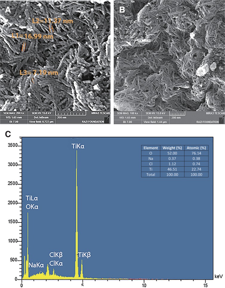
The morphology and elemental analysis of n-TiO2 was performed using a field emission scanning electron microscope (FESEM, ZEISS Co., Germany) at an accelerating voltage of 15 kV equipped with an energy dispersive X-ray (EDX) spectrometer (AURIGA, ZEISS).
In order to perform fracture toughness (KIC), flexural strength, and Vickers microhardness tests, 45 PMMA specimens were fabricated for each phase: the specimens in each project were assigned to three groups (n = 15) according to the percentage of titania nanotubes including TNT 0% (control), TNT 2.5% and TNT 5% by weight. Therefore, 135 specimens (45 specimens for each sub-project) were prepared according to ISO 20795-1:2013 standard and instructions of the manufacturer.36 The size and shape of the specimens differed for each of the 3 sub-projects (they were bar-shaped with 2 sizes for flexural strength and toughness tests, while they were discoid for microhardness tests, detailed below). Wax templates were used to shape the specimens during their heating / fabrication procedure (Fig. 1). There were 3 groups: specimens made of PMMA only (control, n = 45), those made of PMMA + 2.5 wt% TNT (n = 45), and those made of PMMA + 5 wt% TNT (n = 45). To create the experimental groups, the powder of conventional acrylic resin was blended with 2.5 wt% and 5 wt% of synthesized titanium dioxide nanotubes as follows. The TiO2 nanotubes and the powder of a conventional denture base resin (mega CRYL HOT, megadental GmbH, Büdingen, Germany) weighted in an analytical balance (Sartorius, Goettingen, Germany) precisely and divided into equal parts. Then each part of n-TiO2 powder was mixed manually with powder of acrylic resin for 3 minutes, subsequently they mixed with an amalgamator device (Ultramat 2, SDI, Australia) to obtain an adequate particle distribution.
2.3 gr of modified and unmodified powder with 1 mL liquid (monomer) of denture base resin was mixed according the manufacturer's instructions and packed in a dough stage in the dental stone molds (Hydrocal dental stone, Moldano, Bayer Lerekusen, Germany). The two portions of the flask were closed tightly together and placed under the hydraulic press at 40,000 N, and the pressure was slowly applied on the flask so that the dough evenly flowed throughout the mold space. The pressure was then released, the flask was opened, and the excess material was removed with a sharp scalpel. A second trail closure also was performed. Finally, the two portions of the flask were closed and left under pressure (20 bars) for 5 minutes. Subsequently the flask was clamped and maintained under low pressure for 30 minutes, and then transferred to a water bath at room temperature. The temperature was slowly elevated to 73 ± 1℃ for 90 minutes and then raised to the boiling point at 100℃ for 30 minutes. Before opening, the flasks were subjected to bench cooling. All specimens were finished and polished to obtain a smooth and glossy surface.
Before testing the flexural strength and fracture toughness, the specimens were incubated by water storage at 37℃ according to the ISO standard. The durations of water storage were 48 hours and 120 hours for flexural strength and fracture toughness tests, respectively. No water storage was done before Vickers microhardness test.
Bar-shaped specimens with dimensions of 64 × 10 × 3.5 mm3 as stated in ISO 20795-136 were fabricated. There were 45 specimens: 15 controls, 15 PMMA + 2.5 wt% TNT, and 15 PMMA + 5 wt% TNT.
After 2 days of water storage, the specimens were subjected to a three-point bending flexural strength test using a universal testing machine with a 50.0-kg load cell (Bongshin, Bongshin Loadcell Co., Ltd., Seoul, Korea) at a crosshead speed of 5 mm/min. The supporting wedges were placed 50 mm apart from each other, and the specimens centrally located on the device in a way that the loading wedge was applied on the center of the specimens. The flexural strength was calculated in mega Pascal (MPa) from the following formula: S = 3FL/2bd2 where S = flexural strength, F = load at fracture (in N), L= distance between supports (in mm), b = width and d = thickness of sample (in mm).
A total of 45 bar-shaped specimens with dimensions determined according to ISO 20795-1:201336 (length 39 mm, height 8 mm, and width 4 mm) were prepared. There were 45 specimens: 15 controls, 15 PMMA + 2.5 wt% TNT, and 15 PMMA + 5 wt% TNT.
An initial notch with a depth of 3-mm was then cut along the centerline of the specimens, using a 0.5-mm thick diamond blade. Subsequently, another notch of almost 400 µm depth was cut in the middle part of the initial notch. The depth of this notch was checked using a stereomicroscope (EZ4D; Leica Microsystems Ltd., Singapore).
After 5 days of water storage, the specimens were loaded in a 3-point bending configuration (i.e., single edge-notched bend test (SENB)) using a universal testing machine with a 50.0-kg load cell (Bongshin, Bongshin Loadcell Co., Ltd., Seoul, Korea). The crosshead speed of the loading plunger was 1 mm/min and the specimen test span was 32 mm. Eventually, the critical stress intensity factor (KIC) was calculated from the obtained values, as following equation:36
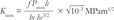
where Pmax is the peak load at fracture (in N), lt is length (in mm), bt is width (in mm), ht is height (in mm), and ƒ is geometrical function, which is calculated as follows:
Fifteen acrylic discs (10 mm in diameter and 5 mm high) from each of the aforementioned 3 material compositions were prepared and hand-polished using silicon carbide papers up to 2000 grit to produce a glossy flat surface. After polishing, the surface microhardness (SMH) of the specimens were measured using a microhardness tester (V-Test II, Baresiss, Germany) with a Vickers diamond indenter (0.8 mm in diameter), which was attached to a digital scale from 0 to 100 units under a load of 50 g force for 10 s.3738 Three measurements of Vickers pyramid numbers (HV) in each specimen were recorded at different sites along with the diametrical line. Ultimately, the average of three readings was calculated as the main finding.
After the confirmation of the normality by the Kolmogorov-Smirnov test, the data were analyzed using one-way analysis of variance (ANOVA), followed by a post-hoc Tukey's test. The correlations between the concentrations of TiO2 nanotubes with each of the 3 physicomechanical properties of PMMA resin were evaluated using a Pearson correlation coefficient. The significance level was set at 5%. The statistical analyses were performed using SPSS 23.0 (IBM, Armonk, NY, USA).
Go to :
RESULTS
The morphology of n-TiO2 was examined by FESEM after calcination. FESEM images of n-TiO2 exhibited the presence of elongated tubular structures with outer diameters of 11.85 nm (Fig. 2A, Fig. 2B). On the basis of the analyses using EDX, Ti (46.51 wt%) and O (52.00 wt%) were the predominant elements with the most abundance among other elements in synthesized powder (Fig. 2C). A low percentage of chloride (1.12 wt%) and trace amount of Na (0.37 wt%) were also detected.
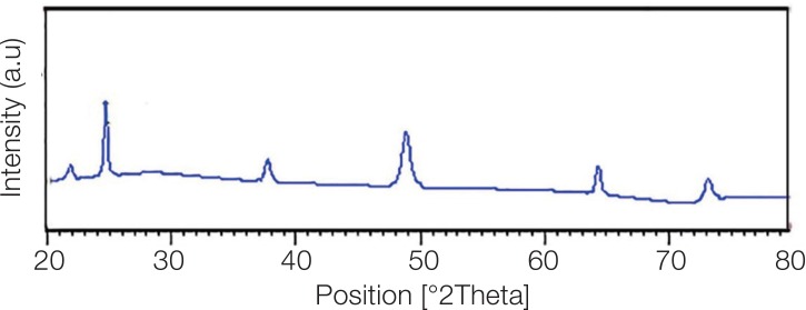
According to International Center for Diffraction Data (ICDD PDF No. 89-4921), a sharp diffraction peak at 24.56° in addition to other peaks at 37.61°, 43.43°, 46.76° and 53.7° and 64.76° (Fig. 3) represent the anatase crystal phase of TiO2.3139
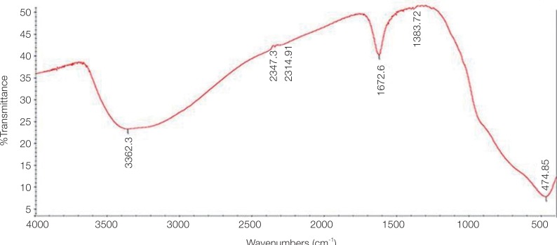
The FTIR spectra of n-TiO2 tubes in Fig. 4 exhibit a broad peak at 3362 cm−1 , which is attributed to O-H bonds associated with hydroxyl groups, and the peak at 474 cm−1 is related to the O-Ti-O bonding of the anatase phase of titania.4041 The sharp peak at 1672 cm−1 is due to the presence of oxygen-containing groups, i.e., C-O, COO, and C=O and hydroxyl groups.4142
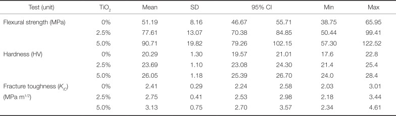
Mechanical properties of reinforced / control PMMA specimens are presented in Table 1 and Fig. 5. The Pearson coefficient indicated moderate to very strong positive associations between the concentration of TNT with PMMA's flexural strength (r = 0.750, P < .001), microhardness (r = 0.894, P < .001), and fracture toughness (r = 0.501, P = .001). ANOVA showed significant differences among the three material compositions (PMMA only vs 2.5% or 5% TNT), regarding their fracture toughness (p = .003), flexural strength (P < .001), and microhardness (P < .001, Table 1, Fig. 5). All Tukey pairwise comparisons among three flexural strength groups were significant (P < .05). All pairwise comparisons among three Vickers hardness groups were significant as well (P < .001). When comparing three fracture toughness averages, only the comparison of the groups “control vs. 5 wt%” became significant (P = .002) and the other two comparisons were not significant (P > 0.1).
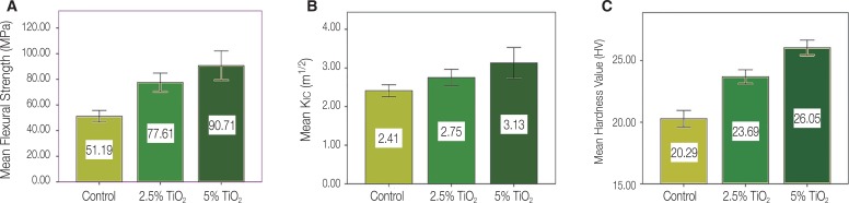 | Fig. 5(A) Mechanical properties of non-modified PPMA, (B) mechanical properties of PPMA modified with 2.5 wt% n-TiO2 tubes, (C) mechanical properties of PPMA modified with 5 wt% n-TiO2 tubes.
|
Table 1
Mechanical properties of control and reinforced PMMA with n-TiO2 tubes
![]()
The values for flexural strength (FS) indicated that the acrylic resin reinforced with 5 wt% n-TiO2 tubes exhibited the highest values for FS. The obtained flexural strength values of both 2.5 and 5 wt% groups were significantly higher than those exhibited by the un-modified group (P < .001). The FS values of 5 wt% were also superior to those modified with 2.5 wt% n-TiO2 tubes (P = .047, Fig. 5, Table 1).
The addition of n-TiO2 significantly influenced the surface hardness of PMMA; the obtained mean Vickers hardness values of both 2.5 and 5 wt% modified groups were significantly higher than the control group's (P < 0.001). Moreover, the statistical analysis showed significant differences between the hardness values of 2.5 and 5 wt% modified groups (P < .001, Fig. 5, Table 1).
The fracture toughness (KIC) values showed that the acrylic resin reinforced with 5 wt% n-TiO2 tubes exhibited significantly higher KIC values compared to the control group (P = .002). However, the KIC values of specimens reinforced with 2.5 wt% were not significantly higher than those of the control group; they were also not significantly lower than the 5 wt% modified one (P > .05, Fig. 5, Table 1).
Go to :
DISCUSSION
In the present study, denture base resins were modified with 2.5% and 5% by weight of synthesized titania nanotubes, and the mechanical properties of the modified and control groups were measured for critical stress intensity factor (KIC), flexural strength, and surface microhardnes. Based on the results, the null hypothesis of this study (i.e., lack of effect of the addition of TiO2 nanotubes on mechanical properties of denture acrylic resin base) was rejected.
SEM was used to observe the morphology of synthesized nanotubes. The obtained SEM images in the current study obviously showed the tubular shapes of synthesized titanate nanotubes. It is claimed that after treating with NaOH, some Ti-O-Ti bonds break down and form the titanate sheets. Under autoclaving and high concentration of NaOH, the produced sheets exfoliate into nanosheets, and then they can scroll or fold into a tubular morphology.23 Different studies have used driving forces for folding these nanosheets.2343 It is likely that the inequality in the width of different layers of multi-walled nanosheets creates a tendency for layers to move within the walls in order to reduce the excess surface energy. Nanotubes are long cylinders with a hollow cavity in the center. The walls of titanate nanotubes are always multilayered and the number of their layers varies from 2 to 10.23 The tubular form and multilayered structure of titania increase its surface area-to-volume ratio, which improves the interfacial interaction and their specific characteristics. In the current study, TiO2 nanotubes were synthesized using an alkaline hydrothermal process. This method was first reported by Kasuga et al.24 as a simple method for preparation of TiO2 nanotubes.
Some studies evaluated the strength of denture base resins using impact tests, which are influenced by the geometry of specimens and loading conditions and therefore may not be the best test for predicting the clinical function of the material.44 Fracture toughness (FT) is considered one of the most important tests for the assessment of mechanical properties of dental polymers.4546 FT determines the maximum energy a material can absorb through crack propagation prior to its failure; it is described by a critical intensity factor (KIC), and processes that can absorb energy at the tip of a crack might improve fracture toughness.4748 FT provides information about crack propagation, which is the main reason of fracture in brittle materials.4748 Various test methods were introduced for the measurement of this property. In this study, we used single-edge-notched (SEN) test as described in ISO36 for assessment of fracture toughness because of its validity, reliability, and simplicity of specimen preparation.444649 In the current study, the specimens reinforced with both 2.5 and 5 wt% titanium nanotubes exhibited greater KIC values than the control group, although only the difference between KIC values of the 5 wt% modified group and the control was statistically significant. For the PMMA reinforced with nanotubes used in this study, absorption of energy at the tip of cracks can be achieved by flexible TNTs, which can absorb energy of forces applied from different directions. The continuous fibrous-like structure of these nanotubes may facilitate the release of the stresses produced in modified denture base resin and enhance FT. Improvement of the fracture toughness property of reinforced acrylic resin polymers can also be associated with the formation of fibrils containing titanium nanotubes, which may hinder crack propagation and increase the stability of the resin structure.29 Pei Feng et al.50 described that when the tip of a crack encounters TiO2 nanotubes, the crack mainly propagates around the nanotubes; this results in additional absorption of fracture energy and improvement of fracture toughness.50 Khaled et al.32 determined the use of the fiber ‘pull out’ phenomenon as the main reason for increased KIC of PMMA-based cements reinforced with TiO2-SrO nanotubes. They reported that this addition reduces the sites of local stress concentration, hence increasing the KIC of the composite.32
The Vickers microhardness measurements obtained in this study in both reinforced groups were significantly higher than that of the control group. As mentioned earlier, the aligned nanotubes may help maintain the stability of reinforced polymer. Porras et al.29 evaluated the hardness and Young's modulus of a composite reinforced with titanate nanotubes. They concluded that well-dispersed nanotubes can improve mechanical properties. They also suggested that in non-agglomerated nanotubes, better mechanical properties of reinforced polymer composites can be achieved with longer nanotubes.29 The high length of synthesized nanotubes used in this study might improve mechanical properties of modified denture base resins. The effect of the length of nanotubes on the mechanical properties was also reported by Arash et al.,51 who observed better mechanical characteristics with nanotubes having higher interfacial regions.
Flexural characteristics of a reinforced composite are affected mostly by interactions between the added fillers and matrix of the composite. In a composite structure, the interfacial adhesion of fillers and matrix indicates how interfacial shear stress transfers from the filler to the matrix.52 The high contact area between reinforced fillers and PMMA resin in acrylates modified with titanium nanotubes increases the load transfer.52 Moreover, the tubular, open-ended structure of nanotubes might cause the methyl methacrylate monomer to pass in nanotubes via capillary action and polymerize there.32 This might increase mechanical interlocking by the engagement of polymers with the internal surface of tubes. Hence, incorporated tubes and polymer molecules might create an improved matrix with a greater extent of cross-linking, leading to increased load transfer within the composites.32 This might justify the superior FS values of nanotube-reinforced acrylic resins.32
This pilot study was limited by some factors. Since there was no previous study in this regard, selecting the percentages of nanotubes and their properties were not optimized. Therefore, due to difficulties in production of titania nanotubes, we were limited to evaluate only the effect of 0%, 2.5%, and 5% concentrations of TiO2 nanotubes on mechanical properties of denture base resins. Further studies are needed to improve TNT's dispersion throughout the polymer matrix. To promote better interfacial interactions between denture base polymer matrix and titania nanotubes, surface modification of titanium oxide nanotubes is suggested. Future studies should be conducted to identify the optimum concentration of titania nanotubes for reaching various physicomechanical and biological properties (e.g., antibacterial effects and biocompatibility) of denture base resins.
Go to :
CONCLUSION
The PMMA denture base resin was successfully reinforced with hydrothermally synthesized n-TiO2 tubes. The fracture toughness, flexural strength, and microhardness of acrylic resin were improved with the addition of nanotubes. These improvements were a linear function of the concentration of added titania nanotubes, and this phenomenon was more vivid in terms of microhardness, followed by flexural strength. Thus, this preliminary study demonstrated for the first time a promising new agent for reinforcing denture base resin materials.
Go to :
 XML Download