

 PDF
PDF ePub
ePub Citation
 Print
Print


Background
An adult mouse normally releases 8-10 oocytes in each cycle [1] which can be increased to 40-50 using different protocols of superovulation [2]. Oocytes obtained from superovulation can be fertilized by in either in vivo or in vitro conditions. A majority of embryos die during early stages of pregnancy and only a few of them can survive until the middle or late stages of pregnancy [3,4]. In superovulation using PMSG and hCG, a mean number of 9.9 fetuses was produced on days 12 to 15th of pregnancy in 10 mature mice [5]. In a study on BALB/c mice superovulated by anti-inhibin serum, the oocytes released from each mouse were ranging from 11 to 30 with a mean (±sem) number of 18±2 oocytes [6]. This paper is to report a case of super pregnancy in a BALB/c mouse pregnant with 30 pups following induction of superovulation using a PMSG-hCG protocol.
Case Presentation
This study has been approved by the Iranian laboratory animal ethics framework under the supervision of the Iranian Society for the Prevention of Cruelty to Animals and Shiraz University Research Council. The present report describes a case of super pregnancy observed during a research project aiming to examine different protocols of superovulation including a new biological source of FSH in BULB/C mice. The female mice of the BALB/C strain were taken from the Center of Shiraz University of Medical Sciences Laboratory Animals. As a routine program, the colony is strictly maintained by inbreeding. Before puberty at the age of 21 days, male mice were separated from female. At 6–7 weeks of age, the female mice were used for this study. They were held in cages (27×21×14 cm) housed in a room with suitable conditions (22℃, 40 to 60 percent humidity, light cycle of 12 h on and 12 h off) and fed with standard pelleted ration (Laboratory animal feed, Javaneh Khorasan, Iran). At 13:00, they received 5 IU of PMSG (Pregnecol PMSG Bioniche, Canada) intraperitoneally for superovulation by an insulin syringe followed by intraperitoneally administration of 5 IU of hCG (Daroupakhsh, Iran) 48 hours later [7,8]. Immediately after hCG administration, the female mice were placed with males at a ratio of 1 to 1 for 24 hours. Male mice ranged from 8–12 weeks in age were maintained in individual cages. We used natural mating for production of embryos and pregnancy rather than in vitro fertilization. On day 14th after mating, animals were killed by cervical dislocation and then ovarian samples were fixed in buffered formalin 10% for histopathological examination and corpora lutea (CLs) counting [9]. In addition, the appearance and contents of uteri were examined for pregnancy and number of fetuses was also counted.
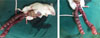
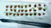
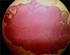
Histologically, the mean (±sem) number of CLs was 23.6±3.97 in all 10 mice (Figure 1). Further, the mean (±sem) number of fetuses obtained was 5.4±3.18. Unexpectedly, we observed a high number of fetuses (30) in one superovulated mouse. Out of 30 fourteen-day fetuses, 28 fetuses were appeared as healthy and the remaining two were observed as mummified (Figure 2,3).
Discussion
This paper reports the occurrence of a super pregnancy in a 6 to 7 weeks of age BALB/c mouse using PMSG-hCG superovulation protocol. To our knowledge, this is the first report of the capability of the BALB/c mice uterus to implant 30 fetuses, 15 fetuses per horn, by day 14 of pregnancy. The ovarian response to superovulation protocols varies depending on many factors including the strain, age, stage of the estrous cycle, nutrition, dose of gonadotrophin, timing of administration and male performance [2,10,11]. Ozgunen et al. (2001) using hMG reported the recovery of 60 mature oocytes from superovulated outbred BLAB/C mice. In random-bred BALB/C mice, the highest reported number of recovered fetuses using PMSG was 24 fetuses on day 13th of pregnancy. Similarly, in golden hamsters, 24 fetuses (9 in one uterus and 15 fetuses in the other uterus) were collected on the day 15th of pregnancy24. The occurrence of super pregnancy in the inbred BALB/C here could be interpreted by the facts that there exists inherent variations in size of the ovarian follicle reserve [12,13] and ovarian sensitivity to FSH from one individual to another. Furthermore, the amount of uterine capacity is one well accepted [14,15,16] factor influencing the number of fetuses in different species including rodents [9,17]. The present observation reports the availability of the uterine capacity for implantation and development of fetuses by day 14 of pregnancy in the inbred BALB/C.
 XML Download