Journal List > Lab Anim Res > v.33(3) > 1053897

Yun, Kim, Kwon, Kim, You, Kim, Che, and Kang: Evaluation of in vitro and in vivo genotoxicity of Angelica acutiloba in a standard battery of assays

Abstract

Among three representative species of Angelica found in Asian countries, including Korea, China, and Japan, Angelica acutiloba (AA) has been used as traditional herbal medicine with antitumor, anti-inflammatory, anti-obesity, and anti-diabetes activities. In this study, the potential genotoxicity and mutagenicity of the AA extract were examined in a battery of in vitro and in vivo tests (bacterial reverse mutation assay, in vitro chromosomal aberrations assay, and in vivo micronucleus assay) in accordance with the test guidelines for toxicity testing developed by the Organization for Economic Cooperation and Development. Upon testing in the bacterial mutation assay (Ames test) using five Salmonella typhimurium TA98, TA100, TA102, TA1535 and TA1537, no significant increase the number of revertant colonies in the metabolic activation system and non-activation system was noted in the AA extract groups. Also, in the chromosome aberration test, the AA extract did not cause chromosomal aberration with or without metabolic activation by S9 mix. A bone marrow micronucleus test of mice demonstrated that the incidence of micronucleated polychromatic erythrocytes in the AA extract groups (500, 1000 and 2000 mg/kg BW) was equivalent to that of the negative control group. Based on these results from a standard battery of assays, the AA extract was concluded to have no genotoxic at the proper dose.

Many species Angelica belonging to the family Umbelliferae has long been known as traditional herbal medicines that can be found throughout Asian countries, including Korea, China, and Japan. In particular, Danggui (root of Angelica) or ‘female ginseng’ is widely used as a typical therapeutic for treating gynecological disorders such as menoxenia, menstrual pain, anemia, and fatigue/low vitality [12345]. Despite similarities in shapes, Danggui has different origin species and scientific names according to geographical locations: Angelica sinensis from China (Chinese Danggui), A. acutiloba (AA) from Japan (Japanese Danggui), and of A. gigas from Korea (Korean Danggui) [6].
Among these three species, AA were reported to exhibit various pharmacological effects including antitumor [7], anti-inflammatory [891011], immunostimulatory [12], anti-complementary [13], and insecticidal [14]. And, the AA could be an effective ethnomedicine for treating diabetic complications and obesity since it has been reported to attenuate fat accumulation in high fat diet-induced obesity through the up-regulation of lipid metabolism [15] and to ameliorate insulin resistance induced by high-fructose diet in rats through the promotion of glucose homeostasis [4]. In addition, it is reported that the AA inhibited the release of histamine from mast cells and the production of pro-inflammatory cytokines, which contributed to the protection against allergic diseases and inflammatory diseases [16171819]. Additional pharmacological effects of the AA include the enhancement of collagen synthesis and suppression of matrix metalloproteinases, resulting in its use as an anti-wrinkle natural agent [1].
The use of medicinal plants as therapeutics, functional foods, or dietary supplements for health-related issues is increasing as self-medication for symptomatic relief because it is believed that these are natural and free from side effects compared with synthetic chemical drugs [2021]. However, concerns associated with the lack of quality control (plant origin and method of preparation) and potential risks of self-medication (treatment duration) have been raised [4]. Recently, we have identified that long-term ingestion of well-known medicinal herbs available in market can induce potential toxicity that may be hepatotoxicity or nephrotoxicity. In particular, Sophorae radix and vinegar-processed Genkwa flos have been found to have clastogenic and mutagenic potentials, respectively, as evidenced by the chromosome aberration assay and the Ames test [222324].
Along with the current use as an herbal medicine available on the market in Asian countries, many studies have reported various beneficial activities of the AA extract, but little information on risk and safety has been reported. Therefore, we carried out the battery of regulatory genotoxicity studies, including in vitro bacterial reverse gene mutation assay (Ames test), in vitro chromosome aberration assay, and in vivo micronucleus assay in mice, to assess the mutagenicity and clastogenicity of the AA extract according to test guidelines for toxicological studies developed by the Organization for Economic Cooperation and Development (OECD) and the Ministry of Food and Drug Safety (MFDS).

Materials and Methods

Test substance and animals

A hot water AA extract was provided by the National Institute of Food and Drug Safety Evaluation (Osong, Korea). AA roots were purchased from an Oriental medicine market in Korea, and an extract of AA was obtained according to a method described previously [25]. In brief, dried AA roots were ground by a mixer, and incubated with distilled water (DW) at 100℃. After filtration through filter paper, the filtrate was freeze-dried and dissolved in DW for oral administration. The extraction yield of the hot water AA extract was 0.153 g of freeze-dried AA extract/g of dried AA root.
ICR mice (Orient Bio, Seongnam, Korea) were used after a week of quarantine and acclimatization. During the studies, the animal facility was maintained under standard conditions (22±2℃, 40-60% humidity, and 12 h light/dark cycle). The animals were fed a rodent diet (LabDiet 5002 Certified Rodent Diet, PMI Nutrition International, St. Louis, MO, USA) and tap water ad libitum. All of the animal experiments were approved by the Institutional Animal Care and Use Committee of the Biomedical Research Institute at the Seoul National University Hospital, and this study was performed in compliance with the guidelines published by the OECD as well as the guidance for the GLP for toxicity tests issued by the Ministry of Food and Drug Safety [26].

In vitro bacterial reverse gene mutation assay (Ames test)

Five characterized histidine-dependent strains of Salmonella typhimurium (TA98, TA100, TA102, TA1535, TA1537; MFDS, Osong, Korea) were utilized for bacterial reverse mutation assay (Ames test) in accordance with OECD guideline 471 [27]. S. typhimurium strains were incubated with the AA extract with or without an S9 mix in the dark at 37℃ for 48 h. The standard mutagens (2-nitrofluorene, sodium azide, mitomycin C, 9-aminoacridine, and 2-aminoanthracene; Sigma-Aldrich, St. Louis, MO, USA) were used as positive controls. The extract was considered to be positive if there was a two-fold increase relative to negative control or a dose-dependent increase in the number of revertant colonies.

In vitro chromosomal aberration assay

An in vitro chromosomal aberration test using Chinese hamster lung (CHL) fibroblast cells was conducted in accordance with OECD guideline 473 [28]. The cells were incubated in a CO2 incubator (5% CO2, 37℃, high humidity) with the AA extract in the presence or absence of an S9 mix for 6 h or 24 h. Mitomycin C and cyclophosphamide (Sigma-Aldrich) were used as positive controls. After colcemid (0.2 µg/mL, GIBCO, Carlsbad, CA, USA) was added for 2 h, the cells were treated with hypotonic solution, fixed in 3:1 methanol/glacial acetic acid, and stained with 4% Giemsa.

In vivo bone marrow micronucleus assay

An in vivo bone marrow micronucleus test was conducted in accordance with OECD guideline 474 [29]. 8-week-old male ICR mice were orally treated with the AA extract at 0, 500, 1000, and 2000 mg/kg of body weight (BW) once daily for 4 d. Mitomycin C (2 mg/kg BW) served as a positive control and was intraperitoneally injected. The clinical signs and body weight were evaluated once daily. The mice were euthanized at 24 h after the last dose. The femoral bone marrow cells were isolated, centrifuged, smeared on the slides, and dried. After fixation with methanol and 5% Giemsa staining, the number of micronucleated polychromatic erythrocytes (MNPCEs) was counted from 2000 PCEs. In addition, the PCE/(PCE+NCE) ratio, where the NCEs indicate the normochromatic erythrocytes, was calculated to detect the possibility of cytotoxicity [30].

Statistical analysis

All of the values are expressed as mean±SD. The statistical analysis was performed using a one-way ANOVA, followed by a multiple comparison procedure with a Tukey/Duncan test using SPSS software version 19 (SPSS Inc., Chicago, IL, USA). P values of less than 0.05 were considered to be statistically significant.

Results and Discussion

In vitro bacterial reverse gene mutation assay (Ames test)

Among the battery of three genotoxicity tests, the Ames test is known as the most accurate and commonly used in vitro method developed by Ames and coworkers in the early 1970s for an initial detection of genotoxicants that induce mutations in the DNA, particularly point mutation induction [3132]. The mutagenic activity of the AA extract was investigated by Ames test using histidine requiring strains of S. typhimurium, such as the frameshift tester strain TA98 and TA1537, the base-pair substitution tester strain TA100 and TA1535, or oxidative and cross-linking tester strain TA102 [33]. The AA extract treatments exerted no significant increases in the number of revertants per plate in the absence and presence of the metabolic activation system at all concentration tested (312.5, 625, 1250, 2500 and 5000 µg/plate) relative to the negative control group (Table 1). In contrast, large increase in the number of revertant colonies were seen for the various mutagens (positive controls) with or without of S9 mix, indicating that the test system conducted in the present study responded appropriately under acceptable experimental condition. Therefore, the results of this assay indicated that the AA extract did not show mutagenicity in the tester strains under these experimental conditions.
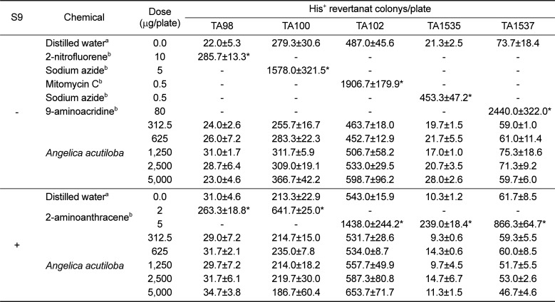
In vitro chromosomal aberration assay

The chromosomal aberration test is an in vitro screening assay that has been used to detect chromosomal damage in cultured mammalian cells [343536]. At first, the effect of the AA extract on the proliferation of CHL cells were analyzed using 3-[4,5-dimethylthiazol-2-yl]-2,5 diphenyl tetrazolium bromide (MTT) assay (Table 2), and we decided to set 5000 µg/mL of the AA extract as the highest exposure level for in vitro chromosomal aberration assay based on the MTT assay resulting to 98.1% viability at 24 h incubation. The positive controls (mitomycin C without the S9 mix and cyclophosphamide with the S9 mix) resulted in significant increases in the incidence of structural chromosome aberrations, supporting the validity of the study (Table 3). In contrast, the number of cells with chromosomal aberrations (breaks, fragments, and exchanges) in the AA extract groups was not significantly different from the negative control group after short-term treatment for 6 h with or without the S9 mix and after continuous treatment for 24 h without the S9 mix. Our current data show that the AA extract did not induce chromosomal aberrations in CHL cells under the conditions used in this assay.
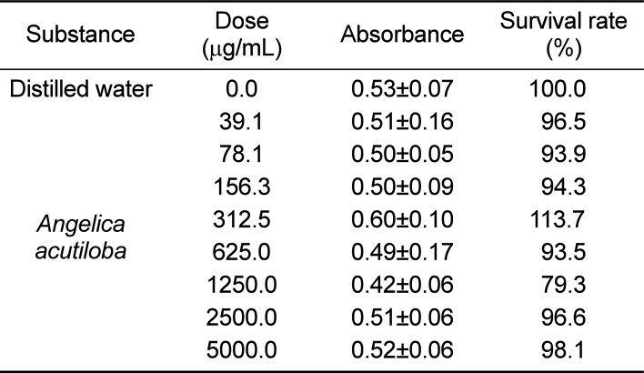
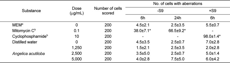
In vivo bone marrow micronucleus assay

Micronuclei, also known as Howell-Jolly bodies, are chromatin particles from damaged chromosome fragments that were not incorporated into the nucleus after mitosis [37]. In this study, the presence of micronuclei, which is regarded as an indirect indicator of quantitative and structural chromosomal aberrations [38], was evaluated to determine the genotoxic property of the AA extract. During the study, no abnormal clinical signs of toxicity in general appearance were detected in any of the mice following the AA extract administration. And, there were no statistically significant differences in body weights across all groups (Table 4). The mean ratio of PCEs to total erythrocytes were 50.9, 51.6, 47.4, 53.9, and 28.5% for the negative control, at 500, 1000, 2000 mg/kg BW of the AA extract, and the positive control, respectively (Table 5), indicating that this study was valid since no decrease in the PCE/(PCE+NCE) ratio (greater than 20%) reflects a lack of toxic effects of the AA extract (Heddle et al., 1984). The incidence of micronuclei showed no significant difference between the negative control group and the AA extract groups (500, 1000, or 2000 mg/kg BW). In contrast, the number of micronuclei in the mitomycin C-treated groups was significantly higher than that of the negative control group.
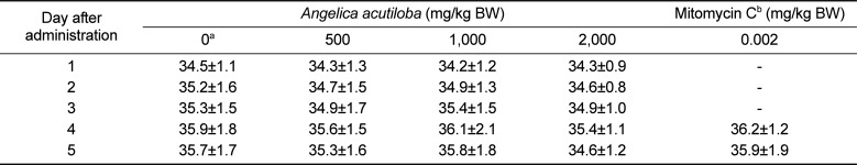
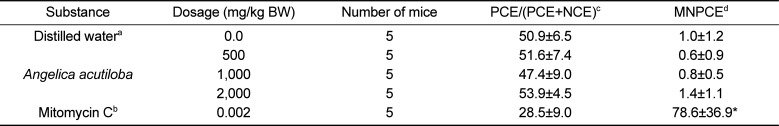
In conclusion, these fundamental toxicology data from the Ames assay, in vitro chromosome aberration assay, and in vivo micronucleus assay clearly suggest that the AA extract is safe in terms of genotoxicity. To our knowledge, this is the first comprehensive study assessing the potential genotoxic effects of the AA extract as a traditional medicine with beneficial activities in accordance with the OECD and the GLP Regulations.

Acknowledgments

This research was supported with a grant (05122MFDS472) from the Ministry of Food and Drug Safety.

Notes

Conflict of interests: The authors declare that there is no financial conflict of interests to publish these results.

References

1. Park MA, Sim MJ, Kim YC. Anti-Photoaging Effects of Angelica acutiloba Root Ethanol Extract in Human Dermal Fibroblasts. Toxicol Res. 2017; 33(2):125–134. PMID: 28503261.
2. Sheng YX, Li L, Wang Q, Guo HZ, Guo DA. Simultaneous determination of gallic acid, albiflorin, paeoniflorin, ferulic acid and benzoic acid in Si-Wu decoction by high-performance liquid chromatography DAD method. J Pharm Biomed Anal. 2005; 37(4):805–810. PMID: 15797805.
crossref
3. Wen KC, Huang CY, Lu FL. Determination of baicalin and puerarin in traditional Chinese medicinal preparations by high-performance liquid chromatography. J Chromatogr A. 1993; 631:241–250.
crossref
4. Liu IM, Tzeng TF, Liou SS, Chang CJ. Angelica acutiloba root attenuates insulin resistance induced by high-fructose diet in rats. Phytother Res. 2011; 25(9):1283–1293. PMID: 21308821.
5. Huntley AL, Ernst E. A systematic review of herbal medicinal products for the treatment of menopausal symptoms. Menopause. 2003; 10(5):465–476. PMID: 14501609.
crossref
6. Jeong SY, Kim HM, Lee KH, Kim KY, Huang DS, Kim JH, Seong RS. Quantitative analysis of marker compounds in Angelica gigas, Angelica sinensis, and Angelica acutiloba by HPLC/DAD. Chem Pharm Bull (Tokyo). 2015; 63(7):504–511. PMID: 25946978.
7. Yamada H, Komiyama K, Kiyohara H, Cyong JC, Hirakawa Y, Otsuka Y. Structural characterization and antitumor activity of a pectic polysaccharide from the roots of Angelica acutiloba. Planta Med. 1990; 56(2):182–186. PMID: 2353065.
8. Yoon TS, Cheon MS, Lee DY, Moon BC, Lee HW, Choo BK, Kim HK. Effects of root extracts from Angelica gigas and Angelica acutiloba on inflammatory mediators in mouse macrophages. J Korean Soc Appl Biol Chem. 2007; 50(4):264–269.
9. Tanaka S, Kano Y, Tabata M, Konoshima M. [Effects of “Toki” (Angelica acutiloba Kitagawa) extracts on writhing and capillary permeability in mice (analgesic and antiinflammatory effects)]. Yakugaku Zasshi. 1971; 91(10):1098–1104. PMID: 5170013.
10. Tanaka S, Ikeshiro Y, Tabata M, Konoshima M. Anti-nociceptive substances from the roots of Angelica acutiloba. Arzneimittelforschung. 1977; 27(11):2039–2045. PMID: 579998.
11. Lee K, Sohn Y, Lee MJ, Cho HS, Jang MH, Han NY, Shin KW, Kim SH, Cho IH, Bu Y, Jung HS. Effects of Angelica acutiloba on mast cell-mediated allergic reactions in vitro and in vivo. Immunopharmacol Immunotoxicol. 2012; 34(4):571–577. PMID: 22129085.
12. Kumazawa Y, Nakatsuru Y, Fujisawa H, Nishimura C, Mizunoe K, Otsuka Y, Nomoto K. Lymphocyte activation by a polysaccharide fraction separated from hot water extracts of Angelica acutiloba Kitagawa. J Pharmacobiodyn. 1985; 8(6):417–424. PMID: 3877157.
crossref
13. Kiyohara H, Yamada H, Cyong JC, Otsuka Y. Studies on polysaccharides from Angelica acutiloba. V. Molecular aggregation and anti-complementary activity of arabinogalactan from Angelica acutiloba. J Pharmacobiodyn. 1986; 9(4):339–346. PMID: 3735056.
crossref
14. Miyazawa M, Tsukamoto T, Anzai J, Ishikawa Y. Insecticidal effect of phthalides and furanocoumarins from Angelica acutiloba against Drosophila melanogaster. J Agric Food Chem. 2004; 52(14):4401–4405. PMID: 15237943.
crossref
15. Liu IM, Tzeng TF, Liou SS, Chang CJ. Regulation of obesity and lipid disorders by extracts from Angelica acutiloba root in high-fat diet-induced obese rats. Phytother Res. 2012; 26(2):223–230. PMID: 21647998.
16. Lee K, Sohn Y, Lee MJ, Cho HS, Jang MH, Han NY, Shin KW, Kim SH, Cho IH, Bu Y, Jung HS. Effects of Angelica acutiloba on mast cell-mediated allergic reactions in vitro and in vivo. Immunopharmacol Immunotoxicol. 2012; 34(4):571–577. PMID: 22129085.
17. Joo SS, Park D, Shin S, Jeon JH, Kim TK, Choi YJ, Lee SH, Kim JS, Park SK, Hwang BY, Lee DI, Kim YB. Anti-allergic effects and mechanisms of action of the ethanolic extract of Angelica gigas in dinitrofluorobenzene-induced inflammation models. Environ Toxicol Pharmacol. 2010; 30(2):127–133. PMID: 21787642.
crossref
18. Sarker SD, Nahar L. Natural medicine: the genus Angelica. Curr Med Chem. 2004; 11(11):1479–1500. PMID: 15180579.
19. Uto T, Tung NH, Taniyama R, Miyanowaki T, Morinaga O, Shoyama Y. Anti-inflammatory Activity of Constituents Isolated from Aerial Part of Angelica acutiloba Kitagawa. Phytother Res. 2015; 29(12):1956–1963. PMID: 26463105.
crossref
20. Markman M. Safety issues in using complementary and alternative medicine. J Clin Oncol. 2002; 20:39S–41S. PMID: 12235223.
21. Shin SH, Koo KH, Bae JS, Cha SB, Kang IS, Kang MS, Kim HS, Heo HS, Park MS, Gil GH, Lee JY, Kim KH, Li Y, Lee HK, Song SW, Choi HS, Kang BH, Kim JC. Single and 90-day repeated oral dose toxicity studies of fermented Rhus verniciflua stem bark extract in Sprague-Dawley rats. Food Chem Toxicol. 2013; 55:617–626. PMID: 23416650.
crossref
22. Che JH, Yun JW, Cho EY, Kim SH, Kim YS, Kim WH, Park JH, Son WC, Kim MK, Kang BC. Toxicologic assessment of Paecilomyces tenuipes in rats: renal toxicity and mutagenic potential. Regul Toxicol Pharmacol. 2014; 70(2):527–534. PMID: 25223566.
crossref
23. Che JH, Yun JW, Kim YS, Kim SH, You JR, Jang JJ, Kim HC, Kim HH, Kang BC. Genotoxicity and subchronic toxicity of Sophorae radix in rats: hepatotoxic and genotoxic potential. Regul Toxicol Pharmacol. 2015; 71(3):379–387. PMID: 25640205.
crossref
24. Yun JW, Kim SH, Kim YS, You JR, Kwon E, Jang JJ, Park IA, Kim HC, Kim HH, Che JH, Kang BC. Evaluation of subchronic (13week) toxicity and genotoxicity potential of vinegar-processed Genkwa Flos. Regul Toxicol Pharmacol. 2015; 72(2):386–393. PMID: 25882305.
crossref
25. Yun JW, Che JH, Kwon E, Kim YS, Kim SH, You JR, Kim WH, Kim HH, Kang BC. Safety evaluation of Angelica gigas: Genotoxicity and 13-weeks oral subchronic toxicity in rats. Regul Toxicol Pharmacol. 2015; 72(3):473–480. PMID: 26032491.
crossref
26. Ministry of Food and Drug Safety (MFDS). Good Laboratory Practice Regulation for Non-clinical Laboratory Studies (Notification No. 2014-67). Korea: MFDS;2014. Available [October 2014]. http://www.mfds.go.kr/.
27. OECD. OECD guideline for testing of chemicals, Test No. 471: Bacterial Reverse Mutation test. 1997.
28. OECD. OECD guideline for testing of chemicals, Test No. 473: in vitro mammalian chromosome aberration test. 1997.
29. OECD. OECD guideline for testing of chemicals, Test No. 474: mammalian erythrocyte micronucleus test. 1997.
30. Heddle JA, Stuart E, Salamone MF. Kilbey BJ, Legator M, Nichols W, Ramel C, editors. The bone marrow micronucleus test in Handbook of mutagenicity test procedures. Amsterdam: Elsevier;1984. p. 441–457.
31. Hakura A, Suzuki S, Satoh T. Advantage of the use of human liver S9 in the Ames test. Mutat Res. 1999; 438(1):29–36. PMID: 9858674.
crossref
32. Maron DM, Ames BN. Revised methods for the Salmonella mutagenicity test. Mutat Res. 1983; 113(3-4):173–215. PMID: 6341825.
crossref
33. Kaleeswaran S, Sriram P, Prabhu D, Mathuram LN. Chinnathambi. Anti- and pro-mutagenic effects of silymarin in the Ames bacterial reverse mutation assay. Phytother Res. 2009; 23(10):1378–1384. PMID: 19277970.
crossref
34. Fenech M, Morley AA. Measurement of micronuclei in lymphocytes. Mutat Res. 1985; 147(1-2):29–36. PMID: 3974610.
crossref
35. Perry P, Evans HJ. Cytological detection of mutagen-carcinogen exposure by sister chromatid exchange. Nature. 1975; 258(5531):121–125. PMID: 52847.
crossref
36. Tucker JD, Auletta A, Cimino MC, Dearfield KL, Jacobson-Kram D, Tice RR, Carrano AV. Sister-chromatid exchange: second report of the Gene-Tox Program. Mutat Res. 1993; 297(2):101–108. PMID: 7687323.
crossref
37. Luzhna L, Kathiria P, Kovalchuk O. Micronuclei in genotoxicity assessment: from genetics to epigenetics and beyond. Front Genet. 2013; 4:131. PMID: 23874352.
crossref
38. Akyıl D, Konuk M. Detection of genotoxicity and mutagenicity of chlorthiophos using micronucleus, chromosome aberration, sister chromatid exchange, and Ames tests. Environ Toxicol. 2015; 30(8):937–945. PMID: 24515492.
crossref
Table 1

Results of S. typhimurium reversion assay with Angelica acutiloba extract

lar-33-231-i001
S9 Chemical Dose (µg/plate) His+ revertanat colonys/plate
TA98 TA100 TA102 TA1535 TA1537
Distilled watera 0.0 22.0±5.3 279.3±30.6 487.0±45.6 21.3±2.5 73.7±18.4
2-nitrofluoreneb 10 285.7±13.3* - - - -
Sodium azideb 5 - 1578.0±321.5* - - -
Mitomycin Cb 0.5 - - 1906.7±179.9* - -
Sodium azideb 0.5 - - - 453.3±47.2* -
9-aminoacridineb 80 - - - - 2440.0±322.0*
312.5 24.0±2.6 255.7±16.7 463.7±18.0 19.7±1.5 59.0±1.0
625 26.0±7.2 283.3±22.3 452.7±12.9 21.7±5.5 61.0±11.4
Angelica acutiloba 1,250 31.0±1.7 311.7±5.9 506.7±58.2 17.0±1.0 75.3±18.6
2,500 28.7±6.4 309.0±19.1 533.0±29.5 20.7±3.5 71.3±9.2
5,000 23.0±4.6 366.7±42.2 598.7±96.2 28.0±2.6 59.7±6.0
+ Distilled watera 0.0 31.0±4.6 213.3±22.9 543.0±15.9 10.3±1.2 61.7±8.5
2-aminoanthraceneb 2 263.3±18.8* 641.7±25.0* - - -
5 - - 1438.0±244.2* 239.0±18.4* 866.3±64.7*
312.5 29.0±7.2 214.7±15.0 531.7±28.6 9.3±0.6 59.3±5.5
625 31.7±2.1 235.0±7.8 534.0±8.7 14.3±0.6 60.0±8.5
Angelica acutiloba 1,250 29.7±7.2 214.0±18.2 557.7±49.9 9.7±4.5 51.7±5.5
2,500 31.7±6.1 219.7±30.0 587.3±80.8 14.7±6.7 53.0±2.6
5,000 34.7±3.8 186.7±60.4 653.7±71.7 11.3±1.5 46.7±4.6

aNegative control

bPositive control

*Significantly different from negative control group (P<0.05).

Table 2

Results of MTT assay in CHL cells treated with Angelica acutiloba extract

lar-33-231-i002
Substance Dose (μg/mL) Absorbance Survival rate (%)
Distilled water 0.0 0.53±0.07 100.0
39.1 0.51±0.16 96.5
78.1 0.50±0.05 93.9
156.3 0.50±0.09 94.3
Angelica acutiloba 312.5 0.60±0.10 113.7
625.0 0.49±0.17 93.5
1250.0 0.42±0.06 79.3
2500.0 0.51±0.06 96.6
5000.0 0.52±0.06 98.1
Table 3

Results of chromosomal aberration induced by Angelica acutiloba extract

lar-33-231-i003
Substance Dose (µg/mL) Number of cells scored No. of cells with aberrations
−S9 +S9
6h 24h 6h
MEMa 0 200 4.5±2.1 2.5±3.5 5.5±0.7
Mitomycin Cb 0.1 200 38.0±7.1* 66.5±9.2* -
Cyclophosphamideb 10 200 - - 98.0±1.4*
Distilled water 0 200 4.5±3.5 2.5±0.7 7.0±2.8
1,250 200 1.5±2.1 2.5±3.5 2.0±2.8
Angelica acutiloba 2,500 200 3.5±5.0 2.5±0.7 5.0±1.4
5,000 200 4.0±2.8 7.5±5.0 6.0±4.2

aMinimum essential medium (negative control)

bPositive control

*Significantly different from negative control group (P<0.05).

Table 4

Body weight changes following treatment with Angelica acutiloba extract

lar-33-231-i004
Day after administration Angelica acutiloba (mg/kg BW) Mitomycin Cb (mg/kg BW)
0a 500 1,000 2,000 0.002
1 34.5±1.1 34.3±1.3 34.2±1.2 34.3±0.9 -
2 35.2±1.6 34.7±1.5 34.9±1.3 34.6±0.8 -
3 35.3±1.5 34.9±1.7 35.4±1.5 34.9±1.0 -
4 35.9±1.8 35.6±1.5 36.1±2.1 35.4±1.1 36.2±1.2
5 35.7±1.7 35.3±1.6 35.8±1.8 34.6±1.2 35.9±1.9

aNegative control

bPositive control

Table 5

Micronucleated polychromatic erythrocytes (MNPCEs) in mice bone marrow following treatment with Angelica acutiloba extract

lar-33-231-i005
Substance Dosage (mg/kg BW) Number of mice PCE/(PCE+NCE)c MNPCEd
Distilled watera 0.0 5 50.9±6.5 1.0±1.2
500 5 51.6±7.4 0.6±0.9
Angelica acutiloba 1,000 5 47.4±9.0 0.8±0.5
2,000 5 53.9±4.5 1.4±1.1
Mitomycin Cb 0.002 5 28.5±9.0 78.6±36.9*

aNegative control

bPositive control

cThe ratio of polychromatic erythrocytes to all erythrocytes (polychromatic+normochromatic) (%)

dPolychromatic erythrocyte with micronuclei was calculated from 2,000 polychromatic erythrocytes (%)

*Significantly different from negative control group (P<0.05).

TOOLS
Similar articles