

 PDF
PDF ePub
ePub Citation
 Print
Print


Danggui, the root of the species Angelica belonging to the family Umbelliferae, has been named ‘female ginseng’ and has been most commonly used traditional herbal medicine for the treatment of gynecological disorders, such as menoxenia and anemia, due to its hemogenic, analgesic, and sedative activities [123]. According to geographical locations, three common species of Danggui are found in Asia: Korean Danggui (Angelica gigas in Korea), Chinese Danggui (A. sinensis in China), and Japanese Danggui (A. acutiloba in Japan). Various chemical constituents and pharmacological effects of these three species have been reported [456].
Various bioactivities of the A. acutiloba (AA) extract were previously reported including antitumor [7], anticomplementary [8], antioxidant [9], and anti-inflammatory activity [10111213]. And, the AA treatment attenuated fat accumulation in high fat diet-induced obesity through the up-regulation of lipid metabolism [14]. Liu et al. [15] have demonstrated that the AA exerted to attenuate insulin resistance and to promote glucose homeostasis as an effective ethnomedicine for treating diabetic complications. Further, that the root of Angelica acutiloba was reported to exert a protective effect against allergic diseases and inflammatory diseases through the inhibition of the release of histamine from mast cells and the production of pro-inflammatory cytokines [13161718]. Also, the AA could be a potent anti-wrinkle agent in the field of traditional medicine through the enhancement of collagen synthesis and suppression of matrix metalloproteinases [1].
The use of natural herbal medicines is increasing for self-medication without supervision because it is believed that these do not have adverse effects compared with synthetic chemical drugs [1920]. However, it is noteworthy that we have recently found the subchronic hepatotoxic or nephrotoxic potentials of various well-known traditional medicines such as Paecilomyces tenuipes, Sophorae radix, and vinegar-processed Genkwa flos although they are currently available on the market [212223]. More importantly, data on the systemic oral toxicity of the AA extract is also lacking in spite of its use as an herbal medicine in East Asia. Therefore, we investigated the subchronic repeated dose oral toxic effects of the AA extract in rats in the present study.
Materials and Methods
Test substance and animals
A hot water AA extract was provided by the National Institute of Food and Drug Safety Evaluation (Osong, Korea). AA roots were purchased from an Oriental medicine market in Korea, and an extract of AA was obtained according to a method described previously [24]. In brief, dried AA roots were ground by a mixer, and incubated with distilled water (DW) at 100℃. After filtration through filter paper, the filtrate was freeze-dried and dissolved in DW for oral administration. The extraction yield of the hot water AA extract was 0.153 g of freeze-dried AA extract/g of dried AA root.
F344 rats (SLC, Hamamatsu, Japan) were used after a week of quarantine and acclimatization. During the studies, the animal facility was maintained under standard conditions (22±2℃, 40-60% humidity, and 12 h light/dark cycle). The animals were fed a rodent diet (LabDiet 5002 Certified Rodent Diet, PMI Nutrition International, St. Louis, MO, USA) and tap water ad libitum. All of the animal experiments were approved by the Institutional Animal Care and Use Committee of the Biomedical Research Institute at the Seoul National University Hospital, and this study was performed in compliance with the guidelines published by the Organization for Economic Cooperation and Development (OECD) as well as the guidance for Good Laboratory Practices for toxicity tests issued by the Ministry of Food and Drug Safety [25].
Experimental design for the oral toxicity study
For the 14-day repeat-dose toxicity study, the hot water AA extract was administered to F344 rats (5/sex/group) by oral gavage at doses of 125, 250, 500, 1000, and 2000 mg/kg of body weight/10 mL DW once daily for 14 days. For the 13-week repeat-dose toxicity study, the hot water AA extract was administered to F344 rats (10/sex/group) by oral gavage at doses of 125, 250, 500, 1000, and 2000 mg/kg of body weight/10 mL DW once daily for 13 weeks in accordance with OECD guideline 408 [26] and the US National Toxicology Program (NTP) protocol (https://ntp.niehs.nih.gov/testing/types/cartox/protocols/13week/index.html). During the administration period, the rats were observed for general appearance daily, and body weights, food intake, and water consumption were recorded weekly. The rats were anesthetized with isoflurane one day after the final gavage.
Hematology and serum biochemistry
Blood samples were collected via the posterior vena cava. The hematology parameters were measured using an automatic hematology analyzer MS9-5 Hematology Counter (Melet Schloesing Laboratories, Osny, France) for the following parameters: total white blood cell (WBC), red blood cell (RBC), hemoglobin (HGB), hematocrit (HCT), platelet (PLT), mean corpuscular volume (MCV), mean corpuscular hemoglobin (MCH), mean corpuscular hemoglobin concentration (MCHC), and differential WBC. And, the standard serum biochemistry parameters were analyzed with an automatic chemistry analyzer 7070 (Hitachi, Tokyo, Japan) to evaluate the following serum biochemistry parameters: blood urea nitrogen (BUN), total cholesterol (TC), total protein (TP), albumin, total bilirubin (TB), alkaline phosphatase (ALP), aspartate transaminase (AST), alanine transaminase (ALT), γ-glutamyl transferase (γGT), creatinine kinase (CK), creatinine, triglyceride (TG), and glucose.
Gross findings, organ weights, and histopathological assessments
At the end of the treatment period, animals were exsanguinated, and organs and tissues were observed macroscopically. Organ weights were obtained for the liver, kidney, testis, thymus, heart, and lung. The eyes with the Harderian glands were fixed in Davidson solution (30 mL 95% ethyl alcohol+20 mL formalin+10 mL glacial acetic acid+30 mL DW). The testis and epididymis were fixed in Bouin's solution. Other organs including the liver, kidney, adrenal gland, urinary bladder, spleen, pancreas, thymus, thyroid gland, parathyroid gland, trachea, esophagus, lung, heart, salivary gland, lymph node, stomach, duodenum, jejunum, ileum, colon, rectum, preputial gland, clitoral gland, skin, brain, pituitary gland, prostate, seminal vesicle, ovary, uterus, and vagina were fixed in 10% neutral buffered formalin. The nasal cavity and femora were treated with a decalcification solution for up to 3 weeks. Tissue samples were embedded in paraffin wax, sectioned and stained with hematoxylin and eosin (H&E). After staining, the histological preparations from animals in the control, 1000, and 2000 mg/kg groups were initially examined via light microscopy. With respect to the organs and tissues showing significant histological changes, preparations of all rats in the other groups were examined microscopically.
Statistical analysis
All of the values are expressed as mean±SD. The statistical analysis was performed using a one-way ANOVA, followed by a multiple comparison procedure with a Tukey/Duncan test using SPSS software version 19 (SPSS Inc., Chicago, IL, USA). P values of less than 0.05 were considered to be statistically significant.
Go to : 
Results and Discussion
14-day repeat-dose oral toxicity study
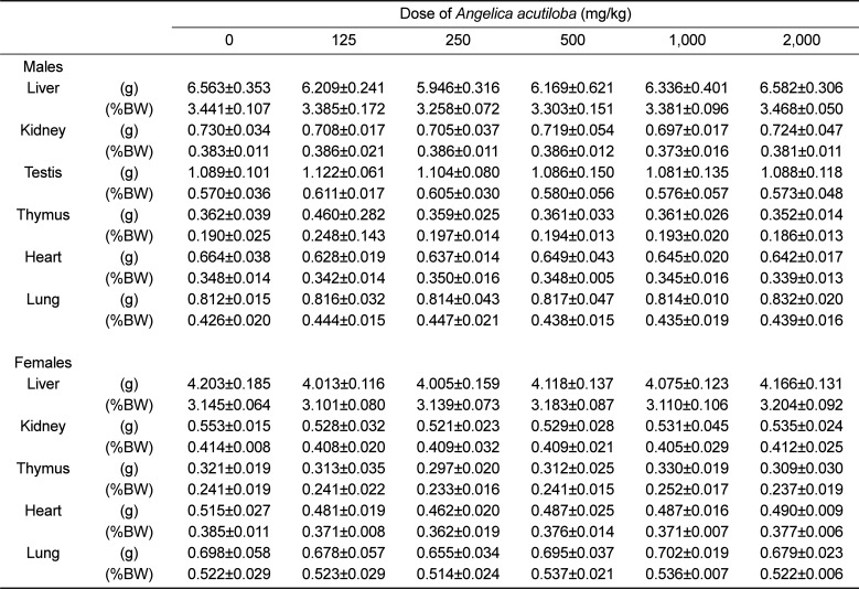
In the 14-day repeat-dose oral toxicity study, the treated animals did not show any abnormal changes in the general appearance and clinical signs, and body weights (Figure 1) throughout the test period at all selected dose levels following the oral administration of the AA extract. Likewise, there was no significant difference in gross macroscopic necropsy findings at all doses at the end of the 14 days of the experimental period. The parameters of absolute and relative organ weights showed no significant differences between the AA extract-treated groups and the control group (Table 1). On a basis of these results, 2000 mg/kg is the highest recommended dose of the AA extract for the 13-week repeat-dose oral toxicity study.
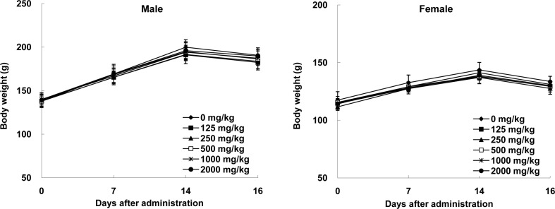 | Figure 1Effects of Angelica acutiloba extract on the body weight changes after oral administration in male and female rats for 14 days. Data expressed as means±SD.
|
Table 1
Organ weights for male and female F344 rats orally administered with Angelica acutiloba extract for 14 days
![]()
General observation, body weight, and feed/water consumption in 13-week repeat-dose oral toxicity study
In all groups, the administration of the AA extract for 13 weeks did not show increases in mortality and abnormal toxic symptoms in males and females. Similarly, the body weight gradually increased for 13 weeks both in the control group and the AA extract-treated groups throughout the study (Figure 2). And, a few incidental significant differences in the food (Figure 3A) and water intake (Figure 3B) of the animals between the AA extract-treated groups and the untreated control group were not dose-dependent. These indicate that the AA extract did not obstruct the normal growth of experimental animals.
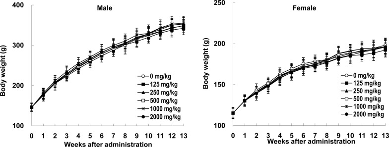 | Figure 2Effects of Angelica acutiloba extract on the body weight changes after oral administration in male and female rats for 13 weeks. Data expressed as means±SD.
|
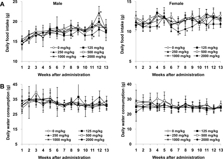 | Figure 3Effects of Angelica acutiloba extract on the daily food intake and water consumption after oral administration in male and female rats for 13 weeks. (A) Daily food intake. (B) Daily water consumption. Data expressed as means±SD. (*) indicates a significant difference relative to the control group (0 mg/kg) (P<0.05).
|
Hematology and clinical chemistry in 13-week repeat-dose oral toxicity study
Table 2 summarizes the results of the effects of the AA extract on different hematological parameters in the 13-week repeat-dose toxicity study. All parameters were not significantly different in the treatment groups from the controls within physiologically normal ranges. It indicates that the AA extract did not affect hematopoiesis and leukopoiesis.
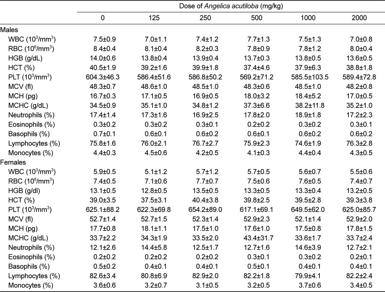
Table 2
Hematological data for male and female F344 rats orally administered with Angelica acutiloba extract for 13 weeks
![]()
Table 3 shows the effects of the AA extract on different biochemical parameters. The TC levels in females treated with 500 (83.9±5.7), 1000 (81.8±7.3), and 2000 mg/kg (83.9±4.8) of the AA extract were significantly lower than that in the control group (93.1±6.8). The albumin levels significantly decreased in females treated with 500 mg/kg (2.8±0.1) of the AA extract relative to the control females (3.0±0.1). The CK levels in males treated with the AA extract with a dose of 1000 mg/kg (290.1±89.2) were significantly lower than those in the control group (446.0±85.0). However, these statistically significant findings were not considered to be test material-related because they were within normal biological variability. In the toxicological research, liver and kidney are the two most important target organs since most of the drugs undergo many interactions such as hepatic metabolism and renal excretion following the oral administration [2728]. Serum levels of creatinine and BUN were used as the primary indicators for kidney function [2930]. The lack of significant changes in the levels of serum BUN and creatinine in the AA extract groups suggests that the repeated administration of the AA extract did not affect kidney function in experimental animals. Liver damages such as hepatocellular damage [31] or biliary obstruction [32] can cause serum hepatic biochemical parameters (ALT, AST, and ALP) to leak into the blood circulation and rise in the serum levels. In the present study, there were no significant differences in these serum liver biomarker enzymes of the groups treated with the AA extract compared to the control, indicating that the repeated administration of the AA extract did not interfere with liver function in experimental animals.
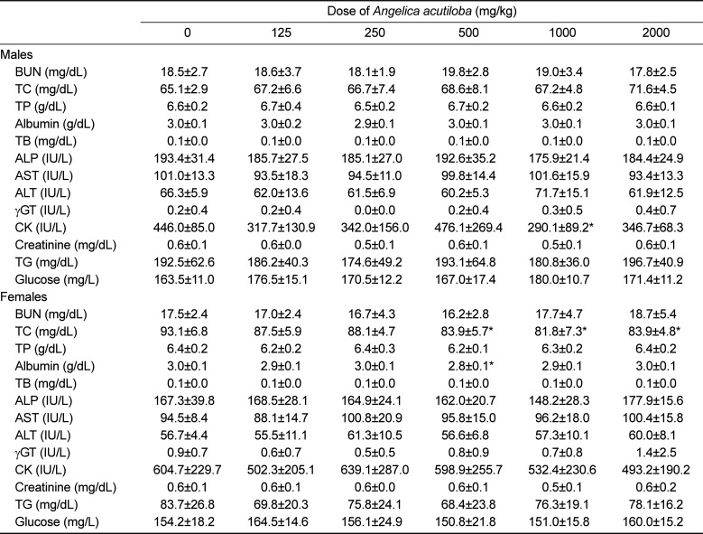
Table 3
Serum biochemistry data for male and female F344 rats orally administered with Angelica acutiloba extract for 13 weeks
![]()
Organ weights and histopathological changes in 13-week repeat-dose oral toxicity study
As shown in Table 4, the AA extract did not appear to affect the absolute and relative (organ-to-body weight ratios) organ weights of the male and female rats at all doses tested. The necropsy showed no significant changes in organ gross anatomy in the AA extract-treated rats when compared with the untreated rats. The histopathological studies of important major organs (i.e. liver, kidney, lung, spleen, pancreas, and brain) indicated no toxic alteration in tissue structures following the longterm administration of the AA extract.
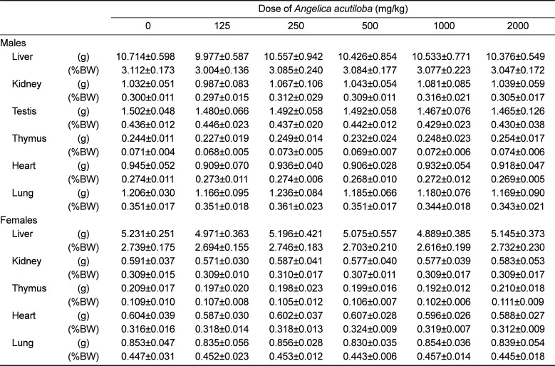
Table 4
Organ weights for male and female F344 rats orally administered with Angelica acutiloba extract for 13 weeks
![]()
Based on the results of the present study, it was concluded that oral administration of the AA extract did not induce any significant adverse toxic reaction when tested for subchronic toxicity. The subchronic NOAEL for the AA extract in both sexes of rat is greater than 2000 mg/kg, which can be extrapolated to the human dose 324 mg/kg for further clinical study by the surface-area-guided dosing adjustment of the US Food and Drug Administration [33]. Among thousands of herbal products consumed in Asia, United States, and United Kingdom, AA is widely available as a functional food and traditional medicine [17]. To the best of our knowledge, there is no data available for potential long-term toxicological concerns of the AA extract, and this study is the first attempt to evaluate the subchronic toxicity in accordance with the OECD and the GLP regulations although further study is needed for major bioactive components of the AA extract.
Go to :
 XML Download