Journal List > Lab Anim Res > v.32(2) > 1053876

Lee, Song, Jang, Kim, Park, Choi, and Han: Anti-inflammatory effect of egg white-chalcanthite and purple bamboo salts mixture on arthritis induced by monosodium iodoacetate in Sprague-Dawley rats

Abstract

The aim of this study is to investigate the potential of anti-osteoarthritis effects on egg white-chalcanthite (EC), purple bamboo salts (PBS), and a mixture of EC and PBS (EC+PBS). EC is a mixture of egg white and pulverized chalcanthite. PBS has been widely used as one of functional foods in Korea and shows unique features compared with common salt. Osteoarthritis was induced by intra-articular injection of monosodium iodoacetate (MIA, 4mg/kg bw) in Sprague-Dawley (SD) rats. Test substances were administered once daily for 6 weeks at doses of 10 mg EC, EC+100 mg PBS, EC+200 mg PBS before and after MIA injection. Each substance was assessed by blood chemistry parameters, and by serum cytokines including IL-1β and IL-6, and nitric oxide (NO) and prostaglandin-E2 (PGE2). Structural changes of articular cartilage were also evaluated by histopathological examination. As a result, body weight and blood chemistry parameter were not different in all experimental groups. EC+PBS mixture reduced the production of PGE2, NO, IL-1β, and IL-6. In histological grade of osteoarthritis, EC+PBS mixture had a tendency to ameliorate damage of articular cartilage induced by MIA in a dose-dependent manner. In conclusion, EC+PBS mixture was demonstrated to have a potential for anti-inflammatory effect against osteoarthritis induced by MIA in a dose-dependent manner.

Osteoarthritis (OA) known as degenerative arthritis is the most common type of joint disease that results from breakdown of articular cartilage and subchondral bone, ligaments, and synovial membrane [1]. The most common symptoms are significant pain and functional disability [2]. The prevalence of OA in South Korea is continuously increased due to the increase of aging population and obesity. In 2008, 10.7 % of Korean adults are suffering from OA and it is estimated to be up to 34.5% in persons over 80 years old [34]. Pathogenesis of OA is not clear. However, mechanical stress, cytokines, nitric oxide, and reactive oxygen species (ROS) have been known to be major factors of OA [567]. Monosodium iodoacetate (MIA), an inhibitor of glyceraldehyde-3-phosphate dehydrogenase activity, is the most common material used to produce animal models of osteoarthritis. Intra-articular injection of MIA caused destruction of the articular cartilage by inhibiting glycolysis and eventually by inducing cell death [8]. OA induced by MIA is very similar to human OA in view of histopathological changes of articular cartilage and chondrocytes [910]. In addition, the model has been used to evaluate the effectiveness of the pharmacologic agents related with arthritis in many previous studies [111213].
Egg white-chalcanthite (EC) is a mixture of egg white and pulverized chalcanthite. In the egg white-chalcanthite prepared as noted above, the toxicity of the chalcanthite is neutralized by egg white, so that the toxicity is reduced or removed and the pharmaceutical properties are increased [14]. Recently, it has been reported that EC inhibited inflammation induced by lipopolysaccharide in BV2 cell and also inhibited the growth of NCI-H460 cells by induction of apoptotic cell death [1415]. Purple bamboo salts (PBS) is widely used functional foods in Korea and shows unique features compared with common salt or sun-dried salt. PBS was produced according to the traditional procedure using a sun-dried salt and bamboo. In this procedure, toxicity of sun-dried salt was decreased and acidity is converting to strong alkalinity [1617]. Also, when compared with sun-dried salt, PBS contained high concentration of iron, potassium, and phosphate, but the concentration of sulfate is low [16]. It has been reported that PBS have protective effects on inflammatory disease [18], oxidative stress [1719], cancer [21] and gastric ulcers [12].
Based on these properties of the EC+PBS mixture, we investigated anti-osteoarthritis effects of EC, PBS and EC+PBS mixture against MIA-induced osteoarthritis in male Sprague-Dawley (SD) rats.

Materials and Methods

Reagent

Monosodium iodoacetate (MIA) was purchased from Sigma Chem. Co. (St. Louis, USA) and purple bamboo salts and egg white-chalcanthite were provided by Insan Bamboo Salt Inc. (Hamyang, Korea). IL-1β, IL-6, NO and PGE2, assay kit were purchased from R&D Systems, Inc. or Cayman Chemical Co. Inc. (Ann Arbor, USA).

Animals

Thirty four male Sprague Dawley (SD) rats weighing 210-240 g were purchased from Daehan-biolink (Eumsung, Korea). The animals were housed two or three per polycarbonate cage in a temperature and humidity controlled room (temperature 20~24℃, humidity 40~70%). Dark/light cycle was controlled for 12 hours. The sterile feed and R/O water were ad libitum. All the experimental procedures were examined and approved by the Animal Research Ethics Committee at the Hoseo Toxicological Research Center of Hoseo University.

Development of osteoarthritis and test substance treatment

For induction of osteoarthritis, rats were anesthetized with ketamine (Yuhan Co. Ltd., Seoul, Korea) and xylazine (Bayerkorea Co. Ltd., Seoul, Korea) and then given single intra-articular injection at dose of 4 mg/kg b.w. monosodium iodoacetate (MIA; Sigma, St. Louis, USA) using 27G needle. MIA was dissolved in physiological saline. Control rats were injected with an equivalent volume of saline. Test substance dissolved in saline was given orally once per day for 6 weeks. MIA was injected in all group except normal control group at the 3 weeks of the experiment (Figure 1).

Measurements of blood chemistry parameter

At the 6 weeks after the experiment, animals were anesthetized by CO2 inhalation. Blood was collected from abdominal aorta and centrifuged at 3,000 rpm for 10 min (2236HR high-speed centrifuge, GYROZEN Co., Ltd, Daejoen, Korea) to obtain serum. The serum were used to measurement of aspartate aminotransferase (AST), alanine aminotransferase (ALT), blood urea nitrogen (BUN), creatinine (CRE) and high-sensitivity C-reactive protein (hs-CRP) using the Automatic Chemistry Analyzer (Hitachi Co. Ltd., Tokyo, Japan).

Measurements of NO, PGE2, IL-1β and IL-6 production

Concentrations of NO, PGE2, IL-1β and IL-6 in the serum were measured by commercial assay kit following the manufacturer's instructions. NO was determined by measuring nitrite, which is a major stable product of NO, using the Nitrite Colorimetric Assay Kit (Cayman, Ann Arbor, USA). PGE2 was determined using the ELISA kit (Cayman, Ann Arbor, USA). IL-1β and IL-6 were determined using the ELISA kit (R&D Systems, Inc. Minneapolis, USA).

Tissue preparation and histological examination

Both knee joints from each animal were dissected and fixed in 10% neutral buffered formalin. Tissues were decalcified in 5% formic acid for 48 hours. After decalcification, the tissues were processed dehydration, clearing, paraffin infiltration step and embedded in paraffin blocks. 5 µm sections were prepared and stained with safranin O-fast green or hematoxylin-eosin. The sections were examined under the light microscope (Olympus BX51, Olympus Co., Ltd., Tokyo, Japan) and evaluated histologically for the severity of lesions according to a modified Mankin grading system [22].

Statistical analysis

Group differences were assessed with a one way ANOVA followed by a Tukey's multiple comparison test. All analyses were conducted using the Statistical Package for Social Sciences for Windows software, version 12.0 (SPSS Inc., Chicago, USA). Statistical significance was assessed at P<0.05. All data were expressed as the mean ± SD.

Results

Effects of EC, PBS and EC-PBS on body weight and serum biochemical parameter

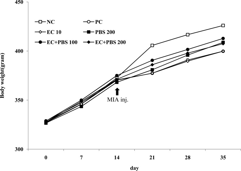
To investigate the effects of EC, PBS and EC+PBS, blood biochemical parameters were measured in the rat serum of MIA-induced OA model. Compared to normal group, all test groups were no significant difference in the levels of blood parameters including aminotransferase (AST), alanine aminotransferase (ALT), blood urea nitrogen (BUN), creatinine (CRE). Although high-sensitivity C-reactive protein(hs-CRP), indicator of inflammation, was increased in normal control group, other groups except normal control group did no significant difference (Table 1). Body weight was not different in all experimental groups (Figure 2).

Effects of EC, PBS and EC+PBS on Nitrite and Prostaglandin E2

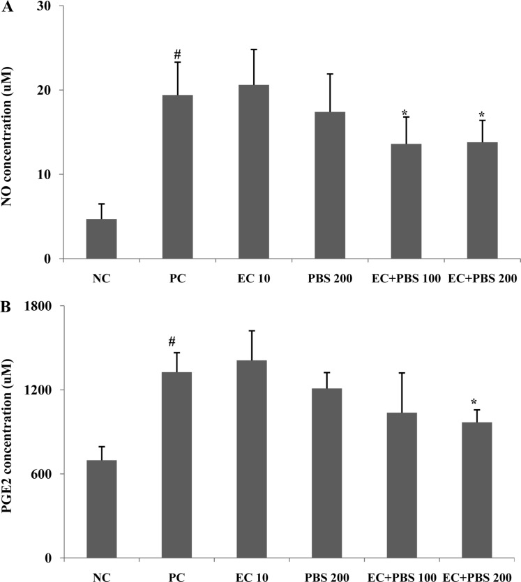
Nitrite, which was major product of nitric oxide assay, was analyzed to investigate the effect of EC, PBS and EC+PBS mixture in MIA-induced osteoarthritis rat. MIA showed significant increase of nitrite concentration when compared to normal control group. EC+PBS mixture induced significant decrease of nitrite at doses of 100 and 200 mg PBS, whereas EC alone and PBS alone did not show a decrease of nitrite (Figure 3A). MIA also showed significant increase of prostaglandin-E2 concentration when compared to normal control group. EC+PBS mixture induced significant decrease of prostaglandin E2 at dose of 200 mg PBS when compared to MIA alone (Figure 3B).

Effects of EC, PBS and EC+PBS on IL-1β and IL-6 production in serum

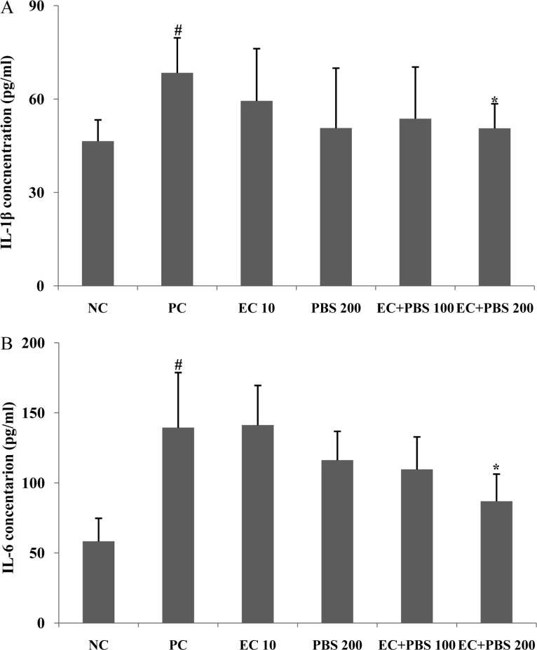
IL-1β and IL-6 assay were performed to investigate the anti-inflammatory effect of EC, PBS and EC+PBS mixture in the MIA-induced osteoarthritis rat. MIA showed significant increase of IL-1β and IL-6 levels when compared to normal control group. EC+PBS induced significant decrease of IL-1β and IL-6 levels at dose of 200 mg PBS when compared to MIA alone. Other groups did no significant difference (Figure 4A, 4B).

Effects of EC, PBS and EC+PBS on histopathological changes of the articular damage

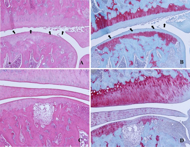
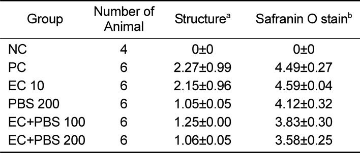
Histopathological examinations were performed to evaluate morphological changes and severity of the articular damage in the MIA-induced osteoarthritis rat. MIA induced surface irregularities and surface cleft, death of chondrocyte and fibrosis in the cartilage. We also measured histopathological findings which indicate severity of surface irregularity and surface cleft by H&E staining and matrix loss and cell death of articular cartilage by Safranin O staining. Histopathological lesions of articular cartilage are summarized in Table 2 and Fig. 5. PBS and EC+PBS mixture ameliorated severity of articular morphological changes such as surface irregularity and surface cleft induced by MIA (Table 2 and Figure 5A). EC+PBS mixture also decreased severity of matrix loss in articular cartilage (Table 2 and Figure 5B).

Discussion

Osteoarthritis (OA) is a degenerative arthritis characterized by defect of articular cartilage and arthralgia. Although the pathogenesis of OA is not fully understood, death of chondrocyte, only cell in articular cartilage, is one of major factor of osteoarthritis [23]. Death of chondrocytes cause disturbance of synthesis and re-absorption in cartilage that result in the loss of cartilage matrix components and defect in the structural and functional properties of the cartilage [24].
Although the results of experimental OA model cannot be correct extrapolation to human OA, a recent study showed that there was a similar correlation between OA in animal model induced by MIA and human OA [39]. The experimental models have often been used for confirm to efficacy in OA [40]. MIA is known to disturb chondrocyte metabolism and to induce cytokine, resulting in increase of NO and PGE2 level that induce chondrocyte death and loss of cartilage matrix. It is not clear whether chondrocyte death cause or consequence of arthritis [25], however it is becoming increasingly apparent that IL-1β and IL-6 is involved in pathogenesis of OA [262728]. IL-1β has an important role in the osteoarthritis. IL-1β induces matrix metalloproteinases, inhibits extracellular matrix synthesis [29] and inhibits chondrocyte proliferation [30]. IL-6 may inhibit the differentiation of mesenchymal cells into chondroblasts [31] and can induce loss of cartilage [32]. In the present study, EC+PBS mixture decreased cytokine level such as IL-1β, IL-6, the MIA increased these parameters. These results suggest that EC+PBS mixture have a potential of prevention against inflammation, and subsequently reduce damage of articular cartilage induced by MIA. These results are consistent with a previous report that PBS reduced production of pro-inflammatory cytokine such as IL-1β, IL-6. [33].
These cytokines can stimulate inflammatory mediators such as NO and PEG2 production [34]. The chondrocytes stimulated with cytokine, especially IL-1β produce NO and PEG2. NO and PGE2 plays a crucial role in the pathogenesis of OA [3536]. High level of NO and PEG2 have been found in cartilage and synovial tissues from OA patients and several models of cartilage degradation [363738].
Rahmati et al. [37] reported that these cytokines are thought to be generated by the synovial membrane and subsequently dispersed by the synovial fluid into cartilage. And these cytokines activate chondrocyte, inducing them to nitric oxide and prostaglandins. Notoya et al. [38] reported that PGE2 enhanced NO mediated cell death in analysis of interactions between PGE2 and the cell death. It means that PGE2 may stimulate human OA chondrocytes to the cell death induced by NO. In the present study, the MIA increased NO and PEG2 production, EC+PBS mixture decreased both of these parameters. These results are considered as decrease of cytokine by EC+PBS mixture. These results are consistent with a previous report that PBS reduced NO and PEG2 production [18].
In this study, MIA induced degenerative joint disease, which showed irregularities of articular surface and microfissures perpendicular to the articular surface. Chondrocytes responded to the loss of matrix by undergoing hypertrophy and proliferated to form clusters or clones. Death of chondrocyte were shown in the damaged cartilage induced by MIA. Sometimes, the epiphyseal marrow lose its hematopoietic elements, which were replaced by fibrous connective tissue.
In summary, EC+PBS mixtures decreased levels of cytokines such as IL-1β, IL-6, and inflammatory mediator such as NO and PGE2 in the OA animal model induced by MIA. EC+PBS mixture protected the articular cartilage damage by reducing IL-1β and IL-6 level, and prevented the loss of cartilage matrix, which is major symptoms of osteoarthritis. Taken together, these findings indicate that EC+PBS mixtures protect the MIA induced osteoarthritis based on anti-inflammatory actions.

Acknowledgments

This study was supported by a grant of the Local-led Industry Development Support program, Korea Institute for Advancement of Technology (R0003771).

Notes

Conflict of interests: The authors declare that there is no financial conflict of interests to publish these results.

References

1. Guzman RE, Evans MG, Bove S, Morenko B, Kilgore K. Monoiodoacetate-induced histologic changes in subchondral bone and articular cartilage of rat femorotibial joints: an animal model of osteoarthritis. Toxicol Pathol. 2003; 31(6):619–624. PMID: 14585729.
crossref
2. Bove SE, Calcaterra SL, Brooker RM, Huber CM, Guzman RE, Juneau PL, Schrier DJ, Kilgore KS. Weight bearing as a measure of disease progression and efficacy of anti-inflammatory compounds in a model of monosodium iodoacetate-induced osteoarthritis. Osteoarthritis Cartilage. 2003; 11(11):821–830. PMID: 14609535.
crossref
3. Studer R, Jaffurs D, Stefanovic-Racic M, Robbins PD, Evans CH. Nitric oxide in osteoarthritis. Osteoarthritis Cartilage. 1999; 7(4):377–379. PMID: 10419772.
crossref
4. Herrington C, Hall PA. Molecular and cellular themes in inflammation and immunology. J Pathol. 2008; 214(2):123–125. PMID: 18161749.
crossref
5. Bove SE, Calcaterra SL, Brooker RM, Huber CM, Guzman RE, Juneau PL, Schrier DJ, Kilgore KS. Weight bearing as a measure of disease progression and efficacy of anti-inflammatory compounds in a model of monosodium iodoacetate-induced osteoarthritis. Osteoarthritis Cartilage. 2003; 11(11):821–830. PMID: 14609535.
crossref
6. Woo YJ, Joo YB, Jung YO, Ju JH, Cho ML, Oh HJ, Jhun JY, Park MK, Park JS, Kang CM, Sung MS, Park SH, Kim HY, Min JK. Grape seed proanthocyanidin extract ameliorates monosodium iodoacetate-induced osteoarthritis. Exp Mol Med. 2011; 43(10):561–570. PMID: 21795829.
crossref
7. Im HJ, Kim JS, Li X, Kotwal N, Sumner DR, van Wijnen AJ, Davis FJ, Yan D, Levine B, Henry JL, Desevre J, Kroin JS. Alteration of sensory neurons and spinal response to an experimental osteoarthritis pain model. Arthritis Rheum. 2010; 62(10):2995–3005. PMID: 20556813.
crossref
8. Kim HY, Lee ES, Jeong JY, Choi JH, Choi YS, Han DJ, Lee MA, Kim SY, Kim CJ. Effect of bamboo salt on the physicochemical properties of meat emulsion systems. Meat Sci. 2010; 86(4):960–965. PMID: 20826069.
crossref
9. Zhao X, Jung OS, Park KY. Alkaline and antioxidant effects of bamboo salt. J Korean Soc Food Sci Nutr. 2012; 41(9):1301–1304.
crossref
10. Shin HY, Lee EH, Kim CY, Shin TY, Kim SD, Song YS, Lee KN, Hong SH, Kim HM. Anti-inflammatory activity of Korean folk medicine purple bamboo salt. Immunopharmacol Immunotoxicol. 2003; 25(3):377–384. PMID: 19180800.
crossref
11. Zhao X, Song JL, Kil JH, Park KY. Bamboo salt attenuates CCl4-induced hepatic damage in Sprague-Dawley rats. Nutr Res Pract. 2013; 7(4):273–280. PMID: 23964314.
crossref
12. Zhao X, Deng X, Park KY, Qiu L, Pang L. Purple bamboo salt has anticancer activity in TCA8113 cells in vitro and preventive effects on buccal mucosa cancer in mice in vivo. Exp Ther Med. 2013; 5(2):549–554. PMID: 23403521.
crossref
13. Kim SH, Kang SY, Jung KK, Kim TG, Han HM, Rheu HM, Moon A. Characterization and anti-gastric ulcer activity of bamboo salt. J Food Hyg Saf. 1998; 13:252–257.
14. Jhun HJ, Ahn K, Lee SC. Estimation of the prevalence of osteoarthritis in Korean adults based on the data from the fourth Korea national health and nutrition examination survey. Anesth Pain Med. 2010; 5(3):201–206.
15. Henrotin Y, Kurz B. Antioxidant to treat osteoarthritis: dream or reality? Curr Drug Targets. 2007; 8(2):347–357. PMID: 17305512.
crossref
16. Dunham J, Hoedt-Schmidt S, Kalbhen DA. Prolonged effect of iodoacetate on articular cartilage and its modification by an anti-rheumatic drug. Int J Exp Pathol. 1993; 74(3):283–289. PMID: 8334078.
17. Choi EA, Park HY, Yoo HS, Choi YH. Anti-inflammatory effects of egg white combined with chalcanthite in lipopolysaccharide-stimulated BV2 microglia through the inhibition of NF-êB, MAPK and PI3K/Akt signaling pathways. Int J Mol Med. 2013; 31(1):154–162. PMID: 23128312.
crossref
18. Janusz MJ, Hookfin EB, Heitmeyer SA, Woessner JF, Freemont AJ, Hoyland JA, Brown KK, Hsieh LC, Almstead NG, De B, Natchus MG, Pikul S, Taiwo YO. Moderation of iodoacetateinduced experimental osteoarthritis in rats by matrix metalloproteinase inhibitors. Osteoarthritis Cartilage. 2001; 9(8):751–760. PMID: 11795995.
crossref
19. Choi EA, Kim KH, Yoo BC, Yoo HS. Induction of apoptotic cell death by egg white combined-chalcanthite on NCI-H460 human lung cancer cells. J Korean Pharmacopuncture Inst. 2009; 12(3):49–59.
crossref
20. Gencosmanoglu BE, Eryavuz M, Dervisoglu S. Effects of some nonsteroidal anti-inflammatory drugs on articular cartilage of rats in an experimental model of osteoarthritis. Res Exp Med (Berl). 2001; 200(3):215–226. PMID: 11426673.
21. Natchus MG, Bookland RG, De B, Almstead NG, Pikul S, Janusz MJ, Heitmeyer SA, Hookfin EB, Hsieh LC, Dowty ME, Dietsch CR, Patel VS, Garver SM, Gu F, Pokross ME, Mieling GE, Baker TR, Foltz DJ, Peng SX, Bornes DM, Strojnowski MJ, Taiwo YO. Development of new hydroxamate matrix metalloproteinase inhibitors derived from functionalized 4-aminoprolines. J Med Chem. 2000; 43(26):4948–4963. PMID: 11150165.
crossref
22. Mankin HJ, Dorfman H, Lippiello L, Zarins A. Biochemical and metabolic abnormalities in articular cartilage from osteo-arthritic human hips. II. Correlation of morphology with biochemical and metabolic data. J Bone Joint Surg Am. 1971; 53(3):523–537. PMID: 5580011.
23. Thomas CM, Fuller CJ, Whittles CE, Sharif M. Chondrocyte death by apoptosis is associated with cartilage matrix degradation. Osteoarthritis Cartilage. 2007; 15(1):27–34. PMID: 16859932.
crossref
24. Goldring SR, Goldring MB. The role of cytokines in cartilage matrix degeneration in osteoarthritis. Clin Orthop Relat Res. 2004; (427 Suppl):S27–S36. PMID: 15480070.
crossref
25. Zamli Z, Sharif M. Chondrocyte apoptosis: a cause or consequence of osteoarthritis? Int J Rheum Dis. 2011; 14(2):159–166. PMID: 21518315.
crossref
26. Goldring MB, Otero M, Tsuchimochi K, Ijiri K, Li Y. Defining the roles of inflammatory and anabolic cytokines in cartilage metabolism. Ann Rheum Dis. 2008; 67(Suppl 3):iii75–iii82. PMID: 19022820.
crossref
27. Carames B, Lopez-Armada MJ, Cillero-Pastor B, Lires-Dean M, Vaamonde C, Galdo F, Blanco FJ. Differential effects of tumor necrosis factor-alpha and interleukin-1beta on cell death in human articular chondrocytes. Osteoarthritis Cartilage. 2008; 16(6):715–722. PMID: 18054255.
28. Abramson SB, Attur M. Developments in the scientific understanding of osteoarthritis. Arthritis Res Ther. 2009; 11(3):227. PMID: 19519925.
crossref
29. Pelletier JP, DiBattista JA, Roughley P, McCollum R, Martel-Pelletier J. Cytokines and inflammation in cartilage degradation. Rheum Dis Clin North Am. 1993; 19(3):545–568. PMID: 8210574.
crossref
30. Guerne PA, Sublet A, Lotz M. Growth factor responsiveness of human articular chondrocytes: distinct profiles in primary chondrocytes, subcultured chondrocytes, and fibroblasts. J Cell Physiol. 1994; 158(3):476–484. PMID: 8126071.
crossref
31. Hashimoto J, Yoshikawa H, Takaoka K, Shimizu N, Masuhara K, Tsuda T, Miyamoto S, Ono K. Inhibitory effects of tumor necrosis factor alpha on fracture healing in rats. Bone. 1989; 10(6):453–457. PMID: 2624827.
crossref
32. Webb GR, Westacott CI, Elson CJ. Chondrocyte tumor necrosis factor receptors and focal loss of cartilage in osteoarthritis. Osteoarthritis Cartilage. 1997; 5(6):427–437. PMID: 9536291.
crossref
33. Nam SY, Oh HA, Choi Y, Park KY, Kim HM, Jeong HJ. Inhibition of IL-32 signaling by bamboo salt decreases proinflammatory responses in cellular models of allergic rhinitis. J Med Food. 2014; 17(9):939–948. PMID: 25089715.
crossref
34. Carames B, Lopez-Armada MJ, Cillero-Pastor B, Lires-Dean M, Vaamonde C, Galdo F, Blanco FJ. Differential effects of tumor necrosis factor-alpha and interleukin-1beta on cell death in human articular chondrocytes. Osteoarthritis Cartilage. 2008; 16(6):715–722. PMID: 18054255.
35. Stadler J, Stefanovic-Racic M, Billiar TR, Curran RD, McIntyre LA, Georgescu HI, Simmons RL, Evans CH. Articular chondrocytes synthesize nitric oxide in response to cytokines and lipopolysaccharide. J Immunol. 1991; 147(11):3915–3920. PMID: 1658153.
36. Amin AR, Attur M, Patel RN, Thakker GD, Marshall PJ, Rediske J, Stuchin SA, Patel IR, Abramson SB. Superinduction of cyclooxygenase-2 activity in human osteoarthritis-affected cartilage. Influence of nitric oxide. J Clin Invest. 1997; 99(6):1231–1237. PMID: 9077531.
crossref
37. Rahmati M, Mobasheri A, Mozafari M. Inflammatory mediators in osteoarthritis: A critical review of the state-of-the-art, current prospects, and future challenges. Bone. 2016; 85:81–90. PMID: 26812612.
crossref
38. Notoya K, Jovanovic DV, Reboul P, Martel-Pelletier J, Mineau F, Pelletier JP. The induction of cell death in human osteoarthritis chondrocytes by nitric oxide is related to the production of prostaglandin E2 via the induction of cyclooxygenase-2. J Immunol. 2000; 165(6):3402–3410. PMID: 10975859.
crossref
39. Barve RA, Minnerly JC, Weiss DJ, Meyer DM, Aguiar DJ, Sullivan PM, Weinrich SL, Head RD. Transcriptional profiling and pathway analysis of monosodium iodoacetate-induced experimental osteoarthritis in rats: relevance to human disease. Osteoarthritis Cartilage. 2007; 15(10):1190–1198. PMID: 17500014.
crossref
40. Lee SW, Song YS, Shin SH, Kim KT, Park YC, Park BS, Yun I, Kim K, Lee SY, Chung WT, Lee HJ, Yoo YH. Cilostazol protects rat chondrocytes against nitric oxide-induced apoptosis in vitro and prevents cartilage destruction in a rat model of osteoarthritis. Arthritis Rheum. 2008; 58(3):790–800. PMID: 18311796.
crossref
Figure 1

Line diagrams showing experimental design for NC, PC, EC 10, PBS 200, EC+PBS 100 and 200 treatment group in MIA treated rats. MIA: monosodium iodoacetate 4 mg/kg b.w. intra-articular injection, NC: normal control, PC: MIA alone, EC 10: egg white-chalcanthite 10 mg/kg b.w, PBS 200: purple bamboo salt 200 mg/kg b.w, EC+PBS 100: EC+PBS 100 mg/kg b.w, EC+PBS 200: EC+PBS 200 mg/kg b.w..

lar-32-91-g001
Figure 2

Effect of EC, PBS and EC+PBS on the body weight in rats. The data were expressed as mean±SD. NC: normal control, PC: MIA alone, EC 10: egg white-chalcanthite 10 mg/kg b.w, PBS 200: purple bamboo salt 200 mg/kg b.w, EC+PBS 100: EC+PBS 100 mg/kg b.w, EC+PBS 200: EC+PBS 200 mg/kg b.w.

lar-32-91-g002
Figure 3

Effects of EC, PBS and EC+PBS on concentration of nitric oxide (A) and prostaglandin E2 (B) The data were expressed as mean ± SD. The data were expressed as mean± SD. NC: Normal control, PC: MIA alone, EC 10: Egg white-chalcanthite 10 mg/kg b.w, PBS 200: Purple bamboo salt 200 mg/kg b.w, EC-PBS 100: EC+PBS 100 mg/kg b.w, EC+PBS 200: EC+PBS 200 mg/kg b.w. #, significantly different from NC at P<0.05.; *, significantly different from PC at P<0.05.

lar-32-91-g003
Figure 4

Effects of EC, PBS and EC+PBS on concentration of IL-1β (A) and IL-6 (B). The results were expressed as mean ± SD. The data were expressed as mean ± SD. NC: normal control, PC:MIA alone, EC 10: egg white-chalcanthite 10 mg/kg b.w, PBS 200: purple bamboo salt 200 mg/kg b.w, EC+PBS 100: EC+PBS 100 mg/kg b.w, EC+PBS 200: EC+PBS 200 mg/kg b.w. #, significantly different from NC at P<0.05; *, significantly different from PC at P<0.05.

lar-32-91-g004
Figure 5

Histopathological findings of articular structure (A) damaging articular structure (surface irregularity and surface cleft, arrows) of rat treated with MIA alone, H&E stain (B) damaging articular structure (matrix loss of cartilage, arrows) of rat treated with MIA alone, Safranin O stain (C) reparing articular structure of rat treated with MIA+EC+200 mg PBS, H&E stain (D) recovering articular structure of rat treated with MIA+EC+200 mg PBS, Safranin O stain.

lar-32-91-g005
Table 1

Changes in blood biochemical parameter of EC, PBS and EC+PBS in MIA-induced rats

lar-32-91-i001
Group AST ALT BUN CRE Hs-CRP
IU/L IU/L mg/dL mg/dL mg/dL
NC 112.4±13.5 53.4±7.4 17.7±2.1 0.4±0.1 14.5±4.3
PC 123.6±23.4 63.4±13.6 18.1±2.5 0.4±0.1 38.4#±8.9
EC 10 129.4±30.5 65.3±16.5 17.8±2.1 0.4±0.0 40.2 ±11.5
PBS 200 115.3±20.4 63.9±10.3 18.2±2.0 0.4±0.1 36.4 ±13.1
EC+PBS 100 118.2±20.5 58.8±9.3 18.1±2.5 0.4±0.1 42.6 ±13.4
EC+PBS 200 110.6±18.4 57.3±11.5 17.9±2.0 0.4±0.0 38.4 ±9.4

AST: aminotransferase, ALT: alanine aminotransferase, BUN: blood urea nitrogen, CRE: creatinine Hs-CRP: high-sensitivity C-reactive protein, NC: normal control, PC: MIA alone, EC 10: egg white-chalcanthite 10 mg/kg b.w, PBS 200: purple bamboo salt 200 mg/kg b.w, EC+PBS 100: EC+PBS 100 mg/kg b.w, EC+PBS 200: EC+PBS 200 mg/kg b.w.

#, Significantly different from the normal control at P<0.05.

Table 2

Cartilage destruction assessment of EC, PBS and EC+PBS mixture in the MIA-induced rats

lar-32-91-i002
Group Number of Animal Structurea Safranin O stainb
NC 4 0±0 0±0
PC 6 2.27±0.99 4.49±0.27
EC 10 6 2.15±0.96 4.59±0.04
PBS 200 6 1.05±0.05 4.12±0.32
EC+PBS 100 6 1.25±0.00 3.83±0.30
EC+PBS 200 6 1.06±0.05 3.58±0.25

NC: normal control, PC:MIA alone, EC 10: egg white-chalcanthite 10 mg/kg b.w, PBS 200: purple bamboo salt 200 mg/kg b.w, EC-PBS 100: EC+PBS 100 mg/kg b.w, EC+PBS 200: EC+PBS 200 mg/kg b.w. a,structure representsmean ± SD of severity of surface irregularity and surface cleft. b,safranin O stain represents mean ± SD of severity of matrix loss in the cartilage.

TOOLS
Similar articles