

 PDF
PDF ePub
ePub Citation
 Print
Print


γ-Secretase activity, which is responsible for generation of Aβ peptides, is associated with a complex that mediates intramembrane proteolytic cleavage [1]. The catalytic core of this enzyme apparently resides on Presenilin (PS) 1 or 2, but amyloidogenic activity requires three other proteins: nicastrin, anterior pharynx-defective phenotypes (APH-1), and Pen-2 (PS-enhancer) [2]. Furthermore, coexpression of all four members is required for full reconstitution of γ-secretase activity in mammalian cells [3456]. Activation of γ-secretase cleaves β-APP, Notch, and other type I transmembrane proteins such as receptor tyrosine kinase ErbB4 [7]. In addtion, the activity and components of the γ-secretase complex are found in various cytosolic systems such as the endoplasmic reticulum-Golgi intermediate compartment, Golgi, trans-Golgi network, plamsa membrane, endosomal-lysosomal, and the mitochondria [8].
Mutations detected in the PS1 and PS2 genes are responsible for the majority of autosomal dominant forms of familial Alzheiemr's disease (FAD) [9]. These proteins undergo physiological endoproteolytic cleavage, yielding N- and C-terminal fragments, under normal conditions. Accumulation of these proteolytic fragments is tightly regulated in vivo, suggesting that constitutive levels of their expression are crucial for cell function [1011]. Especially, several studies reported that PS2 leads to enhanced susceptibility to various apoptotic stimuli. Moreover, FAD-liked mutations of PS2 were shown to potentiate the pro-apoptotic properties of these genes in cultured cells and in neurons from transgenic mice [121314]. However, there has been no study on the effects of mutant PS2 on the activity and components of the γ-secretase complex in mitochondria harvested from brains of NSE/PS2m Tg mice.
As demonstrated by our data, significant changes were observed in the activity and component expression of the γ-secretase complex. These data presented herein provide strong evidence that overexpression of mPS2 could contribute to up-regulation of Aβ peptides in mitochrondria through activation of γ-secretase. Furthermore, these changes may impair function of mitochondria in neuronal cells from subjects with AD.
Materials and Methods
Identification and maintenance of NSE/hPS2m Tg mice
NSE/hPS2m Tg mice were produced by microinjection of the NSE/hPS2m plasmid fragment (Figure 1A), which was kindly provided from Laboratory Animal Resources Division, National Institute of Food and Drug Safety Evaluation (Cheongju, Korea) [12]. In order to identify the NSE/hPS2m Tg mice, the inserted hPS2m transgene was identified by PCR analysis of genomic DNA isolated from the tails of 3-week-old founder mice. The hPS2m genes were synthesized using sense primer (5′-GAGGA AGAAG TGTGT GATGA G-3) and antisense primer (5′-CACGA TGACG CTGAT CATGA TG-3), with complementary hPS2m genes ranging from 817 to 796 nucleotides as the DNA template. After 25 cycles of amplification, levels of hPS2m products (422-bp) were quantified using a Kodak Electrophoresis Documentation and Analysis System 120 on 1% agarose gels.
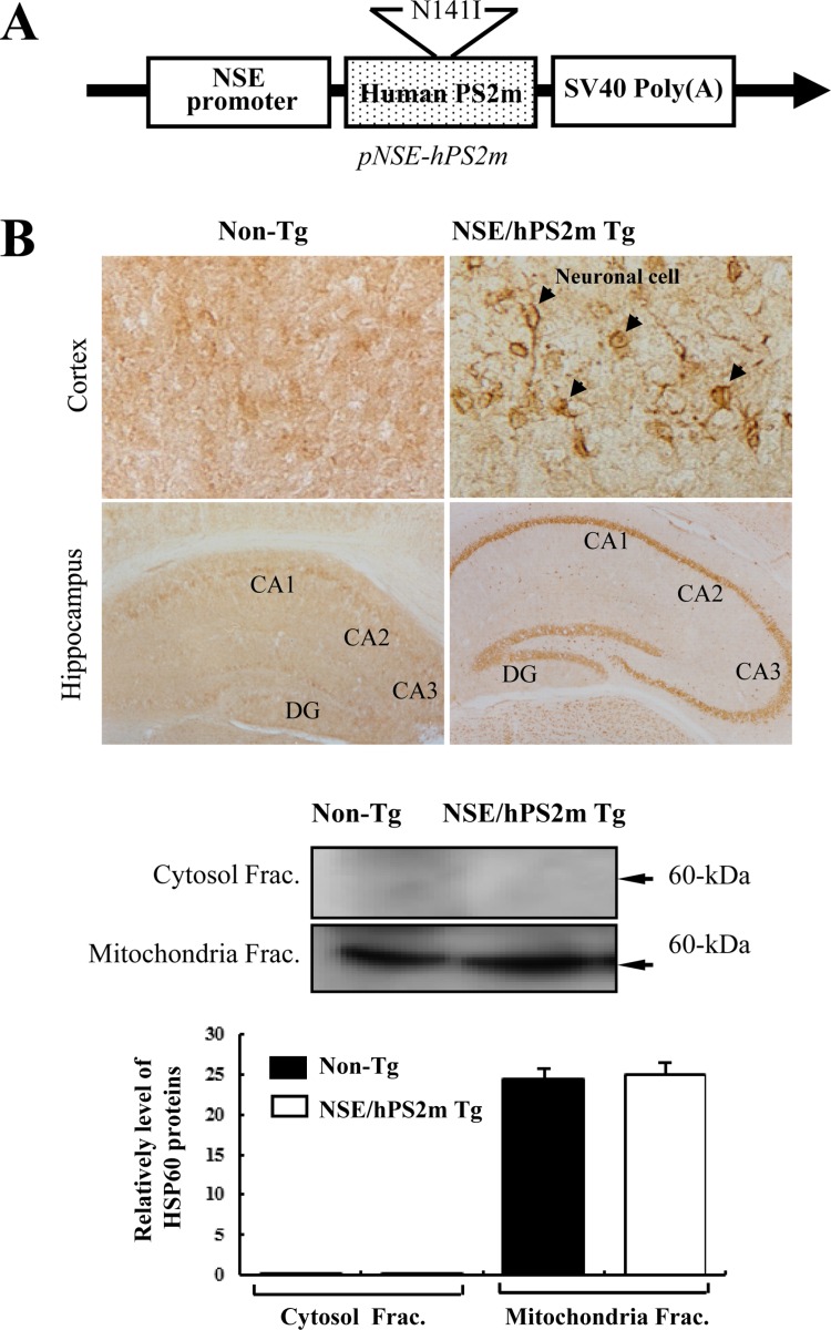 | Figure 1Construction of pNSE/hPS2m expression vector, deposition of Aβ-42 peptides in the brain and identification of the mitochondria fraction. (A) The map of pNSE/hPS2m expression vector. Human PS2 gene containing N141I mutation was inserted into pNSE/SV40 vector under the control of NSE promoter. (B) Immunostaining analysis of Aβ-42 peptide deposition. The deposition profile of Aβ-42 peptides was observed in brains at 12 months of age by immunostaining analysis. Broad distribution and high intensity of Aβ-42 peptide deposition are shown in the hippocampus and cortex of brain tissue derived from NSE/hPS2m Tg mice. CA: cornu ammonis, DG: dentate gyrus. (C) Identification of the mitochondria fraction. Expression of HSP60 protein was detected in the mitochondrial fraction derived from the hippocampus and cortex of brains of by Western blot analysis. Membrane-conjugated mitochondrial proteins were incubated with antibodies for HSP60 and β-actin proteins. Three fractions per group were assayed by Western blotting. The data represent the mean±SD from three replicates.
|
The protocols for the animal experiment were carefully reviewed for ethical and scientific care procedures and approved by the Pusan National University-Institutional Animal Care and Use Committee (PNU-IACUC; Approval Number PNU-2012-0050). All mice were provided with ad libitum access to standard irradiated chow diet (Samtako Inc., Osan, Korea) consisting of moisture (12.5%), crude protein (25.43%), crude fat (6.06%), crude fiber (3.9%), crude ash (5.31%), calcium (1.14%), and phosphorus (0.99%) and water. During the experiment, mice were maintained in a specific pathogen-free state under a strict light cycle (lights on at 08:00 hours and off at 20:00 hrs) at 23±2℃ and 50±10% relative humidity. The mice were housed in the Pusan National University-Laboratory Animal Resources Center accredited by the Korea Ministry of Food and Drug Safety (MFDS) in accordance with the Laboratory Animal Act (Accredited Unit Number-000231).
Immunohistochemistry
NSE/hPS2m Tg and age-matched Non-Tg mice were perfused as described [15]. After perfusion, brain tissue was fixed in 5% formalin at 4℃ for 12 hrs and transferred successively to 10-20 and 30% sucrose solution. Sections (10 µm) were prepared and pretreated at room temperature for 30 min with PBS-blocking buffer containing 10% goat serum (Vector Laboratories Inc. Burlingame, CA, USA) in PBS for 1 hr. These sections were incubated with primary rabbit polyclonal anti-Aβ-42 (Chemicon International, Inc. Billerica, MA, USA) at a dilution of 1:100 in tris-buffered saline (TBS) blocking buffer for 12 hrs. Each complex of antigen-antibody was visualized with biotinylated secondary antibody (goat anti-rabbit)-conjugated HRP streptavidin (Zymed, Histostain-Plus Kit) diluted 1:1,500 in PBS blocking buffer. Aβ peptides were detected using stable 3,3′-diaminobenzidine (DAB; Invitrogen, Carlsbad, CA, USA) and observed with Leica Application Suite (Leica Microsystems).
Purification of cytosol and mitochondrial fractions from brain
The purification of cytosol and mitochondrial fractions from the hippocampus and cortex of brain was performed with a Cytosol/Mitochondria Fraction Kit according to the manufacturer (Calbiochem Inc., San Diego, CA, USA). Brain tissues (200 mg) of mice were chopped with scissors in 1.5 mL of 1× Cytosolic extraction buffer containing DTT (1 µL) and Protease inhibitor cocktail (2 µL). Brain mixtures were homogenated with a glass grinder and incubated on ice for 10 min. The mitochondrial fractions were harvested from the homogenized tissue mixture at 700×g for 10 min at 4℃, after which the supernatant was transferred into a new tube in order to use the cytosolic fraction. Harvested pellets were resuspended with 0.1 mL of mitochondria extraction buffer mix containing DTT (1 µL) and protease inhibitor cocktail (2 µL), after which the protein concentration was detected with a BCA kit.
Western blot analysis
The proteins prepared from the hippocampus and cortex of brain tissues of Non-Tg and NSE/hPS2m Tg mice were separated by electrophoresis on 4-20% SDS-PAGE gels for 2 hrs and then transferred to nitrocellulose membranes for 2 hrs at 40 V. Each membrane was incubated separately with primary anti-PS1 FL (1:1,000, Sigma-Aldrich Co., Missouri, USA), anti-PS1 CTF (1:1,000, Sigma-Aldrich Co.), anti-PS1 NTF (1:1,000, Sigma-Aldrich Co.), anti-PS2 FL (1:1,000, Sigma-Aldrich Co.), anti-PS2 CTF (1:1,000, Sigma-Aldrich Co.), anti-PS2 NTF (1:1,000, Sigma-Aldrich Co.), anti-NCT (1:1,000, Sigma-Aldrich Co.), anti-APH-1 (1:1,000, Sigma-Aldrich Co.), anti-Pen-2 (1:2,000, Calbiochem Inc.), anti-bAPP (1:1,000, Sigma-Aldrich Co.), anti-C-99 (1:2,000, Calbiochem Inc.), anti-Aβ-42 (1:1,000, Calbiochem Inc.), anti-HSP60 (1:1,500, Sigma-Aldrich Co.), and anti-β-actin (1:2,000, Sigma-Aldrich Co.) antibodies overnight at 4℃. The membranes were washed with washing buffer (137 mM NaCl, 2.7 mM KCl, 10 mM Na2HPO4, 2 mM KH2PO4, and 0.05% Tween 20) and incubated with horseradish peroxidase-conjugated goat anti-rabbit IgG (Zymed) at 1:2,000 dilution at room temperature for 2 hrs. Finally, the blots were developed using Chemiluminescence Reagent Plus kit (Pfizer Inc., Gladstone, NJ, USA). The signal image for each protein was acquired using the digital camera (1.92 MP resolution) of the FluorChem® FC2 Imaging system (Alpha Innotech Corporation, San Leandro, CA, USA) and their density was semi-quantified using AlphaView Program version 3.2.2 (Cell Biosciences, Inc., Santa Clara, CA).
Assay of γ-secretase activity in mitochondrial fractions
Activity of γ-secretase in the mitochondrial fraction of brains from Non-Tg and NSE/hPS2m Tg mice was assayed using the γ-secretase activity assay procedure and reagents in the γ-secretase Assay kit (R&D System Inc., Minneapolis, MN, USA). Each mitochondrial fraction prepared using a cytosol/mitochondria fractionation kit was diluted with 1× Cell extraction buffer to yield a final protein concentration of roughly 0.5-2.0 mg/mL. The enzymatic reaction for secretase activity was carried out using the microplate provided by manufacturer and was read with a microplate fluorimeter. A total of 50 µL of 2× Reaction Buffer was added into each well containing about 25-200 µg of total protein (50 µL). Then, 5 µL of substrate was added to each well, and the plate was incubated in the dark at 37℃ for 1-2 hrs. Finally, the absorbance of each well was read by a fluorescent microplate reader using a light filter allowing for EDANS excitation between wavelengths of 335-355 nm, after which emitted light was collected between 495-510 nm.
Statistical analysis
Statistical analyses were performed using SPSS software version 10.10 (SPSS, Inc. Chicago, IL, USA). One-way analysis and post hoc Tukey's test of variance was performed to identify significant differences between Non-Tg and NSE/hPS2m Tg mice. All values are presented as the mean±standard deviation (SD). A P<0.05 was considered to indicate a statistically significant difference.
Go to : 
Results
Deposition of Aβ-42 peptides in brains of NSE/hPS2m Tg mice
Firstly, to detect the localization and deposition of Aβ-42 peptides induced by PS2 expression in brain tissues, Aβ-42 peptide immunoreactivity was analyzed in the hippocampus and cerebral cortex of brains using optical microscopy. These regions were chosen since they are primary target sites for Aβ peptides deposition and neuronal loss induced by γ-secretase activity in AD patients [1011]. As shown in Figure 1B, immunostaining intensity was spread throughout neuronal cells of the cerebral cortex in brain tissue from NSE/hPS2m Tg mice. However, the intensity level in Non-Tg littermates was slightly lower than that in NSE/hPS2m Tg mice. Many cells in the CA1, CA2, and CA3 regions were intensively immunostained with a dense line in the hippocampus of NSE/hPS2m Tg brains, whereas these regions showed low immunoreactivity in Non-Tg mice. Therefore, these results suggest that overexpression of the hPS2m gene could be attributed to the deposition of Aβ-42 peptides in the hippocampus and cortex of NSE/hPS2m Tg mice.
Identification of purity for mitochondrial fraction
Cytochrome c, Tim23, and HSP60 proteins are widely used as mitochondrial markers in various biological studies, whereas GM130, Syntaxin 13, and N-cadherin are used as markers of the ER, cis-Golgi, early endosomes, and plasma membrane [1617]. To examine the purity of the mitochondrial fraction, levels of HSP60 protein, as a mitochondrial marker, were detected in the mitochondrial fraction derived from brain tissue using Western blot analysis. HSP60 protein was detected only in the mitochondrial fraction and not in the cytosolic fraction, and there was no difference on the expression of HSP60 protein between Non-Tg and NSE/hPS2m Tg mice (Figure 1C). These results suggest that the methods applied in this study are very useful tools to harvest the mitochondrial fraction with high purity from brain tissue.
Effects of hPS2m overexpression on component levels of γ-secretase members
To investigate whether or not overexpression of hPS2m protein is associated with component levels of the γ-secretase complex in the mitichondria, protein levels of these components were detected in the mitochondrial fraction purified from brain tissue. For PS protein, which is a main catalytic activator of the γ-secretase complex, full-length PS1 protein was not detected in both mice, whereas PS2 protein was detected at a high level. Furthermore, PS2-FL protein was significantly down-regulated in NSE/hPS2m Tg mice compared to Non-Tg mice (P<0.032). On the other hand, protein level of PS1 and PS2 c-terminal fragment, a main functional protein, was significantly higher in brain tissue of NSE/hPS2m Tg mice compared to Non-Tg mice (P<0.015). Protein levels of NCT and APH-1, a stabilization protein of γ-secretase complex, were not significantly different between Non-Tg and NSE/hPS2m Tg mice, although NCT protein was slightly down-regulated (P<0.071). However, protein level of Pen-2, an activator of the γ-secretase complex, was significantly up-regulated in NSE/hPS2m Tg mice compared to Non-Tg mice (P<0.043) (Figure 2). These results suggest that over-expression of hPS2m protein could induce alterations of main component protein and activator protein of the γ-secretase complex but not stabilization-related protein in the mitochondria of NSE/hPS2m Tg mice.
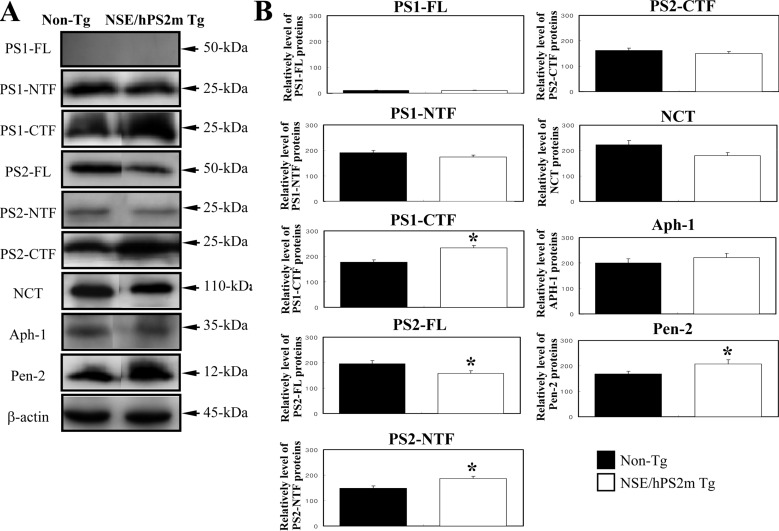 | Figure 2Expression of γ-secretase components in the mitochondrial fraction. The mitochondrial fractions were separated on 4-20% SDS-PAGE, transferred to nitrocellulose membranes, and subsequently incubated with specific antibody against each γ-secretase components. Three fractions per group were assayed by Western blotting. The data represent the mean±SD from three replicates. *, P<0.05; significant difference between the Non-Tg and NSE/hPS2m Tg mice.
|
Effects of hPS2m overexpression on γ-secretase activity
To determine whether or not alteration of γ-secretase components induced by overexpression of hPS2m protein can regulate γ-secretase activity in the mitochondria, γ-secretase activity was measured in the mitochondrial fraction purified from brain tissues of both mice. As shown in Figure 3A, γ-secretase activity was significantly elevated in the mitochondrial fraction of brain tissue derived from NSE/hPS2m Tg mice compared to Non-Tg mice (P<0.013). These results suggest that alteration of γ-secretase components induced by overexpression of hPS2m protein could up-regulate γ-secretase activity in the mitochondrial fraction of NSE/hPS2m Tg mice.
 | Figure 3Activity of γ-secretase complex and production of Aβ-related proteins in the mitochondrial fraction. (A) Activity of γ-secretase complex. The γ-secretase substrate was mixed with the mitochondrial fraction derived from the hippocampus and cortex of either Non-Tg or NSE/PS2m Tg mice. Absorbance of each well was read by a fluorescent microplate reader using a light filter allowing for EDANS excitation between wavelengths of 335-355 nm, after which the emitted light was collected between 495-510 nm. (B) Production of Aβ-related proteins. The mitochondrial fraction derived from the hippocampus and cortex of brains were separated on 4-20% SDS-PAGE, transferred to nitrocellulose membranes, and subsequently incubated with specific antibody for each protein and β-actin protein. Three fractions per group were assayed by Western blotting. The data represent the mean±SD from three replicates. *, P<0.05; significant difference between the Non-Tg and NSE/hPS2m Tg mice.
|
Effect of hPS2m overexpression on production of Aβ-42 peptides
To investigate the effect of γ-secretase changes on production of Aβ-42 peptides in the mitochondria, the peptide level of Aβ-42 was analyzed in the mitochondrial fraction of Non-Tg and NSE/hPS2m Tg mice using a specific antibody. A high level of Aβ-42 peptides was successfully detected in the mitochondrial fraction of NSE/hPS2m Tg mice. Protein levels of bAPP and C-99, which is a C-terminal fragment of APP involving Aβ-42 peptides, were significantly up-regulated in NSE/hPS2m Tg mice compared to Non-Tg mice (P<0.039) (Figure 3B). Therefore, our results suggest that elevation of γ-secretase activity induced by hPS2m overexpression could stimulate production of Aβ-42 peptides in the mitochondrial fraction.
Go to :
Discussion
Mitochondria play an important role in the apoptosis pathway, which is an essential process required for the development and maintenance of normal tissues [11]. Various apoptotic stimuli originating from the extracellular environment cause increased permeability of the outer mitochondrial membrane, resulting in release of mitochondrial apoptogenic proteins such as cytochrome c and Smac/Diablo into the cytosol [18]. This increased permeability is negatively and positively regulated by anti-apoptotic and pro-apoptotic members of the Bcl-2 family, respectively [1920]. Especially, previous reports have demonstrated the potential involvement of Aβ peptides and γ-secretase activity in the induction or regulation of apoptotic neuronal death [172122]. Therefore, in this study, mitochondria were examined as a trigger of abnormal onset of neuronal cell death induced by mPS2 in AD.
Several reports have suggested that all γ-secretase components are located in the mitochondria. Especially, the role and importance of γ-secretase activity to mitochondria were provided from the results that NCT together with APH-1, Pen-2, and PS1 form a high molecular weight complex located in mitochondria [17]. In this study, to investigate the effects of mutant PS2 overexpression on the γ-secretasecomplex in mitochondria, expression levels of γ-secretase components as well as activity of γ-secretase were measured in the mitochondrial extract derived from brain tissue of NSE/hPS2m Tg mice. C-terminal fragments of PS1 and PS2 protein were significantly up-regulated in the mitochondria of NSE/hPS2m Tg mice compared to non-transgenic mice, whereas full-length NTF was unaltered in NSE/hPS2m Tg mice. Furthermore, the level of Pen-2 protein, as an activator of γ-secretase, was higher in NSE/hPS2m Tg mice than in Non-Tg mice. However, there was no difference in NCT or APH-1 protein level between the two mice. These results suggest that mutant PS2 could induce core and activator protein expression of γ-secretase in mitochondria derived from brain tissue of NSE/hPS2m Tg mice.
Activity of γ-secretase was detected in mitochondria using β-APP from BD8 cells or recombinant C100-Flag as substrates. Formation of AICD was abolished in the presence of well characterized γ-secretase inhibitors [17]. These studies demonstrated that γ-secretase complexes located in mitochondria are active and can cleave β-APP. In this study, the activity level of γ-secretase was detected in the mitochondrial fraction derived from brain tissue of NSE/hPS2m Tg mice. mPS2 overexpressed in mitochondria could induce up-regulation of γ-secretase activity in brains of NSE/hPS2m Tg mice compared to Non-Tg mice.
Previous studies have shown that energy depletion and oxidative stress can induce amyloidogenic changes in APP processing [232425]. These results suggested that Aβ production is potentially linked to mitochondrial dysfunction and oxidative stress [26]. Intracellular Aβ-42 peptides produced by APP processing partially colocalize with APP and appear predominantly in the golgi complex and endosomal vesicles. Furthermore, these products such as Aβ-42 peptides, APP, and C-terminal fragments have been shown to accumulate in a detergent-insoluble form in APP-overexpressing cells [27]. Our study showed that protein levels of β-APP, C-99, and Aβ-42 were significantly higher than in NSE/hPS2m Tg mice than in Non-Tg mice. These results suggest that overexpression of the hPS2m gene may induce accumulation of Aβ-related byproducts in hPS2m-overexpressing cells derived from NSE/hPS2m Tg mice similar to APP-overexpressing cells.
Taken together, these results suggest that overexpression of mPS2 gene in mitochondria of brain tissue (hippocampus and cortex) derived from NSE/hPS2m Tg mice may induce changes in the component expression and activity of the γ-secretase complex. A high level of activity may have up-regulated production of Aβ-related products in NSE/hPS2m Tg mice. These results suggest that mutant PS2 plays a critical role in the progression of AD pathology.
Go to :
 XML Download