

 PDF
PDF ePub
ePub Citation
 Print
Print


In modern society, change in the pattern of food consumption increases metabolic syndrome such as obesity, atherosclerosis, hypertension, stroke, diabetes, and cancer [12]. In particular, hyperlipidemia is indicated to be the main cause of health problems in metabolic syndrome [3]. The major risk factors of atherosclerosis and cardiovascular diseases are increased blood levels of total cholesterol (TC) and low-density lipoproteins (LDL), accompanying decrease in high-density lipoproteins (HDL) [45]. It is well known that oxidized LDL (OxLDL) is a triggering molecule of endothelial injury and atheroma formation, whereas HDL is a transporter of cholesterol for metabolism in the liver [6789]. Therefore, it is believed that suppression of blood TC and LDL as well as enhancement of HDL are front-line strategies for the prevention of atherosclerosis [89].
To date, statins has been used to inhibit 3-hydroxy-3-methylglutanyl-coenzyme A (HMG-CoA) reductase, a rate-limiting step enzyme in cholesterol biosynthesis. In addition, statins enhance the activity of LDL receptors on cell membrane, facilitating the metabolism of very low-density lipoproteins (VLDL) remnant and LDL [1011]. In spite of the cholesterol-lowering advantage, however, over-dosage and/or long-term use of statins exert serious adverse-effects like hepatomegaly and renal hypertrophy [1213]. Thus, researches on the novel natural products with minimal adverse-effects to control blood cholesterol level and to improve blood flow are required.
Since the consumption of high-fat diets is the major cause of disturbances in blood flow, selection of fats and oils might be very important. It is well known that unsaturated fatty acids (UFA) properly regulate blood lipid profiles, and thereby improved coronary heart disease [14151617]. Fish oil ω-3 polyunsaturated fatty acids (PUFA) prevented vasoconstriction [18], and suppressed vascular inflammatory response by decreasing production of reactive oxygen species (ROS) [1920]. Notably, α-linolenic acid (ALA), a well-known ω-3 PUFA rich in perilla oil improved insulin sensitivity and hyperlipidemia, and prevented coronary heart disease [2122]. Especially, in recent studies, we demonstrated that perilla oil possessing a low linoleic acid/α-linolenic acid (ω-6/ω-3) ratio not only inhibited platelet aggregation and improved blood flow [23], but also delayed and attenuated brain hemorrhage in stroke-prone spontaneously hypertensive rats (SHR-SP), thereby extending their lifespan [24].
Since PUFA affects both platelets and endothelial cells that play a crucial role in the regulation of thrombosis and haemostasis [25], we investigated the effects of perilla oil on the high-cholesterol diet (HCD)-induced hypercholesterolemia and atherosclerosis.
Materials and Methods
Materials
Perilla oil was obtained from Anydoctor Healthcare Co., Ltd. (Cheonan, Korea). Perilla oil was extracted under a cold-pressed method at 30-48℃, and analyzed with Varian 3800 gas chromatograph (Varian Inc., Walnut Creek, CA, USA) equipped with a Supelcowax 10 fused-silica capillary column (Supelco, Bellefonte, PA, USA). From the fatty acid analysis, it was found that perilla oil contains 72.12% PUFA, 19.1% monounsaturated fatty acids (MUFA), and 8.49% saturated fatty acids (SFA). Especially, among PUFA, 57.47% was ω-3 α-linolenic acid [ALA, 18:2(n-3)] [2324].
Animals
Seven-month-old male New Zealand white rabbits (body weight 2.5 kg) were procured from Samtaco Co. (Osan, Korea), and subjected to the experiment after 2-week acclimation to the laboratory environment. The animals were housed in each cage with free access to feed and water under constant environmental conditions (23±2℃ temperature; 45-65% relative humidity; 12-hour light-dark cycle; 150-300 lux brightness). All the animal experiments were conducted according to the Standard Operation Procedures, and approved by the Institutional Animal Care and Use Committee of Chungbuk National University, Korea.
Induction of hypercholesterolemia and treatment
Acute dietary hypercholesterolemia was induced by feeding rabbits with a powdered HCD containing 0.5% cholesterol and 1% corn oil for 2 weeks, followed by 0.5% cholesterol for additional 10 weeks during treatment period [262728]. After 2-week induction period, the animals were grouped (n=6/group) according to their blood cholesterol levels to adjust to similar mean values. To assess therapeutic efficacy of perilla oil against hypercholesterolemia and atherosclerosis, the rabbits were fed the HCD containing 0.1 or 0.3% perilla oil during the 10-week period.
Blood biochemistry
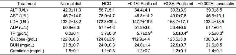
Body weights were recorded every week from immediately before starting the experiment. After 16-hour fasting at the end of 2-week hypercholesterolemia induction period and every 2 week during 10-week treatment period, blood sample was collected from auricular artery, and lipid profiles and parameters of hepatic and renal functions were measured in sera using a blood chemistry analyzer (Hitachi-747; Hitachi Korea, Seoul, Korea). The parameters include TC, LDL, HDL, triglycerides (TG), alanine transaminase (ALT), aspartate transaminase (AST), lactate dehydrogenase (LDH), alkaline phosphatase (ALP), total proteins (TP), glucose, blood urea nitrogen (BUN), and creatinine.
Measurement of atheroma
The aortic arch (10 cm) was dissected from the aortic valve of the heart and washed with saline. The sample was dissected in 5 cm long from the orifice of carotid artery, and opened longitudinally. After removing fats and tissues adhering to the adventitia, the aorta was dehydrated with 100% propylene glycol for 10 min, and stained with 0.7% Sudan IV (in propylene glycol) for 10 min [262728]. Then, it was hydrated with 85% propylene glycol for 5 min, washed with distilled water, and photographed and analyzed with Digital Image Analyzer (Image Inside; Focus, Seoul, Korea) for red atheromatous plaques. The extent of lipid accumulation (atherosclerosis index; AI, %) was calculated as the percent Sudan-positive area to the total area of the aortic wall.
Microscopic examination
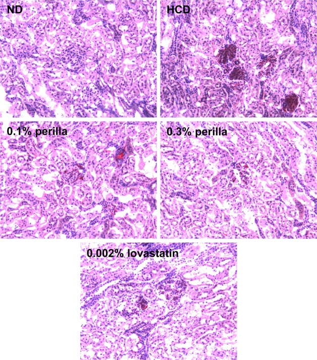
After weighing, liver and kidney tissues were frozen at -70℃, and cryosections (10 µm in thickness) were mounted on gelatin-coated slides. The sections were washed with phosphate-buffered saline (PBS, pH6.5) followed by propylene glycol for 2 min, and stained with 0.5% Oil red O for 1 hour. After washing with 85% propylene glycol, the slides were counterstained with Harris' hematoxylin, washed again with distilled water, and observed under a light microscope.
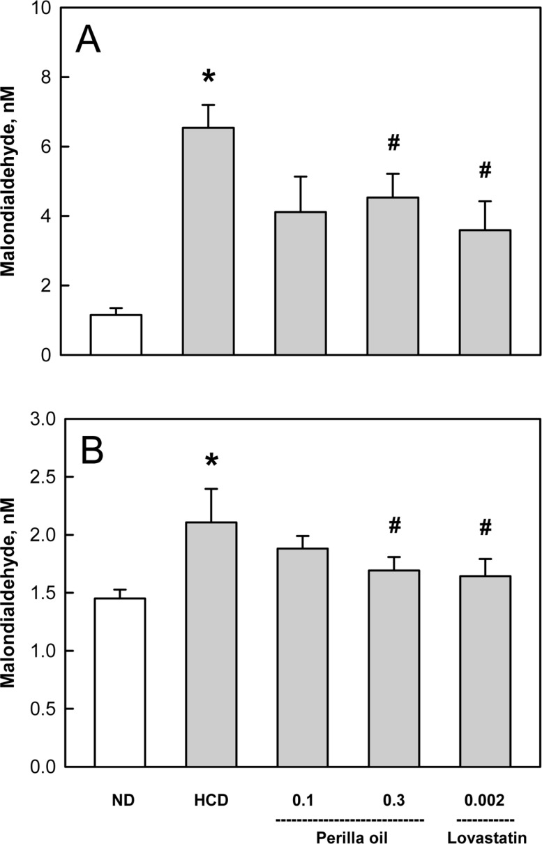
Measurement of lipid peroxidation
The liver and kidney tissues were dissected on an ice block. The tissues were homogenized in 19 volumes of 10 mM PBS (pH7.4) to make a 5% homogenate at 4℃. Into the tissue homogenate (250 µL), 250 µL sodium dodecyl sulphate (SDS, 8.1% solution) and 500 µL 20% acetic acid (adjusted to pH3.5) were added. After adding 250 µL 2-thiobarbituric acid (TBA, 0.75% solution), the mixture was boiled in a glass tube capped with aluminum foil for 30 min [29]. Samples were cooled on ice, centrifuged at 13,000 g for 10 min, and absorbance of the supernatant was read at 532 nm for the quantification of malondialdehyde (MDA).
Statistical analysis
The results are presented as means±standard error. The significance of differences of all results was analyzed by one-way analysis of variance followed by the Dunnett's multiple-range test correction, using SPSS version 12.0 (SPSS Inc., Chicago, IL, USA). Statistical significance was set a priority at P<0.05.
Results
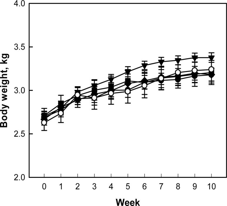
During 10-week treatment period, following 2-week hypercholesterolemia-induction period, there were no abnormal symptoms in control and treated rabbits. The rabbits exhibited a gradual increase in their body weights, although the body weight gain of rabbits fed 0.1% perilla oil was slightly higher than that of control animals without statistical significance (P>0.05) (Figure 1).
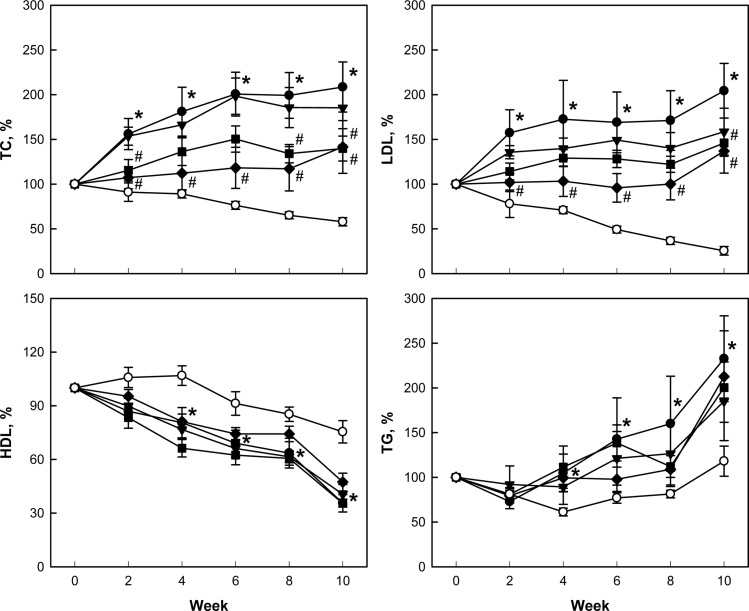
After 2-week feeding of 0.5% cholesterol and 1% corn oil for hypercholesterolemia induction, blood TC was dramatically elevated to 738-754 mg/dL from 59 mg/dL of normal level. LDL also increased to 237 mg/dL from 19 mg/dL in normal animals. In comparison, there were slight changes in blood HDL (from 30 to 38-50 mg/dL) and TG (from 47 to 29-47 mg/dL). During additional 10-week feeding only 0.5% cholesterol, the TC, LDL, and TG levels doubled, but HDL decreased to 40% of initial concentration (Figure 2). Notably, addition of perilla oil attenuated the increases in TC, LDL, and TG in a concentration-dependent manner, without affecting HDL level. Such effects were also achieved with lovastatin.
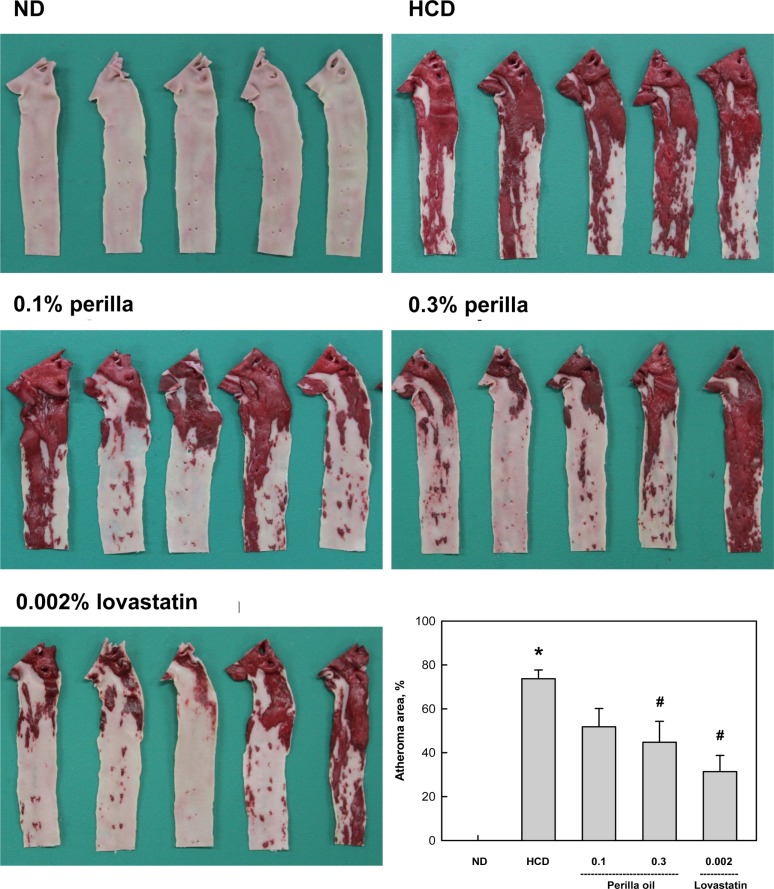
A long-term feeding of HCD caused extensive atheromatous plaques covering 74% of the aortic arch and abdominal artery (Figure 3). The degree of atheromatous plaques formation was markedly attenuated by 10-week treatment with perilla oil to 52% and 45% at 0.1% and 0.3% in diet, respectively. Lovastatin (0.002% in diet) also decreased the atheroma area to 31% of HCD group.
In blood biochemical analysis, a long-term hypercholesterolemia significantly reduced TP level, in comparison with slight increases in ALT, AST, and LDH, indicative of hepatic dysfunction (protein synthesis) rather than acute hepatocytic injury (Table 1). ALP, glucose, BUN, and creatinine levels were not affected by hypercholesterolemia. Notably, perilla oil and lovastatin remarkably restored the blood levels of hepatic dysfunction to normal levels.
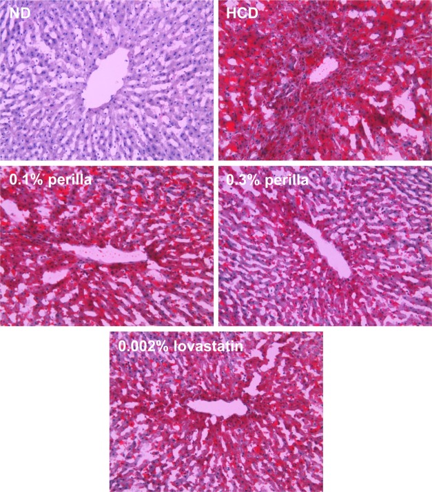
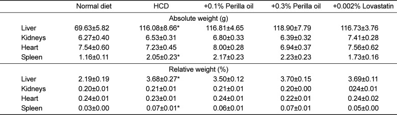
The long-term feeding of HCD significantly increased the weights of the liver and spleen, which were not affected by treatment with perilla oil or lovastatin (Table 2). In microscopic findings, extensive accumulation of lipids were observed in the liver (Figure 4). It is of interest to note that the hepatic steatosis was markedly attenuated by feeding perilla oil in a concentration-dependent manner, in which the effect of 0.3% perilla oil was higher than that of 0.002% lovastatin. Lipid accumulation was also observed focally around the glomerular structures (Figure 5). The glomerular lipid accumulation was remarkably inhibited by perilla oil and lovastatin. Particularly, 0.3% perilla oil fully prevented the renal lipidosis.
In the analysis of lipid peroxidation in the organs showing lipidosis, the MDA concentrations in the liver and kidneys was increased 4 folds and 1.5 folds, respectively, by the long-term HCD feeding (Figure 6). Interestingly, perilla oil (0.1-0.3%) displayed a marked anti-oxidative activity on the MDA formation, comparable with the effect of lovastatin (0.002%).
Discussion
In the present study, perilla oil substantially inhibited hypercholesterolemia-mediated atheroma formation and lipid accumulation in the liver and kidneys. In addition, perilla oil attenuated the oxidative membrane injury (lipid peroxidation) in the organs showing lipidosis.
Actually, statins such as lovastatin is preferentially prescribed to suppress hepatic cholesterol synthesis, remarkably improving blood lipid profiles [3031]. However, it is well known that over-dosage and long-term administration of the statins cause severe hepatotoxicity, and that low doses could not effectively control the blood cholesterol from diets [1227]. Notably, management of risk factors in preventive mode, rather than therapeutic mode, of the cardiovascular and cerebrovascular diseases is extremely important, because the time to death after outbreak of the diseases is very short [32]. Accordingly, dietary restriction and appropriate choice of fats or oils have been recommended.
While excessive consumption of fats and oils, especially containing high levels of saturated fatty acids (SFA), is known to be harmful for vascular diseases, UFA such as α-linolenic acid (ALA, ω-3) are believed to be beneficial. In fact, it has been reported that perilla oil containing a high level ω-3 PUFA decreased blood TG, TC, and LDL in animals and humans [2033]. Also in human studies, perilla oil not only improved blood HDL and recovered the function of arterial endothelial cells [34], but also reduced blood TG and the risk of cardiac infarction [35]. Also, in the present study, perilla oil containing 72.12% PUFA improved the blood lipid profiles and lipid accumulation in tissues, leading to atherosclerosis and hepatic and renal steatosis.
Notably, in our gas chromatographic analysis of perilla oil, 57.47% was ALA out of 72.12% PUFA. Supportively, it was demonstrated that ALA inhibited platelet activation and arterial thrombus formation [36]. In our recent study, perilla oil markedly suppressed platelet aggregation as well as thrombus formation in the FeCl3-induced endothelial injury model, too [23]. Activated platelets attach to vascular endothelial walls injured during oxidative reaction mediated by OxLDL, aggregate there, and form thrombus and atherosclerosis. Therefore, perilla oil has attracted investigators' attention, because a diet rich in PUFA may be helpful in preventing heart diseases [223738] and blood coagulation [17].
More importantly, it was demonstrated that most of the plant oils with high ω-6/ω-3 fatty acid ratios including canola oil, safflower oil, olive oil, corn oil, and soybean oil, increased hemorrhagic stroke in SHR-SP and shortened their life span, except only perilla oil with a low ω-6/ω-3 fatty acid ratio [15163940]. In our previous study, perilla oil with 0.25 ω-6/ω-3 fatty acid ratio delayed and attenuated brain hemorrhagic stroke and renal lesions in SHR-SP, whereas canola oil with 2.63 ω-6/ω-3 ratio aggravated the lesions, advancing time to death [24].
It was reported that ω-3 PUFA has anti-oxidative and anti-inflammatory activities; it inhibited C-reactive protein in an atherosclerosis model [20] and increased mucosal blood flow by inhibiting leukotriene production in an inflammatory bowel disease model [41]. In the present study, the anti-oxidative activity of perilla oil was also confirmed: i.e., feeding perilla oil markedly suppressed lipid peroxidation in the fatty liver and kidneys following HCD-induced hypercholesterolemia.
To date, statins and anti-coagulants have been used to improve hyperlipidemia and blood flow, and thereby to prevent atherosclerosis and cardiovascular diseases [424344]. In fact, it is well known that platelet activation and aggregation is an initial feature of thrombus formation on the injured arterial endothelium undergoing atherosclerosis [45]. However, the most important triggering factor of oxidative injury of arterial intimal layer, leading to platelet aggregation and clonal expansion of vascular smooth muscle cells, is OxLDL containing a high level of cholesterol. By comparison with harmful effects of various fats and plant oils containing high ω-6/ω-3 fatty acid ratios [3940], it was confirmed that perilla oil, only an oil containing a low ω-6/ω-3 ratio, exerted beneficial effects on hemorrhagic stroke and atherosclerosis in our previous and present studies. Therefore, it is strongly recommended that perilla oil could be the first choice on modern food tables consuming high amount of fats and oils.
 XML Download