

 PDF
PDF ePub
ePub Citation
 Print
Print


Helicobacter spp. are gram-negative, microaerobic, motile, fusiform, spiral-shaped bacteria; they may have a curved to spiral rod morphology and move using flagella that vary in number and location among different species [12]. Most of the species are associated with either hepatic or gastric infections [2]. At least 24 Helicobacter spp. have been reported to date and it is likely that several more await discovery [1]. Helicobacter spp. colonize the gastrointestinal tract of humans and several animal species, such as cats, dogs, ferrets, pigs, cheetahs, and monkeys [3]. In humans, Helicobacter pylori is a major cause of chronic diffuse superficial gastritis and peptic ulcers. It is also considered a cofactor in the development of gastric malignancies.
A number of Helicobacter species may confound experimental data because of their association with disease progressing in various kinds of laboratory animals [456]. Thus, Helicobacter infection of laboratory animals may influence the results of research, and it is necessary to clarify the current status of Helicobacter contamination in laboratory animal colonies. Screening of Helicobacter species in laboratory animals is particularly desirable, because they are prevalent in commercial and research animal facilities [789]. The main gastric Helicobacter spp. in dogs are Helicobacter heilmannii and Helicobacter felis [3]. To date, H. heilmannii has not been reliably cultured in vitro [10]. However, both H. heilmannii and H. felis can be identified by electron microscopy or by analysis of their 16S rRNA and urease gene sequences [1112].
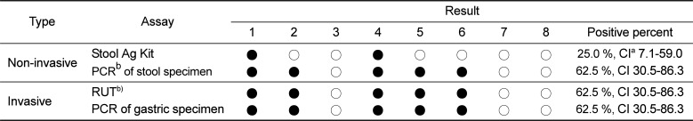
Several methods have been used to diagnose H. pylori infection. There is an increasing interest in non-invasive tests, as they do not require endoscopic assessment [13]. The 13C-urea breath test (UBT) is the most recommended non-invasive test for detecting H. pylori infection and has a high sensitivity and specificity [14]. However, the UBT cannot be applied to animals due to its high cost and the requirement for expensive analytical instruments [15]. For this reason, many researchers have used polymerase chain reaction (PCR) assays to monitor infection in animal stools [1617]. However, PCR assays can be time-consuming and expensive [18]. In addition, DNA extraction and amplification from stool samples can be difficult [19]. Recently, several commercial companies have developed H. pylori stool antigen (HpSA) test kits. HpSA tests are non-invasive diagnostic assays for the detection of H. pylori infection in human stool samples [202122]. Presently, there is little information on the usefulness of the HpSA test in detecting other Helicobacter spp. In this study, Helicobacter spp. infection was detected in dogs with gastric disease by PCR and RUT from stool samples or gastric biopsy specimens. In addition, the ability of the HpSA assay to detect Helicobacter spp. in the stools or gastric biopsies of the infected dogs was compared to that of PCR and RUT.
Materials and Methods
Study animals
Eight dogs displaying symptoms of gastritis (e.g. vomiting) were evaluated in this study. None of the animals had been treated with antibiotics during the four-week period prior to examination. The age of the dogs ranged from six months to eight years (mean 3 years). Five of the dogs were male and three were female. The time interval from the last vomiting episode and the sampling was recorded. All studies were performed in accordance with the Guide for Animal Experimentation by Wonkwang University and approved by the Institutional Animal Care and Use Committee of Wonkwang University (Approval No. WKU11-125.) All efforts were made to minimize pain or discomfort of animals used.
Gastroscopic biopsy and stool specimens
Fasting dogs were anesthetized with diazepam (0.2 mg/kg of body weight) and ketamine (3 to 5 mg/kg given until effective); the dogs were intubated, and anesthesia was maintained with halothane-oxygen. During gastroscopy, the macroscopic appearances of the mucosa were recorded and biopsy samples were taken and then analyzed by the RUT and PCR. Endoscope and biopsy forceps were disinfected with 4% Sekusept Plus solution (Henkel, Muttenz, Switzerland) for 30min and thoroughly flushed with tap water prior to use.
Stool samples were collected in sterile screw-capped containers and stored at room temperature. Half of the sample was used for the HpSA and the other half for PCR analysis. The sample for PCR analysis was processed within 24 h of being collected. Each collection swab was resuspended in 2 mL of 0.1 M phosphate buffered saline (PBS) buffer and vortexed. The PBS suspension was then used to extract genomic DNA for PCR analysis.
Rapid urease test with gastroscopic biopsy specimens
The gastroscopic biopsy specimens were minced and tested for urease activity using a CLO Helicobacter-detection kit (Asan Pharm Co., Ltd., Seoul, Korea). The specimens were incubated at 35℃ for 24 h prior to reading the assay. Negative and positive reactions were indicated as bright yellow and dark red color changes, respectively.
HpSA kit
Stool samples were evaluated using the commercially available SD Bioline HpSA kit following the manufacturer's instructions. Samples (250 mg) were incubated at room temperature for 30 min in the diluents provided in the kit and then 100 µL was transferred to the assay device. The test results were read after 15 min. One red line indicated negative and double red line indicated Helicobacter-positive result.
DNA extraction from gastric biopsies and stool specimens
Gastroscopic biopsy tissues were homogenized and resuspended in PBS for DNA extraction. Briefly, genomic DNA was extracted using an AccuPrep Genomic DNA Extraction kit (Bioneer Corp., Daejeon, Korea) according to the manufacturer's instructions. DNA was eluted in Tris-EDTA buffer (pH 8.0) and was stored at -20℃ until required. Aliquots (x µL) were used for PCR amplification.
Genomic DNA from the stool samples of the dogs was extracted using the AccuPrep Stool DNA Extraction Kit (Bioneer, Korea) according to the manufacturer's instructions. AccuPrep Stool DNA Extraction Kit is designed for the rapid extraction of DNA from fresh or frozen stools containing PCR inhibitors. The kit uses a glass filter fixed in a column tube that can efficiently bind DNA in the presence of chaotropic salts. Using the spin-column method, contaminants and enzyme inhibitors such as heparin, bilirubin bile salts, and porphyrin are eliminated, and after the washing steps, which remove proteins and salt, high-purity DNA is finally eluted using a low-concentration elution buffer, which is ready for use in a variety of applications [18]. It yields between 2 and 5 µg of DNA from 100 mg stool [18].
Consensus polymerase chain reaction to detect Helicobacter genus
A set of primers HF (5'-ACTTTAAACGCATGAA GATAT-3') and HR (5'-ATATTTTGACCTTCTGGGGT-3') was used to amplify the Helicobacter rpoB gene (458 bp) [18]. The template DNA (50 ng) and 20 pmol of each primer were added to a PCR tube (Maxime PCR PreMix; iNtRON Biotechnology, Korea) containing 1 U of Taq DNA polymerase, 250 µM each deoxynucleoside triphosphate, 50 mM Tris-HCl (pH 8.3), 40 mM KCl, and 1.5 mM MgCl2. The volume was adjusted with distilled water to 20 µL. The reaction conditions were 5 min at 95℃ followed by 40 cycles of 30 s at 94℃, 30 s at 52℃, 45 s at 72℃ with a final 5 min extension at 72℃. The PCR products were electrophoresed on a 1.2% (wt/vol) agarose gel. Positive samples were further analyzed using a species-specific PCR and DNA sequencing to identify individual Helicobacter spp.
Multiplex species-specific PCR
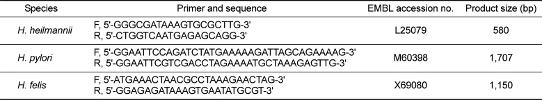
For the multiplex species-specific PCR, primers were designed based on the urease B gene sequence of each Helicobacter spp. Table 1 shows the sequences of the primers, the EMBL accession numbers of the urease B gene, and the expected amplicon lengths [17]. The template DNA (50 ng) and 20 pmol of each primer were added to a PCR tube containing 1 U of Taq DNA polymerase, 250 µM of each deoxynucleoside triphosphate, 50 mM Tris-HCl (pH 8.3), 40 mM KCl, and 1.5 mM MgCl2. The thermocycling parameters were as follows: one cycle of 94℃ for 3 min, 57℃ for 2 min, and 72℃ for 3 min followed by 31 cycles of 94℃ for 30 s, 57℃ for 30 s, and 72℃ for 1 min and a final extension at 72℃ for 5 min. PCR products were analyzed by electrophoresis on a 1.2% agarose gel. The sizes of the expected amplicons were 580, 1,707, and 1,150 bp for H. heilmannii, H. pylori, and H. felis, respectively.
Antibiotic therapy and treatment evaluation
Following a positive diagnosis of Helicobacter spp. infection, the dogs underwent a triple therapy with clarithromycin (0.24 mg/head), metronidazole (0.72 mg/head), and omeprazole (0.021 mg/head). The treatment was administered once a day for two weeks. For the evaluation of the therapy, stool specimens were collected from the dogs and genomic DNA was extracted as previously described. A multiplex Helicobacter speciesspecific PCR to amplify the urease B gene was performed. The dogs were observed for a further three months following the antibiotic therapy.
Results
Consensus PCR of gastric biopsy and stool samples
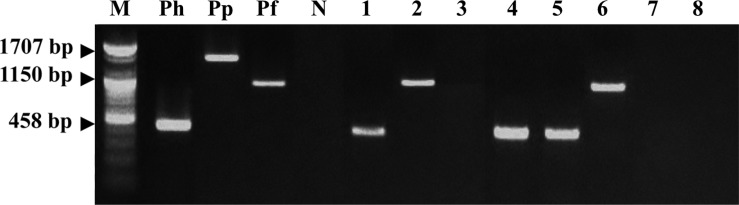
A consensus PCR to amplify the rpoB gene was used to detect Helicobacter spp. in both gastric biopsies and stool samples. The detection limit of the consensus PCR was 0.1 pg of Helicobacter DNA [18]. Five dogs (62.5%) were positive using the consensus PCR using gastric biopsies (Figure 1). Table 2 shows that the consensus PCR detect successfully with both gastric biopsies and stool samples.
Species-specific PCR using gastric biopsies and stool specimens
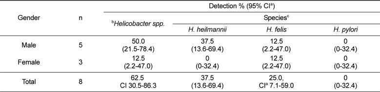
A multiplex species-specific PCR on gastroscopic biopsy and stool specimens was conducted with the primers for the urease B gene. No amplification products corresponding to H. pylori were detected, but some specimens tested were positive for H. felis or H. heilmannii (Figure 2). Table 3 shows the results of the multiplex species-specific PCR which reveal that three (37.5%) and two (25%) of the eight dogs were infected with H. heilmannii and H. felis, respectively. All of the samples that were positive by the consensus PCR were confirmed using the species-specific PCR.
Discussion
In laboratory animals, a lot of Helicobacter species have identified and revealed highly prevalent in animal facilities [789]. Therefore, the monitoring and control of Helicobacter species has been required for better experimental results in laboratory animals. Dogs can be infected with different Helicobacter spp., and coinfections are also know to occur [323]. The presence of Helicobacter spp. in the stools of the dogs meant that they were harboring the bacterium. Therefore, the detection rate of Helicobacter species is very important. This study reports a prevalence of fecal Helicobacter spp. in five (62.5%) of eight pet dogs studied. In addition, three (37.5%) and two (25%) of the dogs were infected with H. heilmannii and H. felis, respectively. A high prevalence of Helicobacter spp. has previously been reported in pet dogs and cats with gastritis and so our present data confirms these findings [1824].
The current gold standard for diagnosing H. pylori infection is endoscopic biopsy of the gastric tissue to be analyzed by RUT, histology, and culture. However, these invasive procedures have major disadvantages, which include discomfort to the patient, the use of anesthesia, and possible ethical issues [25]. In contrast, noninvasive tests are easy to perform and do not produce any significant discomfort for the patient. Such non-invasive tests for H. pylori include serological antibody testing, the UBT, and the HpSA test [25].
The HpSA test and PCR of stool specimens for diagnosing Helicobacter spp. infection offers a useful non-invasive method without having to sacrifice animals. In this study, the HpSA did not efficiently detect Helicobacter spp. in stool samples. On the other hand, the RUT and PCR assay using gastric and stool samples did detect successfully Helicobacter spp. It must be considered, however, that the poor performance of the HpSA may be due to the degradation of the Helicobacter spp. as it passes through the intestine. In addition, excretion of Helicobacter spp. from the intestine of the animal may vary with time. Furthermore, use of Nacetylcysteine-like mucolytic agents may decrease the accuracy of the diagnosis [24]. Cut off titer, though difficult to decide but crucial to reach the conclusion by using antigen detection technique [26]. Several limitations with PCR analysis have also been reported for Helicobacter spp. [1827]. Stool specimens are easy to obtain and are consequently of high interest for the development of direct methods for the detection of Helicobacter spp. [1823]. PCR has been successfully used to detect bacteria in stools even though stool samples remain the most difficult specimens from which DNA can be extracted and amplified [19]. This difficulty is due to the presence of DNA polymerase inhibitors like complex polysaccharides in the stools [19]. In this study, the spincolumn method was used, effectively eliminating contaminants and enzyme inhibitors such as heparin, bilirubin bile salts, and porphyrin. As a result, between 2 and 5 µg of high-purity DNA was obtained.
In conclusion, we have shown that the PCR amplification of stool samples is a useful method for detecting Helicobacter spp. infection in laboratory dogs. This assay may be useful as a screening test for infection and could be used to address questions relevant to pathogenesis and therapy [28].We recommend that the consensus PCR for the urease B gene be used on samples first, followed by the species-specific multiplex assay.
Previously, the consensus PCR has been used to successfully detect Helicobacter spp. This consensus PCR was recommended for diagnosing Helicobacter spp. in dogs. In this study, the consensus PCR was able to successfully detect Helicobacter spp. in the stool specimens of dogs. We suggested that the application of preceding consensus PCR before the species-specific PCRs as shown in Figure 3 might be the most effective strategy for the identification of Helicobacter species in laboratory dogs.


 XML Download