

 PDF
PDF ePub
ePub Citation
 Print
Print


Over the last 10 years, innovative hemostatic devices, such as the electrothermal bipolar vessel sealing system and ultrasonically-activated shears, have been developed as welcome adjuncts to the thyroid surgeon's armamentarium [1]. There were several retrospective comparative studies which demonstrated use of the HS during thyroidectomy significantly reduces operating times and postoperative hypocalcaemia [2-5]. Also, several prospective randomized trials had proved the safety of HS as conventional surgery in terms of voice change and bleeding [6-8]. However, there is no large animal model to determine the clinically acceptable distance safety margin for the application of energy device in the vicinity of the recurrent laryngeal nerve and evaluate the vocal cord. Ong et al. used a canine model to compare the effect on cavernous nerve function of various energy sources and a conventional surgical technique during nerve sparing radical prostatectomy [9].
The aim of this study is to develop a canine model for recurrent laryngeal nerve injury by Harmonic scalpel (HS) device and to evaluate feasibility of using this model for evaluating the safety use of an energy device during thyroid surgery.
Materials and Methods
Operative procedures
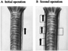
The study was approved by the Institutional Animal Care and Use Committee of the Clinical Research Institute of Seoul National University Hospital (approval number 07156, 06-2006-210-9). Nine male dogs weighing 17 to 21.5 kg were assigned to one of 3 groups according to the distance that was to be maintained between the site of Harmonic scalpel (HS) application and the recurrent laryngeal nerve (RLN): (i) group 1 (1 mm); (ii) group 2 (2 mm); and (iii) group 3 (3 mm). The animals were administered 3 to 5mg/kg of intramuscular zolazepam as a premedication followed by 25 mg/kg of intramuscular cefazolin (Chong Kun Dang pharmaceutical corp., Seoul, Korea). Direct laryngoscopy was performed to assess vocal cord function. The animals were then intubated and administered 1% to 3% isoflurane as a general anesthetic. A midline skin incision of approximately 10 cm was made in the neck, and the left lobe of the thyroid and the left RLN were identified. Left thyroid lobectomy was performed. The HS (Harmonic Ace 36P; Johnson & Johnson Medical, Cincinnati, OH, USA) was applied near the RLN at the assigned distance. Low power HS (70 µm vibration) was applied for 10 seconds (Figure 1). A nonabsorbable suture was applied to facilitate subsequent harvesting of the nerve. The wounds were closed in multiple layers. To limit variability, all procedures were performed by the same surgeon, who was experienced in thyroid surgery. An aseptic technique was used throughout all procedures. An intramuscular injection of 0.4 mL/kg meloxicam was administered for postoperative pain control.
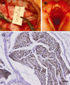
One week later, the animals were administered 3 to 5 mg/kg of intramuscular zolazepam as a premedication, and repeat laryngoscopy was performed. Two weeks after the initial operation, the animals were returned to the operating theater for re-exploration. The neck was reopened and the left RLN was harvested. This was placed in formalin for histological evaluation of subacute morphological changes. The right lobe of the thyroid and the RLN were then identified. Right thyroid lobectomy was performed, and HS was applied to the tissues adjacent to the right RLN at the assigned distance (Figure 1). All three fragments of right RLN were immediately harvested and placed in formalin for histological evaluation of acute morphological changes.
Histological evaluation
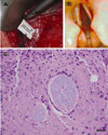
One left RLN could not be located during reexploration. Thus, 3 left RLNs from groups 1 and 2, and 2 left RLNs from group 3 (a total of 8 nerves), were examined for the presence of subacute morphological changes. Nine right RLNs from each group (a total of 27 nerves) were evaluated for the presence of acute morphological changes. The specimens were embedded in paraffin, sliced to a thickness of 3-4 mm, and stained with hematoxylin and eosin. Immunohistochemical (IHC) analyses were performed using 4-µm-thick sections and the DAKO Autostainer Plus Link (Dako, Glostrup, Denmark). With the exception of primary antibodies, all IHC components were taken from the EnVision FLEX High pH kit (K8000; Dako, Glostrup, Denmark). Antigen retrieval was achieved by treating the tissue sections with EnVision FLEX Target Retrieval Solution (DM812; Dako, Glostrup, Denmark; Tris/EDTA buffer, pH 9) for 45 min at 95℃. The tissue sections were then incubated with mouse antihuman neurofilament monoclonal antibodies (NF; Dako, Glostrup, Denmark) at a dilution of 1:2,000 for 20 min at room temperature. EnVision FLEX Peroxidase-Blocking Reagent (SM801; Dako, Glostrup, Denmark) was used as the blocking buffer. Antibody binding was visualized with a peroxidaseconjugated polymer backbone (EnVision FLEX; SM802; Dako, Glostrup, Denmark) and the use of diaminobenzidine as a chromogen. The sections were then counterstained with hematoxylin. Appropriate positive control-tissues were used, and the omission of primary antigen was used as a negative control condition. Luxol fast blue (LFB) staining was used to identify myelinated white matter [10]. All histological analyses were performed by a single neuropathologist who was blind to the study group of the specimens. The neurovascular bundles were evaluated for the presence of swelling, vacuolar change, and atrophy, and for myelin and axonal loss.
Results
Functional outcome
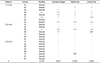
Normal vocal cord movement was observed in all animals prior to the initial operation. One week postsurgery, 2 cases of a fixed vocal cord and 1 case of decreased vocal cord motility were observed in group 1, 1 fixed vocal cord was observed in group 2, and no abnormalities in vocal cord mobility were observed in group 3 (P=0.020) (Table 1).
Histological outcome
A total of 8 left RLNs were examined for the presence of subacute damage. Subacute morphological changes such as swelling, vacuolar change, and myelin and axonal loss were observed in the nerves of animals with abnormal vocal cord function (Table 1, Figure 2 and 3). Nine right RLNs from each group (a total of 27 nerves) were evaluated for acute damage. Acute morphological changes such as swelling, vacuolar change, and myelin and axonal loss were observed in 5 of the 8 nerves from group 1, and in 1 of the 7 nerves from group 2, whereas no acute morphological changes were observed in any of the nerves from group 3 (Table 2).
Discussion
Although many clinical studies for the usefulness of the HS during thyroidectomy speculated upon the safety of HS, it did not propose a safe distance for the application of HS in the vicinity of the RLN during thyroid surgery. This study aimed to develop a large animal model for recurrent laryngeal nerve injury by HS.
The majority of nerve injury studies have used a rodent sciatic nerve model as this is inexpensive, convenient, and involves straightforward histological analyses. Although the HS causes an increase in the temperature of adjacent tissues, the spread of heat is less than that associated with bipolar or monopolar coagulation [11]. A study of pig arteries reported that the mean length of thermal spread was 1.6~2.4 mm [12]. A study of rat sciatic nerves found that the length of damage caused by ultrasonically activated scalpels was 0.9~1.1 mm [13]. In a further study of rat sciatic nerves, Owaki et al. concluded that use of the ultrasonic shears at a distance of 3 mm from the RLN for less than 10 seconds at level 3 was safe [14]. However, there is no large animal model to determine the clinically acceptable distance safety margin for the application of energy device in the vicinity of the recurrent laryngeal nerve. To our knowledge, the present study is the first to have examined the effect of HS application in the vicinity of the RLN during thyroidectomy in large animal. There is a study using the rabbit but it did not used laryngoscopy to evaluate the vocal cord function [15].
The canine model was selected since the vocal cords of dogs are more accessible than those of pigs, and are thus easier to evaluate the movement of the vocal cords by laryngoscope examination. Also, the anatomy of the thyroid gland is similar in dogs and humans, which facilitates the surgeons to mimic the real thyroid surgery as in human. In both species it is a dark red, elongated structure that is attached to fascia along the ventrolateral surfaces of the proximal trachea. The right thyroid lobe extends between the cricoid cartilage and the 5th tracheal ring, and the left thyroid lobe extends from the 3rd to the 8th tracheal ring. The gland is covered by the sternocephalicus and the sternohyoideus muscles ventrally and by the sternothyroideus muscle laterally. The common carotid artery, the internal jugular vein, and the vagosympathetic trunk are situated on the dorsolateral surface of the right thyroid lobe, and the esophagus lies over the dorsolateral surface of the left thyroid lobe. The caudal laryngeal nerves, termed the RLNs in humans, are dorsal to the thyroid lobes [16].
In this study we used 9 dogs for evaluation of both functional and histological outcomes. For histological outcomes both acute and subacute changes were evaluated using both sides of recurrent laryngeal nerves. As depicted in the results, there were functional abnormalities in the group 1 and 2, which were the groups of dogs with HS application in vicinity of less than 3 mm. In contrast, the group 3 with HS application with safety margin of 3 mm, there were no adverse outcomes. This is small sample size, however, conjunction with the study by Owaki et al. we could postulate a safety margin of 3 mm could be used [14].
Subacute morphologic changes were evaluated using left recurrent laryngeal nerves harvested 2 weeks after initial energy source application. Subacute changes were observed in the nerves of animals with abnormal vocal cord function. Acute morphological changes were evaluated using right recurrent laryngeal nerves harvested immediate after energy source application during second operation. Acute changes were observed in group 1 and 2, which were the groups of dogs with HS application in vicinity of less than 3 mm. In contrast, the group 3 with HS application with safety margin of 3 mm, there were no adverse acute morphologic changes. We could depict from these results that the histologic changes could be matched with the functional outcomes. The anatomical and functional correlation of this canine model might provide a rationale to evaluate the safety margin of an energy device during thyroid surgery.
This study has some limitations including; (1) have small sample size, (2) did not evaluated other energy sources, (3) have other ways to evaluate the function, such as electromyography, and (4) have short period observation after application of energy source. However, this study would warrant further study to determine the clinically acceptable distance safety margin for the application of an energy device in the vicinity of the recurrent laryngeal nerve.

 XML Download