

 PDF
PDF ePub
ePub Citation
 Print
Print


It has been reported that Houttuynia (H.) cordata extracts have diverse pharmacological effects including antiviral and antibacterial [1,2], antiallergic [3,4], antioxidant [5,6] and antimutagenic activities [5]. Thus, H. cordata Thunb has been used as an Oriental medicine for the therapy of inflammatory diseases such as ulcerative colitis [7].
Recent studies revealed that the constituents of H. cordata essential oil are methyl nonyl ketone, β-myrcene, β-pinene, α-pinene, α-terpineol and n-decanoic acid, and that the anti-inflammatory effects of this oil were also demonstrated [8]. Notably, the essential oil constituents and their concentrations varied with different extraction methods [2]. Recently, supercritical extraction technology has been adopted for the total extraction of constituents from natural plant sources.
In a previous study, we demonstrated the anti-inflammatory activity of H. cordata supercritical extract (HSE) in both macrophage cell line and a mouse model of carrageenan-induced air pouch inflammation [9], an in vivo model suitable for the analysis of diverse biochemical and pathological parameters [10,11]. In the present study, we evaluated the effectiveness of HSE in a rat model of air pouch inflammation, since the change in the inflammatory parameters was found to be different in mouse and rat models.
The aerial part of H. cordata was extracted for 2 hours under CO2 supercritical conditions (temperature 60℃, pressure 400 bar). After separating the CO2 solvent by reduced pressure, the extract (yield=1.5%) was collected [12]. The extract was dissolved in soybean oil, and orally administered at 4 mL/kg.
Six-week-old male Sprague-Dawley rats (body weight 200-220 g; n=8/group) (Orient-Bio, Seongnam, Korea) were subcutaneously injected with 20 mL of sterile air into the back side to form a pouch [10,11]. After 2 and 5 days, the pouch was re-injected with 10 mL of air. Twenty four hours after the final air injection, HSE (20, 65 or 200 mg/kg) was orally administered, followed 30 min later by injection with 2 mL of lambda carrageenan (1% in saline; Sigma-Aldrich, St. Louis, USA) into the pouch. For comparisons, additional rats were given intraperitoneal injections of either dexamethasone (2 mg/kg; Sigma) or indomethacin (2 mg/kg; Sigma). The animal experiments were approved by the Institutional Animal Care and Use Committee of the Laboratory Animal Research Center, Chungbuk National University, Korea.
The pouch was washed with 2 mL of cold saline after 6 hours, and the net volume of lavage fluid was recorded. Total numbers of inflammatory cells and albumin, a marker of vascular leakage, were determined using a coulter counter and a blood biochemistry analyzer, respectively. Tumor-necrosis factor-α (TNF-α) and interleukin-6 (IL-6) were analyzed using enzyme-linked immunosorbent assay (ELISA) kits (Komabiotech, Seoul, Korea). The concentrations of NO and PGE2 were determined by Griess reagent (Sigma) and enzyme immunoassay (EIA) using a Correlate-EIA kit (Assay Designs, Ann Arbor, Ann ArboUSA), respectively. The results were expressed as the mean±SD. Tests of significance were performed using Duncan's multiple-range test after one-way analysis of variance, with P<0.05 as a criterion of difference.
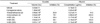
Injection of carrageenan into rat air pouches significantly increased the exudate volume in the pouches and the albumin contents in the exudate (Table 1). However, oral treatment with HSE suppressed the carrageenan-induced increases in both the exudate volume and albumin leakage in a dose-dependent manner, inhibiting by 28.4-43.2% and 71.4-128.6% at 20-200 mg/kg, respectively. For comparison, intraperitoneal administration of indomethacin (2 mg/kg) also reduced both the exudate volume (78.1%) and albumin content (200%). In contrast, dexamethasone (2mg/kg) specifically suppressed exudates volume (83.8%), but not albumin leakage (14.3%).
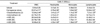
Total white blood cells (WBC) in the exudate, including neutrophils, monocytes and lymphocytes, were greatly increased by carrageenan (Table 2). Interestingly, infiltrating inflammatory cells were suppressed to by a high dose of HSE (200 mg/kg) and dexamethasone, but not by indomethacin.
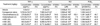
Carrageenan enormously enhanced major inflammatory cytokines TNF-α and IL-6 in the exudate (Table 3). Administration of HSE (65-200mg/kg) and dexamethasone, significantly lowered the carrageenan-induced increases in TNF-α, while indomethacin was ineffective. In comparison, the increased IL-6 level was significantly attenuated only by dexamethasone. Interestingly, HSE (65-200mg/kg) markedly blocked the carrageenan-induced increases in both NO and PGE2. Dexamethasone specifically inhibited NO production, while indomethacin reversed PGE2 concentration to the control level.
In comparison with the mouse air-pouch model [9], higher and lower responses to carrageenan exposure in exudate volume and albumin leakage, respectively, were observed in the rat model. That is, 1.80- and 3.88-fold increases in exudate volumes and 2.64- and 1.47-fold increases in albumin contents in mice and rats, respectively, were observed following carrageenan injection. An extract of H. cordata obtained under a supercritical condition markedly attenuated the secretion of both major inflammatory mediators, NO and PGE2. The numbers of neutrophils, monocytes and lymphocytes increased by carrageenan were reduced following treatment with HSE (200 mg/kg). This was an effect obtained by dexamethasone, but not by indomethacin (1-2 mg/kg) [11,13]. Although migration of monocytes was suppressed only by a high dose (200 mg/kg) of HSE, the corticosteroid-like effect of HSE was confirmed by its inhibitory action on TNF-α and NO, which are the main inflammatory mediators from macrophages. Therefore, the effect of HSE on macrophages may come from mainly the suppression of signaling pathways, as supported by the highly-sensitive inhibition by HSE of NO secretion from RAW 264.7 cells [9].
In addition to the TNF-α-NO pathway, HSE exerted inhibitory activity on the in vivo release of PGE2, which is produced from arachidonic acid via cyclooxygenase-2 (COX-2). It is assumed that HSE directly inhibits COX-2 or deactivates inflammatory cells expressing COX-2 [9]. Although there was a different sensitivity to NO and PGE2, the production of PGE2 was also markedly suppressed by HSE treatment in RAW 264.7 cells, mice and rats [9, the present study]. Such a difference between in vitro and in vivo or between mice and rats studies may be due to the different animals and stimulators, i.e., lipopolysaccharide (LPS) and carrageenan.
There are few reports demonstrating the anti-inflammatory effects of H. cordata. Intravenous injection of H. cordata essential oils (including 22 constituents) reduced inflammatory responses in a carrageenan-induced pleurisy model, and attenuated xylene-induced ear edema [8]. Notably, our previous result shows that supercritical extract of H. cordata (IC50<0.001%) is much superior to an aqueous extract (IC50≒0.1%) in the inhibition of NO production by RAW 264.7 cells [9,14].
Notably, supercritical extraction technology has been adopted for the total extraction of constituents from natural plant sources. We assessed the anti-inflammatory effects of HSE by analyzing mediators in the two major pathways of inflammation, TNF-α-NO and COX-2-PGE2, in comparison with the synthetic steroid dexamethasone and a non-steroidal anti-inflammatory drug (NSAID) indomethacin. We demonstrated dual actions of HSE in the inflammatory process, dexamethasone- and indomethacin-like effects. Therefore, a supercritical extract of H. cordata could be a good drug candidate for the relief of various types of inflammation responsive to corticosteroids or NSAIDs.
 XML Download