

 PDF
PDF ePub
ePub Citation
 Print
Print


Canine hemangiosarcoma is a common type of cancer that originates from blood vessels [1-3]. It can occur in any bodily blood vessels and can be categorized into two groups: cutaneous and visceral hemangiosarcoma. In cutaneous hemangiosarcoma, a neoplasm develops on the skin while in visceral hemangiosarcoma a neoplasm commonly occurs in the canine spleen, heart, or kidneys. Visceral hemangiosarcoma is the more aggressive type and has a higher chance of metastasis, with poor prognosis [4,5]. The cause of hemangiosarcoma has not yet been clearly defined. It is more prevalent in elder dogs, especially spayed females between 8 and 13 years, and in large breed dogs such as German Shepherds and Golden Retrievers [6,7].
Before surgery, serum chemistry results showed the dog had anemia with hematocrit 11.3% and hemoglobin 3.6 g/dL, inflammation with increased white blood cells 45.3×10⁹/L (reference range: 6.0-16.9×10⁹/L), and a high level of alkaline phosphatase, 419 U/L (reference range: 23-212 U/L). Radiography revealed a circular mass on the spleen posterior.
The mass was surgically removed and then fixed in 10% neutral buffered formalin for 24 h, before being processed and embedded in paraffin wax. After being sectioned at 4 µm thickness, the sections were stained with hematoxylin and eosin for microscopic analysis.
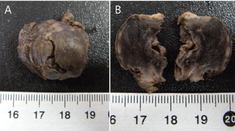
The mass was approximately 3×2.5×1.5 cm in size. Because it developed in spleen, where there are many red blood cells exposed to oxygen, it was mostly brown with white spots of necrosis appearing on the outer side of the spherical mass (Figure 1).
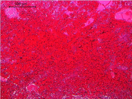
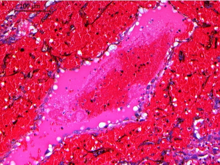
Tissue showed signs of hemorrhage, with a significant amount of blood outside the blood vessels throughout the sample. Both the rupture and poor blood vessel formation in the neoplastic cells could have been responsible for the extensive hemorrhage. The tumor was poorly circumscribed and the normal morphological features of the spleen were hard to find due to both the hemorrhage and the infiltration of neoplastic cells (Figure 2). The mass also showed inflammation, with clusters of neutrophils and a large necrotic area on the outer side that was devoid of cells (Figure 3). Large numbers of pleomorphic cells, both polygonal and round, with eosinophilic cytoplasm and vesicular nuclei were observed, some of which had occasionally formed channels filled with red blood cells, plasma, and a small number of white blood cells (Figure 4).
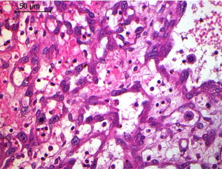
Mitotic figures indicating rapid growth were frequently found where the cell population was relatively high (Figure 5). Moderate anisocytosis and anisokaryosis were also found, which could have resulted from the uncontrolled and uneven development of neoplastic cells.
Based on these findings, the Jindo dog was diagnosed with canine splenic hemangiosarcoma. Hemangiosarcoma is a malignant neoplasm that arises from vascular endothelial tissue [1,2,5]. According to past study, the majority of canine hemangiosarcoma cases occur in the spleen [5,6]. In this research, more than 55% of dogs were diagnosed due to a rupture of the primary or metastatic tumor [6]. Of the 104 dogs considered, 100 were dead within four months of diagnosis and the remaining four barely survived a year on average [6]. Although canine hemangiosarcoma is considered fairly common, it is an aggressive sarcoma type that often leads to metastasis. It has a poor prognosis once discovered because a dog with hemangiosarcoma will show little or no clinical signs until a rupture of the mass leads to hemoabdomen [1,4]. However, close clinical attention and observation, combined with various diagnostic techniques such as ultrasonography and biopsy could help with early diagnosis and thus improve survival rates.

 XML Download