Abstract
Feline endometrial adenocarcinomas are uncommon malignant neoplasms that have been poorly characterized to date. In this study, we describe a uterine adenocarcinoma in a Persian cat with feline leukemia virus infection. At the time of presentation, the cat, a female Persian chinchilla, was 2 years old. The cat underwent surgical ovariohystectomy. A cross-section of the uterine wall revealed a thickened uterine horn. The cat tested positive for feline leukemia virus as detected by polymerase chain reaction. Histopathological examination revealed uterine adenocarcinoma that had metastasized to the omentum, resulting in thickening and the formation of inflammatory lesions. Based on the histopathological findings, this case was diagnosed as a uterine adenocarcinoma with abdominal metastasis. To the best of our knowledge, this is the first report of a uterine adenocarcinoma with feline leukemia virus infection.
Uterine adenocarcinomas have been reported in most domestic species but are considered rare except in rabbits and cows [1]. The prevalence of uterine adenocarcinoma in rabbits after 5 years of age has been reported to be 79% [2]. Because rabbits are induced ovulators that are often housed individually, laboratory rabbit and possibly many pet rabbits are under almost constant estrogen stimulation. Virgin rats have a similar high incidence of endometrial adenocarcinomas [3]. Cats, another species that exhibit induced ovulation, may also be subjected to long periods of unopposed estrogen stimulation, but the frequent practice of neutering queens, and the tendency to periodically breed or induce ovulation in sexually intact queens, may explain the low prevalence of endometrial adenocarcinoma in cats [4]. In women, postmenopausal hormone replacement therapy, estrogen in particular, is considered a risk factor for endometrial adenocarcinoma; both estrogen-dependent and higher-grade estrogen-independent adenocarcinomas are recognized [5].
Viruses cause cancer in amphibians, fish, fowl, rodents, cats, cattle, and subhuman primates, and if humans were to be exempt from such a general biological phenomenon, it would be a circumstance unparalleled in the history of parasitism. In fact, evidence is accumulating that some human cancers are virally induced. For example, Burkitt's lymphoma, nasopharyngeal carcinoma, cervical cancer, and Kaposi's sarcoma are thought to be caused by herpesviruses, which are able to remain latent in the host for many years [6]. Feline leukemia virus (FeLV) is an exogenous retrovirus, belonging to the genus Gammaretrovirus, which infects domestic cats and, sporadically, wild cats as well [7,8]. FeLV causes a wide range of proliferative diseases in cats, including lymphoid and myeloid leukemia [9].
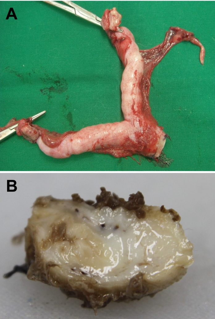
Feline endometrial adenocarcinomas are uncommon malignant neoplasms that have to date been poorly characterized. In this study, a case of uterine adenocarcinoma case is described in a Persian cat with FeLV infection. At the time of presentation, the cat was a 2-year-old female Persian chinchilla (Figure 1A). She presented with vaginal discharge and interrupted vomiting, and was given a health examination. Abdominal sonography and radiography demonstrated abnormal enlargement of the uterus in the abdominal cavity. The blood analysis revealed neutrophilia and lymphopenia. The concentrations of progesterone and estrogen in blood were evaluated and the results revealed normal ranges. The cat underwent surgical ovariohystectomy. During the laparotomy, thickening and inflammatory lesions of the omentum and spleen were observed (Figure 1B). The results of bacterial culture with omentum and spleen samples were negative for pathogenic bacteria. The ovariouterus and the omentum were removed surgically, submitted to gross examination, and trimmed. Cross-sections of the uterine wall revealed a thickening of uterine horn (Figure 2). Additionally, yellowish contents were observed in the lumen of the uterus. The trimmed tissues were fixed in 10% neutral buffered formalin and embedded in paraffin. Four-micrometer sections were made and stained with hematoxylin and eosin (H&E) for histopathological examination. The histopathological analysis revealed adenocarcinoma in the uterus and omentum (Figure 3), but no pathological changes were observed in the ovary. The adenocarcinoma had metastasized to the omentum, resulting in thickening and the formation of inflammatory lesions (Figure 4). Based on these histopathological findings, the case was diagnosed as uterine adenocarcinoma with abdominal metastasis. The cat made a complete recovery following an ovariohysterectomy and chemotherapy.
The blood samples were tested for FeLV, feline infectious peritonitis (FIP), feline immunodeficiency virus (FIV), feline panleukopenia virus (FPV), feline herpesvirus (FHV), feline calicivirus (FCV), heart worm, feline chlamydia, Toxoplasma, Babesia spp., Ehrlichia spp., Haemobartonella felis, Rickettsia spp., and Brucella spp. using a polymerase chain reaction (PCR) assay. The following primer sequences were used for PCR amplification: 5'-CTACCCCAAAATTTAGCCAGCTACT-3' and 5'-AAGACCCCCGAACTAGGTCTTC-3' for the amplification of the 468-bp segment of FeLV [10]; 5'-TAATGCCATACACGAA CCAGCT-3' and 5'-GTGCTAGATTTGTCTTCGGACACC-3' for the amplification of the 295-bp segment of FIP [11]; 5'- CCACAATATGTAGCACTTGACC-3' and 5'-GGGTACTTTCTG GCTTAAGGTG-3' for the amplification of the 583-bp segment of FIV [12]; 5'-CATTGGGCTTACCACCATTT-3' and 5'-GGTGC ACTATAACCAACCTCAGC-3' for the amplification of the 172- bp segment of FPV [13]; 5'-CGGGAAAATCCAGTACGAGT-3' and 5'-AGGAAGAGTTCGGCGGTATT-3' for the amplification of the 383-bp segment of FHV [14]; 5'-TTCGGCCTTTTGTG TTCC-3' and 5'-TTGAGAATTGAACACATCAATAGATC-3' for the amplification of the 673-bp segment of FCV [15]; 5'- AGTGCGAATTGCAGACGCATTGAG-3' and 5'-AGCGGGTA ATCACGACTGAGTTGA-3' for the amplification of the 542- bp segment of heartworm [16]; 5'-ATGAAAAAACTCTTGA AATCGG-3' and 5'-CAAGATTTTCTAGACTTCATTTTGTT-3' for the amplification of the 1069-bp segment of feline chlamydia [15]; 5'-AGTTTAGGAAGCAATCTGAAAGCACAT C-3' and 5'-GATTTGCATTCAAGAAGCGTGATAGTAT-3' for the amplification of the 529-bp segment of Toxoplasma [16]; 5'-ATAACCGTGCTAATTGTAGG-3' and 5'-TGTTATTTCTTGT CACTACC-3' for the amplification of the 327-bp segment of Babesia spp. [17]; 5'-GGAATTCAGAGTTGGATCATGGCTC AG-3' and 5'-CGGGATCCCGAGTTTGCCGGGACTTCTTCT- 3' for the amplification of the 492-bp segment of Ehrlichia spp. [18]; 5-AGCAGCAGTAGGGAATCTTCCAC-3' and '5- TGCACCACCTGTCACCTCGATAAC-3 for the amplification of the 674-bp segment of Haemobartonella felis [19]; 5'- AGAGTTTGATCCTGGCTCAGAAC-3' and 5'-CCTACGGCTA CCTTGTTACGACTT-3' for the amplification of the 356-bp segment of Rickettsia spp. [20]; 5'-TCGAGCGCCCGCA AGGGG-3' and 5'-AACCATAGTGTCTCCACTAA-3' for the amplification of the 905-bp segment of Brucella spp. [21].
Genomic DNA was isolated from blood using an AccuPrep Genomic DNA extraction kit (Bioneer, Daejeon, Korea) according to the manufacturer's instructions. The DNA was eluted in Tris-EDTA buffer (pH8.0), and an aliquot was used for PCR amplification. All DNA samples were stored at -20℃ until the PCR assays were performed. The template DNA (50 ng) and 20 pmol of each primer were added to a PCR mixture tube (AccuPower PCR PreMix; Bioneer) containing 2.5 U of Taq DNA polymerase, 250 µM each deoxynucleoside triphosphate, 10 mM Tris-HCl (pH8.3), 40 mM KCl, 1.5 mM MgCl2, and the gel loading dye. The final volume was adjusted to 20 µL with distilled water. The reaction mixture was subjected to denaturation at 95℃ for 2 min, followed by 40 cycles of 95℃ for 30 sec (denaturation), 54℃ for 30 sec (annealing), and 72℃ for 30 sec (extension), and a final extension step of 72℃ for 5 min, and samples were kept at 4℃ until analysis. Reactions were conducted using My Genie 32 Thermal Block PCR (Bioneer). Eight microliters of each sample were mixed with 2 µL of loading buffer, and electrophoretically separated on 1.2% agarose gels stained with 0.5 µg/mL ethidium bromide. DNA bands were observed under ultraviolet light.
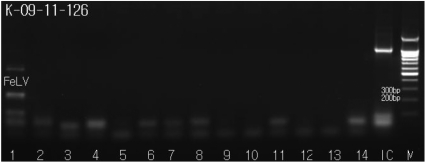
The PCR-amplified samples were evaluated for the presence of PCR amplicons corresponding to FeLV, FIP, FIV, FPV, FHV, FCV, Heart worm, Feline Chlamydia, Toxoplasma, Babesia spp., Ehrlichia spp., Haemobartonella felis, Rickettsia spp. and Brucella spp. We were able to visualize bands that were 529 base pairs in length, corresponding to the FeLV amplicon (Figure 5); other feline pathogens tested negative in the PCR assay (Figure 5).
Significant changes in the structure of the endometrium are associated with degeneration of luminal epithelium, cystic endometrial hyperplasia, pyometra, and adenocarcinoma [22]. Endometrial tumors of the uterus are rare in domestic animals [23]. Adenocarcinoma of the uterus is very rare in animals, in marked contrast to the prevalence of this tumor in human females [23]. The basic mechanisms associated with these changes are poorly understood. Prolonged exposure to megestrol acetate has been associated with endometrial hyperplasia and pyometra in domestic cats, and progestin contraceptives may have similar effects on zoo felids [24,25]. Megestrol acetate is a derivative of progesterone and used commonly both in contraception and hormone replacement therapy [26]. Based on available clinical and epidemiological data, it has been long suspected that tumors of the canine female genital tract develop under the influence of ovarian hormones [22]. In this case, however, the animal had not been given a progesterone derivative such as megestrol acetate.
FeLV is an exogenous retrovirus causing several diseases in domestic cats, including tumors of most hematopoietic cells, aplastic anemia, myeloproliferative disorders, and immunosuppression [27]. The FeLVs that primarily cause T-cell tumors in cats are transmitted in a horizontal manner [28], particularly through saliva [29]. The T-cell tropism of these agents is observed even when the virus infects human cells [30]. Although many free-roaming cats become persistently infected with FeLV, only a small fraction develops leukemia or lymphoma. A large number develop lethal opportunistic infections, particularly coronavirus-induced peritonitis [31]. FeLV causes lymphopenia, depressed cell-mediated immune responses, thymic atrophy, and depressed humoral responses to T-cell dependent antigens despite the presence of hypergammaglobulinemia [31].
In this study, the cat had uterine adenocarcinoma without pathognomic ovarian lesions or significant hormonal imbalances. The cat also suffered from FeLV infection and lymphopenia. We believe that the uterine adenocarcinoma in this case was probably related to immunogenic depression by FeLV infection, although we cannot rule out the contributions of other factors, such as old age, to the induction of this tumor. To the best of our knowledge, this is the first report of feline uterine adenocarcinoma with feline leukemia virus infection.
Acknowledgments
This was supported by Basic Science Research Program through the National Research Foundation of Korea (NRF) funded by the Ministry of Education, Science and Technology (2010-0021940).
References
1. Kennedy PC, Cullen JM, Edwards JF, Goldschmidt MH, Larsen S, Munson L, Nielsen S. Cullen JM, editor. Tumors of the uterus. Histological Classification of Tumors of the Genital System of Domestic Animals. 1998. 2nd ed. Washington DC: Armed Forces Institute of Pathology;p. 32–33.
2. Elsinghorst TA, Timmermans HJ, Hendriks HG. Comparative pathology of endometrial carcinoma. Vet Q. 1984; 6(4):200–208. PMID: 6388139.

3. Deerberg F, Rehm S, Pitterman W. Uncommon frequency of adenocarcinomas of the uterus in virgin Han:Wistar rats. Vet Pathol. 1981; 18(6):707–713. PMID: 7292893.

4. Stabenfeldt GH, Pedersen NC. Pedersen NC, editor. Reproduction and reproductive disorders. Feline Husbandry: Diseases and Management in the Multiple-Cat Environment. 1991. Goleta: American Veterinary Publications Inc;p. 129–162.
5. Anderson MC, Robboy SJ, Russell P, Morse A. Robboy SJ, editor. Endometrial carcinoma. Pathology of the Female Reproductive Tract. 2002. London: Churchill Livingstone;p. 331–359.
6. De-The G. Hiatt HH, editor. Viruses as causes of some human tumors? Results and prospectives of the epidemiologic approach. Origins of Human Cancer. 1977. Cold Spring Harbor: Cold Spring Harbor Laboratory;p. 1113–1131.
7. Arjona A, Barquero N, Domenech A, Tejerizo G, Collado VM, Toural DM, Gomez-Lucia E. Evaluation of a novel nested PCR for the routine diagnosis of feline leukemia virus (FeLV) and feline immunodeficiency virus (FIV). J Feline Med Surg. 2007; 9(1):14–22. PMID: 16863698.
8. Daniels MJ, Golder MC, Jarret O, MacDonald DW. Feline viruses in wildcats from Scotland. J Wildl Dis. 1999; 35(1):121–124. PMID: 10073361.

9. Khan KNM, Kociba GJ, Wellman ML. Macrophage tropism of feline leukemia virus (FeLV) of subgroup-C and increased production of tumor necrosis factor-α by FeLV-infected macrophages. Blood. 1993; 81(10):2585–2590. PMID: 8387834.
10. Cattori V, Tandon R, Pepin A, Lutz H, Hofmann-Lehmann R. Rapid detection of feline leukemia virus provirus integration into feline genomic DNA. Mol Cell Probes. 2006; 20(3-4):172–181. PMID: 16488115.

11. Simons FA, Vennemac H, Rofinab JE, Pold JM, Horzineke MC, Rottiera PJM, Egberinka HF. A mRNA PCR for the diagnosis of feline infectious peritonitis. J Virol Methods. 2005; 124(1-2):111–116. PMID: 15664058.

12. English RV, Kershaw O, Kempf VA, Gruber AD. In vivo lymphocyte tropism of feline immunodeficiency virus. J Virol. 1993; 67(9):5175–5186. PMID: 7688819.
13. Buchmann AU, Kershaw O, Kempf VAJ, Gruber AD. Does a feline leukemia virus infection pave the way for Bartonella henselae infection in cats? J Clin Microbiol. 2010; 48(9):3295–3300. PMID: 20610682.
14. Reubel GH, Ramos RA, Hickman MA, Rimstad E, Hoffmann DE, Pedersen NC. Detection of active and latent feline herpesvirus 1 infections using the polymerase chain reaction. Arch Virol. 1993; 132(3-4):409–420. PMID: 8397503.

15. Sykes JE, Allen JL, Studdert VP, Browning GF. Detection of feline calicivirus, feline herpesvirus 1 and Chlamydia psittaci mucosal swabs by multiplex RT-PCR/PCR. Vet Microbiol. 2001; 81(2):95–108. PMID: 11376956.

16. Xie DH, Zhu XQ, Cui HL, Qiu CJ, Fan WH, Liao SQ, Zhai ML, Lin RQ, Weng YB. Development of a PCR assay for diagnosing swine toxoplasmosis. Chin J Vet Sci Technol. 2005; 35(4):289–293.
17. Simking P, Wongnakphet S, Stich RW, Jittapalapong S. Detection of Babesia vogeli in stray cats of metropolitan Bangkok, Thailand. Vet Parasitol. 2010; 173(1-2):70–75. PMID: 20638794.
18. Aktas M, Altay K, Dumanli N, Kalkan A. Molecular detection and identification of Ehrlichia and Anaplasma species in ixodid ticks. Parasitol Res. 2009; 104(5):1243–1248. PMID: 19247690.

19. Cooper SK, Berent LM, Messick JB. Competitive, quantitative PCR analysis of Haemobartonella felis in the blood of experimentally infected cats. J Microbiol Methods. 1999; 34(3):235–244.
20. Tijsse-Klasen E, Fonville M, Reimerink JH, Spitzen-van der Sluijs A, Sprong H. Role of sand lizards in the ecology of Lyme and other tick-borne diseases in the Netherlands. Parasit Vectors. 2010; 3:42. PMID: 20470386.

21. Romero C, Gamazo C, Pardo M, López-Goñi I. Specific detection of Brucella DNA by PCR. J Clin Microbiol. 1995; 33(3):615–617. PMID: 7538508.

22. Klein MK. Withrow SJ, editor. Tumors of the female reproductive system. Small Animal Clinical Oncology. 2001. Philadelphia: WB Saunders;p. 445–454.

23. MacLachlan NJ, Kennedy PC. Meuten DJ. Tumors of the genital system. Tumors in Domestic Animals. 2002. Ames: Iowa State Press;p. 547–573.
24. Kim KS, Kim O. Cystic endometrial hyperplasia and endometritis in a dog following prolonged treatment of medroxyprogesterone acetate. J Vet Sci. 2005; 6(1):81–82. PMID: 15785129.

25. Munson L, Gardner IA, Mason RJ, Chassy LM, Seal US. Endometrial hyperplasia and mineralization in zoo felids treated with melengestrol acetate contraceptives. Vet Pathol. 2002; 39(4):419–427. PMID: 12126144.

26. Pazol K, Wilson ME, Wallen K. Medroxyprogesterone acetate antagonizes the effects of estrogen treatment on social and sexual behavior in female macaques. J Clin Endocrinol Metab. 2004; 89(6):2998–3006. PMID: 15181090.

27. Cotter SM. Feline leukemia virus: Pathophysiology, prevention, and treatment. Cancer Invest. 1992; 10(2):173–181. PMID: 1312884.

28. Hardy WD, Old LJ, Hess PW, Essex M, Cotter SM. Horizontal transmission of feline leukemia virus. Nature. 1973; 244(5414):266–269. PMID: 4147636.
29. Francis DP, Essex M, Hardy WD. Excretion of feline leukemia virus by naturally infected pet cats. Nature. 1977; 269(5625):252–254. PMID: 201852.
30. Azocar J, Essex M. Susceptibility of human cell lines to feline leukemia virus and sarcoma virus. J Nat Cancer Inst. 1979; 63(5):1179–1184. PMID: 228103.
31. Essex M, McLane MF, Kanki P, Allan J, Kitchen L, Lee TH. Retroviruses associated with leukemia and ablative syndromes in animals and in human beings. Cancer Res. 1985; 45(9 Suppl):4534s–4538s. PMID: 2990682.
Figure 1
Photographs of gross findings. (A) Client's cat. (B) Thickness and inflammatory lesions in omentum and spleen.
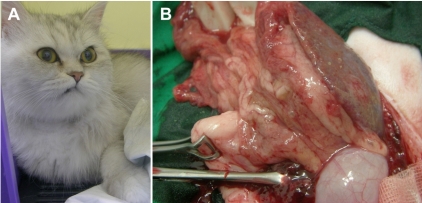
Figure 3
Histopathological findings of the uterus. (A) Neoplastic cell growth in the lumen of uterus. H&E stain, ×100. (B) Adenocarcinoma lesion in the uterus. H&E stain, ×400.
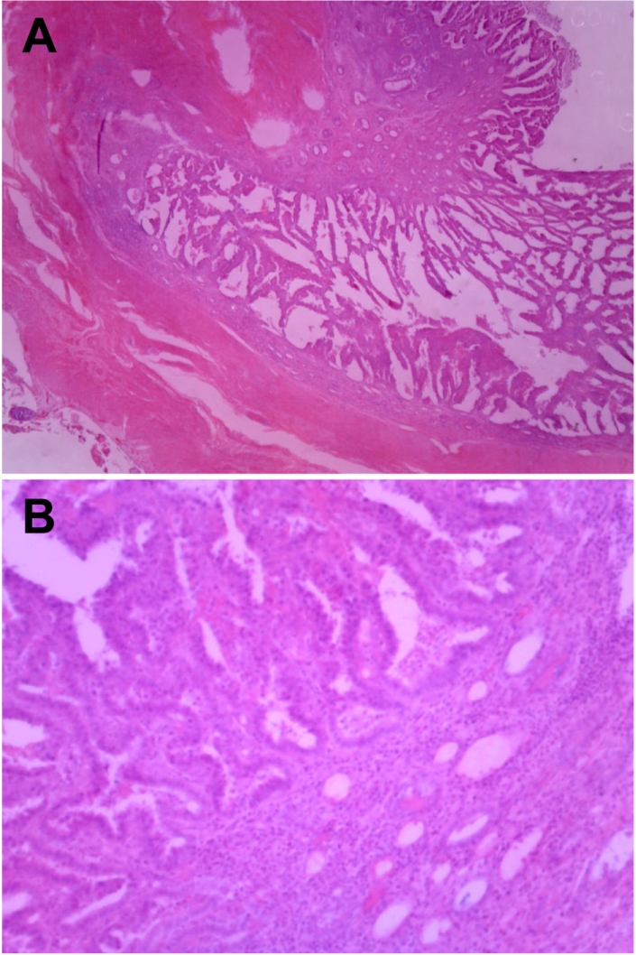
Figure 4
Histopathological findings of the omentum. (A) Neoplastic cell infiltration in the omentum. H&E stain, ×400. (B) metastatic adenocarcinoma tumor cells in the omentum. H&E stain, ×400.
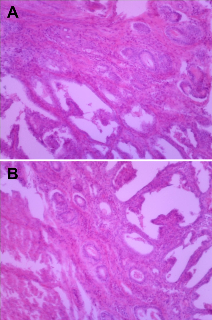
Figure 5
Amplicons from sample DNAs by species-specific PCRs for suspected pathogens were identified by electrophoresis on a 1.2% agarose gel. Lane 1, feline leukemia virus (+); 2, feline infectious peritonitis (-); 3, feline immunodeficiency virus (-); 4, feline panleukopenia virus (-); 5, feline herpesvirus (-); 6, feline calicivirus (-); 7, heart worm (-); 8, feline chlamydia (-); 9, Toxoplasma (-); 10, Babesia spp. (-); 11, Ehrlichia spp. (-); 12, Haemobartonella felis (-); 13, Rickettsia spp. (-); 14, Brucella spp. (-); IC, internal control; M, size marker.


 PDF
PDF ePub
ePub Citation
 Print
Print


 XML Download